Marcin Słojewski
1, 4, Krzysztof Safranow
2, Katarzyna Jakubowska
2,
Maria Olszewska
2, Andrzej Pawlik
3, Zygmunt Juzyszyn
3, Marek Droździk
3,
Dariusz Chlubek
2, Peter Hammerer
4, Andrzej Sikorski
1Can We Predict Urinary Stone Composition Based
on an Analysis of Microelement Concentration
in the Hair and Urine?*
Czy można przewidzieć skład kamieni moczowych na podstawie analizy
zawartości mikroelementów we włosach i w moczu?
1 Department of Urology and Urological Oncology, Pomeranian Medical University, Szczecin, Poland 2 Department of Biochemistry and Medical Chemistry, Pomeranian Medical University, Szczecin, Poland 3 Department of Pharmacokinetics and Therapeutic Drug Monitoring, Pomeranian Medical University, Szczecin, Poland 4 Urologische Klinik, Braunschweig, Germany
Abstract
Background. In recent years the role of trace elements in lithogenesis has received steadily increasing attention.
Objectives. This study was aimed to attempt to find the correlations between the chemical content of the stones and the concentration of chosen elements in the urine and hair of stone formers.
Material and Methods. The proposal for the study was approved by the local ethics committee. Specimens were taken from 219 consecutive stone-formers. The content of the stone was evaluated using atomic absorption spec-trometry, spectrophotometry, and colorimetric methods. An analysis of 29 elements in hair and 21 elements in urine was performed using inductively coupled plasma-atomic emission spectrometry.
Results. Only a few correlations between the composition of stones and the distribution of elements in urine and in hair were found. All were considered incidental.
Conclusions. The data obtained did not allow for the creation of a proper and practical algorithm to predict stone chemical composition based on hair and urine analysis (Adv Clin Exp Med 2012, 21, 4, 469–475).
Key words: urinary stone, urine, hair, analysis, trace elements.
Streszczenie
Wprowadzenie. W ostatnich latach obserwuje się rosnące naukowe zainteresowanie rolą mikroelementów w pro-cesie tworzenia kamieni moczowych.
Cel pracy. Niniejsze badanie zostało podjęte w celu oceny potencjalnej korelacji między składem chemicznym kamieni a zawartością wybranych pierwiastków w moczu i we włosach chorych na kamicę dróg moczowych.
Materiał i metody. Projekt badania został zaakceptowany przez lokalny komitet etyczny. Próbki pobierano od kolej-nych 219 chorych na kamicę, kwalifikowakolej-nych do leczenia zabiegowego. Skład kamieni badano z zastosowaniem metod spektrometrii absorpcji atomowej, spektrofotometrii i kolorymetrii. Analiza zawartości 29 pierwiastków we włosach i 21 w moczu została wykonana z użyciem spektrometru emisji atomowej z plazmą wzbudzoną indukcyjnie.
Wyniki. Wśród badanych zależności stwierdzono jedynie pojedyncze, uznane za incydentalne, korelacje między składem kamieni a zawartością pierwiastków w badanych materiałach biologicznych.
Wnioski. Uzyskane dane nie pozwalają na stworzenie odpowiedniego, praktycznego algorytmu, na podstawie którego można byłoby przewidzieć lub odtworzyć skład chemiczny kamieni moczowych (Adv Clin Exp Med 2012, 21, 4, 469–475).
Słowa kluczowe: kamienie moczowe, mocz, włosy, analiza, pierwiastki śladowe.
Adv Clin Exp Med 2012, 21, 4, 469–475 ISSN 1899–5276
OrIGINAl PAPErS
© Copyright by Wroclaw Medical University
M. Słojewski et al.
470
Urinary stones affect 2–20% of the population in industrialized countries, their prevalence is ris-ing and they are considered a serious socio-med-ical problem [1–3]. Although important advances have been made in understanding the multifacto-rial pathophysiology of stone formation, there is not yet a complex and satisfactory explanation for this process. The process of crystallization of su-persaturated urine components and the formation of solid concretions can be modified by the activity of promoters and inhibitors, and by some morpho-anatomic, dietary, and environmental factors. The role of trace elements in lithogenesis is still unclear and under debate. Some trace elements have an effect on the crystallization of stone components; they act at the surface of the crystals, as their con-centration in urine is too small to affect the lattice ions in solution [4, 5]. It has also been documented that some trace elements influence the external morphology of growing crystals, and may increase or decrease the speed of the crystallization process [6, 7]. Some heavy metal ions, e.g. zinc and stron-tium, can substitute for calcium in crystals due to the similarity between the ions’ charge and size. It has been demonstrated that metals such as mag-nesium, zinc, aluminum, iron and copper may act as inhibitors of calcium oxalate growth at very low concentrations [4, 8, 9]. It is probable that some other elements also promote or, conversely, inhibit crystal nucleation of the organic or mineral species involved in lithogenesis, but the reports are often conflicting. Some studies focus on determining the total levels of elements in studied materials; others focus on the elements’ interactions with promoters or inhibitors such as citrate, glycosaminoglycans, pyrophosphate, and Tamm-Horsfall protein [6, 7, 10–14]. Many researchers have found correlations between essential elements in hair and metabolic disorders, diseases, nutritional status, and envi-ronmental exposures [15, 16]. Using hair to assess trace elements is controversial, but it has advan-tages over using blood or urine. Blood and urine represent current or recent body status, whereas hair is a reliable and convenient biological indica-tor reflecting long-term exposure [17, 18].
The query included in the title of this paper may seem to be weird. In a fact, if the answer is positive, we would have a practical tool for pa-tients who, for instance, expelled their stones so it was lost for biochemical analysis.
Material and Methods
The proposal for the study was approved by the local ethics committee (No. BN-001/14/07, 28 Feb. 2007). All patients signed informed consent
Urinary Stone Composition Based on Analysis of Hair and Urine 471
(Cd), boron (B), barium (Ba), mercury (Hg), sulfur (S), germanium (Ge), silicone (Si), iodine (I), arse-nium (As), and tin (Sn) were determined. Multi-elemental analysis was performed with inductively coupled plasma-atomic emission spectrometry (ICP-OES Optima 5300DV, Perkin Elmer, Massa-chusetts, USA). Using the same method, the level of 21 elements (Ca, Na, K, P, Zn, Mg, Fe, Cu, Sr, Ni, Mn, Cr, Mo, Co, li, V, Al, Pb, Cd, B, and Hg) was determined in urine. The limits of detection were < 1 μg/l for Zn, Sr, Ni, Mn, Se, Mo, Co, li, V, Cd, B, Ba, Hg, Ge, As and Sn, 1–10 μg/l for K, Mg, Cu, Cr, Pb, Si and I and 10–50 μg/l for Ca, Na, P, Fe, Al, and S. The calibration curves were linear up to 100 μg/l for Cu, Sr, Ni, Se, Cr, Mo, Co, li, V, Pb, Cd, Hg, Ge, Si, I, As and Sn, up to 1000 μg/l for Mn, B and Ba, and up to 10000 μg/l for Ca, Na, K, P, Zn, Mg, Fe, Al, and S. relative standard de-viation ranged from 0.32% to 2.19% for individual elements. Accuracy of the method was controlled with two certified materials (ICP Multi-Element Standard Solutions IV and VI) which were ana-lyzed before each series of measurements and re-peated every 30 samples. The strength of correla-tion was described with the value of a Spearman’s rank correlation coefficient (rs). Statistical signifi-cance was determined at p < 0.05. Statistica 7.1 software (Statsoft Inc., Oklahoma, USA) was used for the calculations. The methods were previously described in detail [19, 20].
Results
The mean (± standard deviation) age of the first episode of kidney stone disease in the studied group was 45.3 ± 16.1 years. The mean weight of the stone sent for analysis was 16.5 g (minimum 3, maximum 381.5). The mean volume of the 24-h urine sample was 1669 ± 336 ml. The most com-mon components of all 219 analyzed stones were calcium oxalate (58.6 ± 37.62%) and calcium phos-phate (25.33 ± 27.99%). Other components seen were uric acid (11.76 ± 29.95%) and magnesium phosphate (4.28 ± 12.19%). None of the stones contained cysteine, xanthine or 2,8-dihydroxyade-nine.
The tables show the analysis of the correla-tions between the composition of the stones and the distribution of the elements in urine (Table 1) and between the composition of the stones and the distribution of the elements in hair (Table 2). The elements in the tables are listed according to their position in the periodic table. Only a few cor-relations at a moderate level of significance were found. They all were considered incidental. The authors also analyzed the correlations between the
levels of microelements in three biological materi-als, e.g. stones, urine and hair, and we made the following observations. The more that elements such as V, Pb, Hg, Ge, Si, I, and Sn were seen in the stones, the higher their level in hair; the more that Zn, Ni, li, V, Al, and Pb were seen in urine, the higher their concentration in hair; and the higher the level of Mo, Co, V, Al, and Pb in urine, the more of these elements were found in the stones (data not shown). There were also 109 positive two-element correlations between two materials, i.e. when one element in one material correlated significantly positively (rs > 0, p < 0.05) with an-other element in anan-other material. Among them, the most common (more than 15 correlations) were observed for six elements listed according to the number of correlations: V, Al, li, Pb, Co, and Mo. The content of the following pairs of elements in stones–hair was correlated with high statistical significance (p < 0.001): Fe–V, Fe–Ni, Cu–Ni, Cr– V, Mo–Co, Co–Ni, Co–V, li–Mn, li–V, V–Ni, V–Al, Cd–Ni, Cd–V, B–V, Hg–V, and Hg–Pb. Positive correlations for all samples (stones, urine, and hair) were established for only three elements: V–V, Pb–Pb, Pb–V, and Al–Pb (data not shown).
Discussion
Although the initial papers on trace elements in urinary stones were published in 1963 [21], so far little data has been presented that would link the presence or absence of certain trace elements in the urine of stone formers to the pathogenesis of this disease [4, 14, 22]. The results of many studies suggest that some elements including Ni, Mg, Al, Pb, Cd, and Cu together with other factors may affect the process of stone precipitation [12, 23]. On the other hand, most theories explaining the pathogenesis of stones formation do not include any role of trace elements. Special attention is gen-erally paid to environmental, alimentary, occupa-tional and socioeconomic factors [13, 24–26].
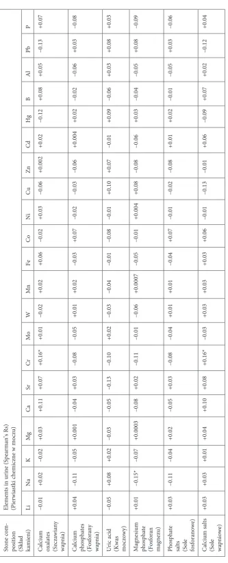
study-Table 1. Correlation analysis between mineral composition of stones and distribution of elements in urine Tabela 1. Analiza korelacji składu kamieni i składu pierwiastków chemicznych w moczu Stone com
-position (Skład kamieni)
Elements in urine (Spearman’s rs) (Pierwiastki chemiczne w moczu) li Na K Mg Ca Sr Cr Mo W Mn Fe Co Ni Cu Zn Cd Hg B Al Pb P
Calcium oxalates (Szczawiany wapnia)
–0.01 +0.02 –0.02 +0.03 +0.11 +0.07 +0.16* +0.01 –0.02 +0.02 +0.06 –0.02 +0.03 –0.06 +0.002 +0.02 –0.12 +0.08 +0.05 –0.13 +0.07
Calcium phosphates (Fosforany wapnia)
+0.04 –0.11 –0.05 +0.001 –0.04 +0.03 –0.08 –0.05 +0.01 +0.02 –0.03 +0.07 –0.02 –0.03 –0.06 +0.004 +0.02 –0.02 –0.06 +0.03 –0.08 Uric acid
(Kwas moczowy)
–0.05 +0.08 +0.02 –0.03 –0.05 –0.13 –0.10 +0.02 –0.03 –0.04 –0.01 –0.08 –0.01 +0.10 +0.07 –0.01 +0.09 –0.06 +0.03 +0.08 +0.03
Magnesium phosphate (Fosforan magnezu)
+0.01 –0.15* –0.07 +0.0003 –0.08 +0.02 –0.11 –0.01 –0.06 +0.0007 –0.05 –0.01 +0.004 +0.08 –0.08 –0.06 +0.03 –0.04 –0.05 +0.08 –0.09
Phosphate salts (Sole fosforanowe)
+0.03 –0.11 –0.04 +0.02 –0.05 +0.03 –0.08 –0.04 +0.01 +0.01 –0.04 +0.07 –0.01 –0.02 –0.08 +0.01 +0.02 –0.01 –0.05 +0.03 –0.06 Calcium salts
(Sole wapniowe)
+0.03 +0.03 +0.01 +0.04 +0.10 +0.08 +0.16* –0.03 +0.03 +0.03 +0.03 +0.06 –0.01 –0.13 –0.01 +0.06 –0.09 +0.07 +0.02 –0.12 +0.04 Spearman’s rank correlation coefficient (r s) is calculated for each pair of stone component correlation and element; * p < 0.05;
# p
<
0.01;
+ p
<
Tabela 2. Correlation analysis between mineral composition of stones and distribution of elements in hair Tabela 2. Analiza korelacji składu kamieni i składu pierwiastków chemicznych we włosach
Stone composition (Skład kamieni)
Elements in hair (Spearman’s rs) (Pierwiastki chemiczne we włosach) li Na K Mg Ca Sr Ba Cr Mo W Mn Fe Co Ni Cu Zn Cd Hg B Al Si Ge Sn Pb P As S Se I
Calcium oxalates (Szczawiany wapnia)
–0.01 +0.05 +0.06 +0.01 –0.06 –0.12 –0.07 –0.10 –0.05 –0.03 –0.04 +0.08 +0.08 –0.03 –0.10 –0.06 +0.03 –0.11 –0.06 –0.004 –0.06 –0.06 +0.08 +0.05 +0.02 –0.05 –0.001 +0.001 –0.09
Calcium phosphates (Fosforany wapnia)
–0.02 –0.08 –0.13 +0.04 +0.07 +0.08 +0.09 +0.06 +0.11 +0.003 +0.06 +0.003 –0.04 –0.01 +0.09 –0.02 –0.02 –0.05 +0.02 +0.05 +0.02 +0.05 –0.04 –0.04 –0.03 +0.03 +0.03 –0.12 +0.07 Uric acid
(Kwas moczowy)
+0.04 +0.03 +0.09 –0.03 –0.01 +0.06 –0.05 +0.004 –0.06 +0.01 –0.04 –0.07 –0.03 +0.03 –0.01 +0.06 –0.02 +0.17* +0.11 –0.06 –0.01 +0.04 –0.001 +0.03 +0.002 +0.02 –0.08 +0.13* –0.01
Magnesium phosphate (Fosforan magnezu)
–0.03 +0.04 –0.03 +0.004 +0.04 +0.08 +0.10 +0.12 +0.11 +0.04 –0.01 –0.13 –0.06 –0.10 +0.07 +0.002 –0.04 +0.02 +0.10 +0.04 +0.09 –0.02 –0.09 –0.02 –0.03 –0.03 +0.01 –0.07 –0.04
Phosphate salts (Sole
fos -foranowe) –0.03 –0.07 –0.12 +0.03 +0.06 +0.07 +0.09 +0.06 +0.11 +0.004 +0.05 –0.01 –0.06 –0.02 +0.08 –0.02 –0.03 –0.04 +0.03 +0.06 +0.03 +0.03 –0.06 –0.04 –0.03 +0.02 +0.02 –0.12 +0.05
Calcium salts
(Sole wapniowe) –0.03 –0.05 –0.04 +0.03 –0.02 –0.10 –0.04 –0.10 –0.03 –0.06 +0.02 +0.13 +0.05 +0.005 –0.05 –0.07 +0.04 –0.17* –0.14* +0.04 –0.08 –0.02 +0.04 +0.01 +0.03 –0.01 +0.01 –0.08 +0.01 Spearman’s rank correlation coefficient (r s) is calculated for each pair of stone component correlation and element; * p < 0.05;
# p
<
0.01;
+ p
<
M. Słojewski et al.
474
ing the distribution of Fe, Cu, Cd, Zn, and Mg in 47 stones and hair, found significant differences among the element levels in stones, patient hair, and control hair [14].
One of the most common trace element in the human body is Zn but the data concerning its role in lithogenesis is divergent. Some studies [22, 28] showed that low Zn levels in the urine of stone formers suggest its potential inhibiting action. Other data, however, shows increased excretion of Zn and Cu in stone formers or even no differ-ence between stone formers and healthy popula-tions [8, 29, 30]. There are studies showing that low concentrations of some elements like Mg, Mn and Zn in stones make them resistant to extra-corporeal shock wave lithotripsy [31, 32]. The au-thors observed a negative correlation between Mg level and the content of calcium oxalate and uric acid (rs values were −0.29, p < 0.001; and −0.34, p < 0.001, respectively). This finding supports the conclusions of some authors that Mg may act as an inhibitor of calcium oxalate stones [22]. This study also confirmed in a much larger group (5 vs. 219) the observation of Scott et al. about relatively low concentrations of Na in calcium oxalate stones and a high concentration of K and M in phosphate stones [12]. The analysis in Table 1 shows, that on-ly one statisticalon-ly significant positive correlation was observed between the concentration of Cr in urine and the content of calcium oxalate and cal-cium salts in stones (p < 0.05). This finding could prove the possible influence of Cr on lithogenesis; however the authors did not find supportive data in the literature, and it was therefore considered incidental and meaningless. The authors also ob-served a negative association between the concen-tration of Na in urine and magnesium phosphates in stones (p < 0.05), but the concentration of this element is highly diet-dependent because of its presence in food, table salt, and preservatives.
Increased scientific interest in trace elements has led to a search for reliable methods of quanti-fying and monitoring their levels in human body
tissues. Hair can be collected easily, non-invasively, and painlessly, and it accurately reflects body loads and human exposure to different contaminants, es-pecially metals [17, 18, 33]. Hair structure contains a significant amount of cysteine, and some heavy metals (such as Pb and Hg) have a high affinity to sulfhydryl groups in this amino acid [17]. The main purpose of this study was to find the correla-tion between the levels of microelements in urine, stones, and hair and the mineral composition of the stones. Apart from the cognitive aspect, this correlation could also have practical application in cases in which the stone has been expelled and lost and cannot be examined. In cases such as this, an analysis of hair and/or urine could reveal, with some probability, the content of the stone and help in the prophylaxis of recurrence. Unfortunately, the obtained data (Table 2) does not allow for the creation of a proper and practical algorithm. With respect to hair, only Hg, B, and Se showed some weak correlation with uric acid and calcium salts. The analysis of the level of elements in urine and hair shows that when diet-dependent elements (Na, K) and those that are included in stones in high amounts (Ca, P) are excluded from investiga-tion, the rest of the elements, mostly metals, can be detected in hair in several to several hundred times higher concentrations than they can be detected in urine (data not shown). However, significant variability in the profile of elements in hair is seen depending on the subject’s age, sex, dietary hab-its, geochemical environment, hair color, smoking habits, and lifestyle [15, 16, 18]. Some authors even found significant inter-laboratory variations and concluded that standardization of testing methods is needed [34].
The authors concluded that although this in-vestigation showed a correlation between the lev-els of some elements in the urine and hair and the chemical composition of stones, the authors were not able to create any practical algorithm to pre-dict stone composition based on hair and urine analysis.
References
Trinchieri A:
[1] Epidemiological trends in urolithiasis: impact on our health care systems. Urol res 2006, 34, 151–156.
Bartoletti R, Cai T, Mondaini N, Melone F, Travaglini F, Carini M, Rizzo M:
[2] Epidemiology and risk factors in urolithiasis. Urol Int 2007, 79, Suppl. 1, 3–7.
Pak CYC:
[3] Kidney stone. lancet 1998, 251, 1797–1801.
Bazin D, Chevallier P, Matzen G, Jungers P, Daudon M:
[4] Heavy elements in urinary stones. Urol res 2007, 35, 179–184.
Komleh K, Hada P, Pendse AK, Singh PP:
[5] Zinc, copper and manganese in serum, urine and stones. Int Urol Nephrol 1990, 22, 113–118.
Meyer JL, Angino EE:
[6] The role of trace metals in calcium urolithiasis. Invest Urol 1977, 14, 347–350.
Munoz JA, Valiente M:
[7] Effects of trace metals on the inhibition of calcium oxalate crystallization. Urol res 2005, 33, 267–272.
Joost J, Tessadri R:
Urinary Stone Composition Based on Analysis of Hair and Urine 475
Welshman SG, McGeown MG:
[9] A quantitative investigation of the effects on the growth of calcium oxalate crystals on potential inhibitors. Br J Urol 1972, 44, 677–680.
Sutor DJ:
[10] Growth studies of calcium oxalates in the presence of various ions and compounds. Br J Urol 1969, 41, 171–178.
Meyer JL, Thomas WC Jr:
[11] Trace metal-citric acid complexes as inhibitors of calcification and crystal growth. II. Effects of Fe(III), Cr(III) and Al(III) complexes on calcium oxalate crystal growth. J Urol 1982, 128, 1376–1378.
Scott R, East BW, Janczyszyn J, Boddy K, Yates AJ:
[12] Concentration of some minor and trace elements in urinary tract stones: a preliminary study. Urol res 1980, 8, 167–169.
Li MK, Blacklock NJ, Garside J:
[13] Effects of magnesium on calcium oxalate crystallization. J Urol 1985, 133, 123–125.
Durak I, Kilic Z, Sahin A, Akpoyraz M:
[14] Analysis of calcium, iron, copper and zinc contents of nucleus and crust parts of urinary calculi. Urol res 1992, 20, 23–26.
Chojnacka K, Górecka H, Chojnacki A, Górecki H:
[15] Inter-element interactions in human hair. Envir Toxicol Pharm 2005, 20, 368–374.
Steindel SJ, Howanitz PJ:
[16] The uncertainty of hair analysis for trace metals. JAMA 2001, 285, 83–85.
Rodrigues JL, Batista BL, Nunes JA, Passos CJS, Barbosa F Jr:
[17] Evaluation of the use of human hair for biomoni-toring the deficiency of essential and exposure to toxic elements. Sci Total Environ 2008, 405, 370–376.
Bass DA, Hockok D, Quig D, Urek K:
[18] Trace elements in hair: factors determining accuracy, precision and reali-ability. Alt Med rev 2001, 5, 472–481.
Słojewski M, Czerny B, Safranow K, Jakubowska K, Olszewska M, Pawlik A, Gołąb A, Droździk M, Chlubek [19]
D, Sikorski A: Microelements in stones, urine, and hair of stone formers: a new key to the puzzle of lithogenesis? Biol Trace Elem res 2010, 137, 301–316.
Słojewski M, Czerny B, Safranow K, Droździk M, Pawlik A, Jakubowska K, Olszewska M, Gołąb A, Byra E, [20]
Chlubek D, Sikorski A: Does smoking have any effect on urinary stone composition and the distribution of trace elements in urine and stones? Urol res 2009, 373, 17–22.
Nagy Z, Szabó E, Kelenhegyi M:
[21] Spektralanalytische untersuchung von nierensteien auf metallische spurenele-mente. Z Urol 1963, 56, 185–190.
Atakan IH, Kaplan M, Seren G, Aktoz T, Gül H, Inci O:
[22] Serum, urinary and stone zinc, iron, magnesium and copper levels in idiopathic calcium oxalate stone patients. Int Urol Nephrol 2007, 39, 351–356.
Perk H, Serel TA, Kosar A, Deniz N, Sayin A:
[23] Analysis of the trace element contents of inner nucleus and outer crust parts of urinary calculi. Urol Int 2002, 68, 286–290.
Pak CY, Poindexter JR, Adams-Huet B, Pearle MS:
[24] Predictive value of kidney stone composition in the detection of metabolic abnormalities. Am J Med 2003, 115, 26–32.
Stoller ML, Meng MV, Abrahams HM, Kane JP:
[25] The primary stone event: a new hypothesis involving a vascular etiology. J Urol 2004, 171, 1920–1924.
Coe FL, Bushinsky DA:
[26] Pathophysiology of hypercalciuria (review). Am J Physiol 1984, 247, 1–13.
Miekeley N, Dias Carneiro MTW, Porto da Silveira CL:
[27] How reliable are human hair reference intervals for trace elements. Sci Total Environ 1998, 218, 9–17.
Bird ED, Thomas WC:
[28] Effect of various metals on mineralization in vitro. Proc. Soc Exp Biol Med 1963, 112, 640–643.
Trinchieri A, Castelnuovo C, Lizzano R, Zanetti G:
[29] Calcium stone disease: a multiform reality. Urol res 2005, 33, 194–198.
Rangnekar GV, Gaur MS:
[30] Serum and urinary zinc levels in urolithiasis. Br J Urol 1993, 71, 527–529.
Küpeli S, Arikan N, Durak I, Sarica K, Akpoyraz M, Karalezli G:
[31] Efficiency of extracorporeal shockwave litho-tripsy on calcium-oxalate stones: role of copper, iron, magnesium and zinc concentrations on disintegration of the stones. Eur Urol 1993, 23, 409–412.
Turgut M, Unal I, Berber A, Demir TA, Mutlu F, Aydar Y:
[32] The concentration of Zn, Mg and Mn in calcium oxalate monohydrate stones appears to interfere with their fragility. Urol res 2008, 36, 31–38.
Morley N, Ford RPK:
[33] Hair-element analysis – still on the fringe. Child Care Health Dev 2002, 28, Suppl 1, 31–34.
Seidel S, Kreutzer R, Smith D, McNeel S, Gilliss D:
[34] Assessment of commercial laboratories performing hair mineral analysis. JAMA 2001, 285, 67–72.
Address for correspondence:
Marcin Słojewski
Department of Urology and Urological Oncology Pomeranian Medical University
Powstańców Wlkp. 72 70-111 Szczecin Poland
Tel. +48 91 466 11 01 E-mail: [email protected]
Conflict of interest: None declared