Introduction
Arthritis is an umbrella term used for various different arthritic conditions that are chronic, painful and debilitating. Arthritic diseases include six main types: rheumatoid arthritis, juvenile arthritis, psoriatic arthritis, gouty arthritis, ankylosing spondylitis and osteoarthritis.1 These musculoskeletal disorders contribute
significantly to the global disability burden.2,3 Osteoarthritis, also
known as degenerative joint disease or degenerative arthritis is the most common chronic condition of the joints. The joints most commonly affected include the knees, hips, lower back and neck, small joints of the fingers, base of the thumb and big toe.4 Osteoarthritis occurs mainly later in life,5 and according to
epidemiological studies, affects an estimated 15% of the world population.2 It is a debilitating progressive disease, affecting
60% of men and 70% of women over the age of 65.6 The level of
disability depends on several factors and the effect that OA has on a given individual does vary.5
Pathophysiology of osteoarthritis
Osteoarthritis is a complex, chronic inflammatory disease of synovial joints,7 involving the articular cartilage, a unique tissue
between the ends of bones in the joints.5,8 Below the cartilage is a
layer of bone, the subchondral bone that acts as a shock absorber in weight-bearing joints (e.g. hips and knees). Synovial fluid fills the joint space and contains abundant hyaluronic acid (HA) that acts as a lubricant. In OA patients, hyaluronan is both smaller in size (referring to its molecular weight) and lower in concentration,
providing less efficient lubrication. The subchondral bone plate thickens in patients suffering from OA9,10 causing the joint space
to narrow.11,12
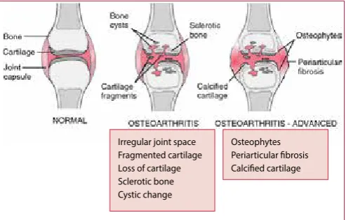
Recent research eluded that OA involves the entire joint and not only the articular cartilage, as initially thought – refer to Figure 1. A combination of cellular changes and biomechanical stresses cause several secondary changes. The latter include subchondral bone remodelling, formation of osteophytes, development of bone marrow lesions, changes in the synovium, joint capsule, ligaments and periarticular muscles, and meniscal tears and extrusion.2,13-15
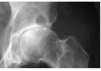
Figure 2 depicts a radiograph of the hip, revealing severe superior migration of the femoral head, subchondral sclerosis, prominent osteophytes, and a large Egger cyst in the superior acetabulum. The femoral head also reflects loss of articular cartilage.16
Osteoarthritis
Engler D1, BPharm, BSc Hons (Pharmacology), MSc Med (Clinical Pharmacy), Lecturer Grootboom W2, BPharm, MSc Med (Clinical Pharmacy), Lecturer
Mogale K1, BPharm, Academic Intern (MPharm) 1School of Pharmacy, Sefako Makgatho Health Sciences University
2Dr George Mukhari Academic Hospital
Correspondence to: Deirdré Engler, e-mail: [email protected]
Keywords: osteoarthritis, cartilage, subchondral bone, joint disease, chondrocytes
Abstract
Osteoarthritis (OA) is a chronic disease involving the entire joint, although the main tissue implicated in osteoarthritis is the cartilage. The most common joints affected include the knees, hips, lower back and neck, small joints of the fingers, base of the thumb and big toe. Progressive degeneration, chronic pain, stiffness, joint instability and joint space narrowing are clinical features associated with the condition. Osteoarthritis develops and progresses due to risk factors such as advancing age, female gender, and excessive mechanical stress, affecting mainly weight bearing joints of the knee, hip and spine. Thorough history taking, physical examination for signs and symptoms as well as appropriate imaging examinations and laboratory markers have to be considered during diagnosis and monitoring. The disease pathology is complex and current therapy does not prevent initiation or progression of osteoarthritis. Appropriate management combines non-pharmacological and pharmacological strategies aimed at alleviating symptoms and improving a patients’ quality of life.
© Medpharm S Afr Pharm J 2018;85(5):43-50
Irregular joint space Fragmented cartilage Loss of cartilage Sclerotic bone Cystic change
Osteophytes Periarticular fibrosis Calcified cartilage
Figure 2. OA of the hip16
Cartilage is the main tissue afflicted by OA.17 Chondrocytes are the
only cells found in healthy cartilage and are responsible for the production and maintenance of the cartilage over the lifetime of an individual.18 During the progression of OA, chondrocytes
actively remodel the extracellular matrix (ECM) of cartilage under inflammatory conditions – the latter due to wear and tear or trauma. In the presence of inflammation, the disease progresses further due to changes in the biomechanical environment of chondrocytes, brought about by alteration of the ECM. The changes in ECM composition and structure, further prevent mesenchymal stem cells (that migrate from bone marrow) to partake in the repair process by inhibiting their chondrogenic differentiation. The continuous interplay between changes in ECM and changes in cellular function under inflammation, drives the pathology of this chronic degenerative joint disease.18 Figure 3 depicts this interplay.
Aetiology and risk factors of osteoarthritis
Idiopathic OA is the most common form of arthritis6 as the
root cause of the disease is unknown.7 The development and
progression of OA is multifactorial, precipitating from interplay between various risk factors:4,15,19, 20-24
• Advancing age – incidence of OA does increase with age, although not a definite consequence of aging.
• Female gender – women after menopause are more susceptible to knee arthritis because of an increasing level of osteocalcin and bone resorption.25,26
Osteocalcin is a noncollagenous protein hormone found in bone and dentin, also known as bone gamma-carboxyglutamic acid-containing protein (BGLAP)
• High bone density • Reduced muscle strength • Malalignment of joints • Excessive mechanical stress • Obesity
• Sports and other trauma to the joints
The normal joint is well adapted to withstand physiological loads, but abnormal loading can increase the risk of OA. Trauma, heavy manual labour, and obesity all carry an increased risk of OA:
- Workers in certain occupations, such as coal miners, dockyard workers, and farmers have an increased risk of hip and knee OA
- Obesity is a well-established risk factor in the development of knee OA
• Genetic predisposition – a study of monozygotic twins aged 48 to 70 years, having identical genes, showed 65% influence of genetic factors in developing osteoarthritis.27 Between 39%
and 65% of osteoarthritis in the general population can be attributed to genetic factors.25
• Meniscus deficient knees – the prevalence of OA seems to be high among former athletes from team and individual sports when compared to the general population and other occupational sectors.28
• Childhood hip disorders e.g. acetabular dysplasia, congenital hip dislocation, epiphysiolysis (abnormal separation of an epiphysis from the bone shaft), and Legg-Calve’-Perthes (a disorder initiated by a disruption of blood flow to the head of the femur ) have been implicated in the development of early onset OA of the hip.
• Coxa valga (a deformity of the hip) with acetabular dysplasia. • Degeneration of the acetabular rim triggered by
femoroacetabular impingement (the ball shaped femoral head rubs abnormally or does not permit a normal range of motion in the acetabular socket).
Clinical presentation of osteoarthritis
The main clinical symptoms of osteoarthritis are chronic pain, stiffness, joint instability and joint space narrowing (seen radiographically).22 Exertion exacerbates the pain associated with
osteoarthritis, and may recur following rest. Joint stiffness may occur during morning hours, lasting up to 30 minutes. Locking or instability of joints may also be present.23 Table I depicts the
different signs and symptoms depending on the joints affected.
Figure 3. Changes in ECM of cartilage during inflammation17
Diagnosis
Osteoarthritis is one of the most common, costly and disabling forms of joint disease, and far more common than rheumatoid arthritis. The latter is an autoimmune disease that does not only affect the small joints, but also presents with extra-articular manifestations
e.g. anaemia and cardiovascular diseases. Figure 8 depicts the differences between these two diseases.
Diagnosis of OA has been widely based on clinical features, imaging examinations and laboratory findings.15,20, 22-24 The
American Geriatrics Society has provided clinical guidelines that have guided diagnostic practice in osteoarthritis.30,31 The clinical
approach takes into consideration thorough history taking and physical examination for signs and symptoms. Imaging examinations used diagnostically, to help establish prognosis and monitor effects of therapy, include radiography, computed tomography, positron tomography, ultrasonography and magnetic resonance imaging.32
Laboratory indication of chronic inflammatory changes, with production of pro-inflammatory cytokines, is apparent during early development of osteoarthritis. Increases in circulating levels of C-reactive protein (CRP), interleukin (IL)-6 and IL-4, and tumour necrosis factor alpha (TNF-α) have been linked to OA and used as possible biomarkers. Interleukin-6 and IL-4 identify radiologically
Table I. Signs and symptoms of osteoarthritis23,29 Joints affected Signs and symptoms
Hand Heberden nodes (Fig. 4) at distal interphalangeal joints Bouchard’s nodes (Fig. 4) at proximal interphalangeal joints
Osteophytes at first metacarpal joint give characteristic square appearance to hands
Tenderness over carpometacarpal joint of the thumb Hip Pain in groin during weight lifting exercises
Stiffness, pronounced following activity Pain in gluteal region
Limited joint movement, especially internal rotation Knee Pain associated with climbing stairs
Transient joint effusion Lateral instability
Genu varum (outward bowing of the knees, bandi-leg) Popliteal cyst (Baker’s cyst – Fig. 5)
Crepitus associated with range of motion
Foot Usually involves the first metatarsal-phalangeal joint Pain on ambulation
Hallux rigidus (Fig. 6) – a limitation to normal movements of flexion and extension
Hallux valgus (bunion – Fig. 7) – a deformity of the first metatarsophalangeal joint
Spine Typical lumbar involvement at L3 and L4 Paraesthesia
Loss of sensation at lower extremities
Motor weakness from compression of nerve root Loss of reflexes
Pseudoclaudication from spinal stenosis
Shoulder Limited range of motion, especially external rotation Crepitus associated with range of motion
Figure 5. A Baker’s cyst Figure 6. Hallux rigidus
Figure 7. Hallux valgus
severe osteoarthritis with low-grade inflammation.24,33
Chemotactic cytokine ligands and chemokine (C–C motif) receptor 2-expressing cells appear to be increased during joint injury and osteoarthritis.22,33 Other inflammatory cytokines, such
as IL-1b and TNF-α, may be used to indicate degenerative events in cartilage. Elevated serum concentrations of cartilage oligomeric matrix protein (COMP) is another biomarker proposed to have potential use diagnostically.24
Management of osteoarthritis
Due to poor understanding of the disease pathology, no current treatment can prevent either initiation or progression of OA.17 Osteoarthritis cannot be cured,4 and appropriate
management includes a combination of non-pharmacological and pharmacological measures with the ultimate goal to alleviate pain and improve functional status.16
Non-pharmacological management
Daily use of the joints actually preserves rather than “wears out” articular cartilage and inadequate use is the commonest cause of cartilage degeneration, as noted more than 50 years ago! An OA-patient should agree to manage themselves by ensuring positive behavioural changes e.g. exercise, weight loss, and use of suitable footwear (including those with shock-absorbing properties).19 Physical activity, e.g. walking, can reduce pain and
help to maintain (or attain) a healthy weight. Excess weight adds additional stress to weight-bearing joints and by losing weight, it can reduce OA pain and limit further joint damage. Strengthening exercises build muscle around the OA-affected joints – this can ease the burden on the affected joints. Exercise also improves joint flexibility and reduces joint stiffness. Gentle stretching of joints can further improve flexibility, decrease stiffness and lessen pain.4 Other non-pharmacological interventions include patient
education, occupational therapy, and heat and cold therapies.4,16
Pharmacological management
Analgesics e.g. paracetamol are indicated for mild to moderate OA pain without apparent inflammation. Should the pain relief be insufficient or the clinical presentation of OA is inflammatory, a nonsteroidal anti-inflammatory medicine (NSAID) can be added to the regimen. A topical NSAID preparation can be particularly useful in patients who are at increased risk for adverse events with systemic NSAIDs. Patients with an elevated risk for gastro-intestinal toxicity can be managed with a selective cyclooxygenase (COX)-2 inhibitor e.g. celecoxib. Should a patient’s pain be highly resistant, tramadol is another option. Judicious use of narcotics e.g. oxycodone and paracetamol with codeine, is reserved for patients presenting with severe OA.16 Other pharmacological
therapy to reduce pain and inflammation includes intra-articular injections of corticosteroid or sodium hyaluronate (also referred to as viscosupplementation e.g. hyaluronan). In patients with OA of the knee, these injections can reduce pain as soon as one week post-injection and last, on average, four to six weeks.16
Intra-articular corticosteroid injections should be considered as
an adjunct to core treatments for the relief of moderate to severe pain in people with OA.19
Patients with OA who experience joint symptoms that have a substantial impact on their quality of life and are refractory to non-surgical interventions are considered for referral for surgery.19
Conclusion
Effective management of OA is multimodal and involves the participation of both the patient and a multidisciplinary healthcare team. Patient education plays a pivotal role in the success of therapy. Pharmacotherapy must be supported by relevant strengthening physical activity and weight reduction measures to conserve joint integrity. Occupational therapy, and surgical interventions as a last resort, form an important part of the treatment regimen to ensure a better quality of life for the patient.
References
1. Usenbo A, Kramer V, Young T, Musekiwa A. 2015. Prevalence of arthritis in Africa: A sys-tematic review and meta-analysis. PLoS ONE. 10(8): e0133858. doi:10.1371/journal. pone.0133858. Available at https://www.ncbi.nlm.nih.gov/pmc/articles/PMC4524637/ pdf/pone.0133858.pdf [Accessed 12 August 2018]
2. Man GS, Mologhianu G, Davila C. 2014. Osteoarthritis pathogenesis – a complex process that involves the entire joint. Journal of Medicine and Life. 7(1): 37-41. Available at https:// www.ncbi.nlm.nih.gov/pmc/articles/PMC3956093/pdf/JMedLife-07-37.pdf [Accessed 12 August 2018]
3. Brennan-Olsen SL, Cook S, Leech MT, et al. 2017. Prevalence of arthritis according to age, sex and socioeconomic status in six low and middle-income countries: analysis of data from the WHO study on global AGEing and adult health (SAGE) Wave 1. BMC Musculo-skeletal Disorders. 18:271 doi 10.1186/s12891-017-1624-z. Available at https://www.ncbi. nlm.nih.gov/pmc/articles/PMC5479046/pdf/12891_2017_Article_1624.pdf [Accessed 12 August 2018]
4. Arthritis Foundation®. Osteoarthritis. Available at https://www.arthritis.org/about-arthri-tis/types/osteoarthritis/ [Accessed 12 August 2018]
5. Litwic AE, Edwards M, Dennison E, Cooper C. 2013. Epidemiology and burden of osteoar-thritis. Br Med Bull. 105: 185-199. doi: 10.1093/bmb/ids038 Available at https://www.ncbi. nlm.nih.gov/pmc/articles/PMC3690438/pdf/emss-53077.pdf [Accessed 12 August 2018] 6. Sarzi-Puttini P, Cimmino MA, Scarpa R, et al. 2005. Osteoarthritis: an overview of the disease
and its treatment strategies. Semin Arthritis Rheum 35:1–10. Available at: https://www. ncbi.nlm.nih.gov/pubmed/16084227 [Accessed 18 August 2019]
7. Ashkavand Z, Malekinejad H, Vishwanath BS. 2013. The pathophysiology of osteoarthritis. J of Pharmacy Research. 7(1): 132-138. Available at https://www.researchgate.net/publica-tion/257434938_The_pathophysiology_of_osteoarthritis [Accessed 19 August 2018] 8. Tiku ML, Sabaawy HE. 2015. Cartilage regeneration for treatment of
osteoarthri-tis: a paradigm for nonsurgical intervention. Therapeutic advances in musculoskel-etal disease. Available at https://www.ncbi.nlm.nih.gov/pmc/articles/PMC4426098/ pdf/10.1177_1759720X15576866.pdf [Accessed 12 August 2018]
9. Bailey AJ, Mansell JP, Sims TJ, Banse X. 2004. Biochemical and mechanical properties of subchondral bone in osteoarthritis. Biorheology 41(3-4):349–358 Available at https:// www.ncbi.nlm.nih.gov/pubmed/15299267 [Accessed 20 August 2018]
10. Burr DB. 2005. Increased biological activity of subchondral-mineralized tissues underlies the progressive deterioration of articular cartilage in osteoarthritis. J Rheumatol 32:1156– 1158.
11. Bonnet CS, Walsh DA. 2005. Osteoarthritis, angiogenesis and inflammation. Rheumatol-ogy (Oxford). 44(1):7–16 Available on https://www.ncbi.nlm.nih.gov/pubmed/15292527 {Accessed 18 August 2018]
12. Pelletier JP, Martel-Pelletier J, Abramson SB. 2001. Osteoarthritis, an inflammatory dis-ease: potential implication for the selection of new therapeutic targets. Arthritis Rheum 44:1237–1247.
13. Mobasheri A, Batt M. 2016. An update on the pathophysiology of osteoarthritis. Annals of Physical and Rehabilitation Medicine. 59(2016): 333-339. Available at https://ac.els-cdn. com/S1877065716300847/1-s2.0-S1877065716300847-main.pdf?_tid=0d2f69dd-2456-4157-a7d2-157c1e3ad4f7&acdnat=1534094496_be8c1337080590730c524ac538be6f18 [Accessed 12 August 2018]
15. De Rezende MU, de Campos GC, Pailo AF. 2013. Current concepts in osteoarthritis: Acta Ortopedica Brasileira, 21(2):120-122. Available at: https://www.ncbi.nlm.nih.gov/pmc/arti-cles/PMC3861968/ [Accessed 26 July 2018]
16. Lozada CJ, Culpepper Pace SS. 2018. Osteoarthritis. Medscape. Available at: https://emedi-cine.medscape.com/article/330487-overview [Accessed 16 August 2018]
17. Maldonado M, Nam J. 2013. The role of changes in extracellular matrix of cartilage in the presence of inflammation on the pathology of osteoarthritis. BioMed Research Int. Article ID 284873. Available at https://www.ncbi.nlm.nih.gov/pubmed/24069595 [Accessed 19 August 2018]
18. Del Carlo M, Loeser RF. 2008. Cell death in osteoarthritis. Curr Rheumatol Rep. 10(1): 37-42. Available at https://www.ncbi.nlm.nih.gov/pubmed/18457610 [Accessed 18 August 2018] 19. National Institute for Health and Clinical Excellence (NICE). 2017. Management of osteo-arthritis. Available at https://pathways.nice.org.uk/pathways/osteoarthritis/management-of-osteoarthritis.pdf [Accessed 3 August 2018]
20. Anderson AS, Loeser RF. 2010. Why is osteoarthritis an age-related disease?: Best practice and research. Clinical Rheumatology, 24(1):15. Available at: https://www.ncbi.nlm.nih.gov/ pmc/articles/PMC2818253/ [Accessed 26 July 2018]
21. Lane NE, Shidara K, Wise BL. 2017. Osteoarthritis year in review 2016: clinical: Osteoarthritis Research Society International, 25(2):209-215. Available at: https://www.ncbi.nlm.nih.gov/ pubmed/28100423 [Accessed 01 August 2018]
22. Chen D, Shen J, Zhao W, et al. 2017. Osteoarthritis: toward a comprehensive understanding of pathological mechanism: Bone Research, 5:16044. Available at: https://www.ncbi.nlm. nih.gov/pmc/articles/PMC5240031/#!po=0.308642 [Accessed 26 July 2018]
23. Sinusas K. 2012. Osteoarthritis: Diagnosis and Treatment: American Academy of Family Physicians, 85(1):49-56. Available at: https://www.aafp.org/afp/2012/0101/p49.html [Ac-cessed 26 July 2018]
24. Iolascon G, Gimigliano F, Moretti A, et al. 2017. Early osteoarthritis: How to define, diag-nose, and manage. A systematic review: European Geriatric Medicine, 8(5): 383-396. Avail-able at: http://www.em-consulte.com/en/article/1183117 [Accessed 02 August 2018] 25. Hunter D, De Lange M, Snieder H, et al. 2001. Genetic contribution to bone metabolism,
calcium excretion, and vitamin D and parathyroid hormone regulation. J Bone Miner Res.
16(2):371-8. Available at: https://www.ncbi.nlm.nih.gov/pubmed/11204437 [Accessed 18 August 2018]
26. Reginato AM, Olsen BR, 2002. The role of structural genes in the pathogenesis of osteoar-thritic disorders. Arthritis Res. 4:337–345 Available at: https://www.ncbi.nlm.nih.gov/pmc/ articles/PMC153840/pdf/ar595.pdf [Accessed 18 August 2018]
27. Spector TD, MacGregor AJ. 2004. Risk factors for osteoarthritis: genetics. Osteoarthritis Car-tilage. 12 (Suppl A): 39-44. Available at: https://www.ncbi.nlm.nih.gov/pubmed/14698640 [Accessed 18 August 2018]
28. Gouttebarge V, Inklaar H, Backx F, Kerkhoffs G. 2014. Prevalence of osteoarthritis in former elite athletes: overview of the recent literature. Rheumatol Int. 35(3): 405-418. doi: 10.1007/ s00296-014-3093-0. Available at https://www.researchgate.net/publication/264053494_ Prevalence_of_osteoarthritis_in_former_elite_athletes_a_systematic_overview_of_the_ recent_literature [Accessed 18 August 2018]
29. DiPiro JT, Talbert RL, Yee GC, et al. 2017. Osteoarthritis. Pharmacotherapy: A Pathophysi-ologic Approach. 10th ed. New York: McGraw-Hill Education.
30. Salehi-Abari I. 2016. 2016 ACR revised criteria for early diagnosis of knee osteoarthritis: Au-toimmune Diseases an Therapeutic Approaches Open Access, 3(1): 118-122. Available at: https://www.google.co.za/url?sa=t&source=web&rct=j&url=http://aperito.org/uploads/ pdf/ADTOA-3-118.pdf&ved=2ahUKEwjV9dbo5NPcAKHUpGAEAQFjABegQICBAB&usg=AO vVaw1X4jVgB-4jZFQAyCD2XVc2 [Accessed 01 August 2018]
31. Johns Hopkins Medicine. 2017. Osteoarthritis: ACR Diagnostic Guidelines: Johns Hopkins Arthritis Center. Available at: https://www.Hopkinsarthritis.org/physician-corner/educa-tion/arthritis-education-diagnostic-guidelines/ [Accessed 01 August 2018]
32. Hafezi-Nejad N, Demehri S, Guermazi A, Carrino JA. 2018. Osteoarthritis year in review 2017: updates on imaging advancements: Osteoarthritis Research Society International, 26(): 341-349. Available at: https://0-www.sciencedirect.com.innopac.smu.ac.za/science/ article/pii/S1063458418300220 [Accessed 01 August 2018]