R E V I E W
Open Access
The impact of transposable element
activity on therapeutically relevant
human stem cells
Gerald G. Schumann
1*, Nina V. Fuchs
2, Pablo Tristán-Ramos
3,4, Attila Sebe
1, Zoltán Ivics
1and Sara R. Heras
3,4Abstract
Human stem cells harbor significant potential for basic and clinical translational research as well as regenerative medicine. Currently ~ 3000 adult and ~ 30 pluripotent stem cell-based, interventional clinical trials are ongoing worldwide, and numbers are increasing continuously. Although stem cells are promising cell sources to treat a wide range of human diseases, there are also concerns regarding potential risks associated with their clinical use, including genomic instability and tumorigenesis concerns. Thus, a deeper understanding of the factors and molecular mechanisms contributing to stem cell genome stability are a prerequisite to harnessing their therapeutic potential for degenerative diseases. Chemical and physical factors are known to influence the stability of stem cell genomes, together with random mutations and Copy Number Variants (CNVs) that accumulated in cultured human stem cells. Here we review the activity of endogenous transposable elements (TEs) in human multipotent and pluripotent stem cells, and the consequences of their mobility for genomic integrity and host gene expression. We describe transcriptional and post-transcriptional mechanisms antagonizing the spread of TEs in the human genome, and highlight those that are more prevalent in multipotent and pluripotent stem cells. Notably, TEs do not only represent a source of mutations/CNVs in genomes, but are also often harnessed as tools to engineer the stem cell genome; thus, we also describe and discuss the most widely applied transposon-based tools and highlight the most relevant areas of their biomedical applications in stem cells. Taken together, this review will contribute to the assessment of the risk that endogenous TE activity and the application of genetically engineered TEs constitute for the biosafety of stem cells to be used for substitutive and regenerative cell therapies.
Keywords:Adult stem cells, Genomic destabilization, LINE-1, Methylation, Pluripotent stem cells, Restriction, Regenerative medicine, Transposable elements
Background
Origin of stem cell types
Regenerative medicine is a recent and emerging branch of medical science, addressing functional restoration of spe-cific tissues and/or organs of patients suffering from severe injuries or chronic diseases in a condition where the organ-ism’s own regenerative responses do not suffice [1]. Stem cells are defined by their ability to regenerate multiple dif-ferentiated cell types, while retaining the capacity to self-replicate and self-renew. Those found in vivo have dif-ferent origins and can be divided into three broad categories
accordingly: embryonic (ESCs), foetal (FSCs) and adult stem cells (ASCs). Among the latter are hematopoietic stem cells (HSCs) and mesenchymal stem cells (MSCs), which repre-sent the most used stem cell types in current clinical trials.
ESCs are derived from the inner cell mass of human blastocysts, have the potential for self-renewal, and are considered pluripotent: they maintain the ability to dif-ferentiate into cells and tissues from the three main germ layers [2] and generate all the tissues found in an organism. Human embryonic stem cells (hESCs) emerged as a primary cell source for regenerative medi-cine in recent years, to repair tissue and organ anomalies that resulted from congenital defects, disease, age and environment-associated effects. Similar characteristics are associated with induced pluripotent stem cells * Correspondence:[email protected]
1Division of Medical Biotechnology, Paul-Ehrlich-Institut,
Paul-Ehrlich-Str.51-59, 63225 Langen, Germany
Full list of author information is available at the end of the article
(iPSCs), although these are generated in vitro by ectopic expression of endogenous pluripotency factors which epigenetically transform terminally differentiated cells into ESC-like cells [3,4].
FSCs,the second category of stem cells, are located in foetal tissues and embryonic annexes and are multipo-tent: they can differentiate into cell types from some, but not all of the three main germ layers [5]. FSCs have been subdivided into hematopoietic (blood, liver, bone mar-row), mesenchymal (blood, liver, bone marrow, lung, kidney and pancreas), endothelial (bone marrow, pla-centa), epithelial (liver, pancreas) and neuronal stem cells (neurons, glia and oligodendrocytes [6].
The third category, termed ASCs or progenitor cells, comprises multipotent tissue-resident stem cells found in fully developed tissues. They reside in niches that cre-ate a special microenvironment for their replication and self-renewal. Domiciled in most tissues of the human body, discrete populations of ASCs generate cells to re-place those that are lost through normal repair, disease, or injury. ASCs are found throughout the lifetime of the organism and were identified in tissues such as the um-bilical cord, placenta, bone marrow, muscle, brain, fat tissue, skin, gut, etc. ASCs are thought to act to repair and regenerate tissues in which they reside, helping to maintain tissue homeostasis.
Clinical application of adult and pluripotent stem cells Adult stem cellssuch as HSCs, MSCs, and neural stem cells (NSCs) are the most frequently used cell types in interventional clinical trials (www.clinicaltrials.gov) (Fig. 1a). HSCs are essential for the generation and homeostasis of the blood system, and give rise to all blood cell types, including lymphocytes, erythrocytes, monocytes, granulocytes, and platelets [7]. Indeed, HSC transplantation is the accepted therapy of choice for a variety of malignant and non-malignant blood-related diseases in children and adults. Initially developed as rescue therapy for a patient with cancer after high doses of chemotherapy and radiation, as well as the correction of severe deficiencies in the hematopoietic system, it has evolved into an adoptive immune therapy for malignan-cies and autoimmune disorders [8]. Human MSCs are multipotent stem cells with the capacity to differentiate into the mesodermal lineage such as osteocytes, adipo-cytes and chondroadipo-cytes, as well as some ectodermal (neurocytes) and endodermal lineages (hepatocytes) [9]. MSCs have been isolated from various tissues including bone marrow, adipose tissue, amniotic fluid, endomet-rium, dental tissues, umbilical cord and Wharton’s jelly [9]. MSCs have immunomodulatory features and se-crete cytokines and immune receptors which regulate the microenvironment in the host tissue [10]. Multili-neage potential, immunomodulation and secretion of
anti-inflammatory molecules makes MSCs an effective tool in the treatment of chronic diseases. Consequently, there are several clinical trials harnessing their innate characteristics or other in vitro observations in a series of diseases (for a review see [11]). NSCs are a group of ectodermal progenitor cells which can differentiate into neural subtypes, such as neurons, astrocytes or oligo-dendrocytes. Neural stem cell derivatives are used in a number of clinical trial applications including treat-ments of ALS and Parkinson’s Disease [12–14] and cur-rently ongoing interventional clinical trials (Fig.1a).
In contrast to MSCs and other types of tissue-specific stem cells, pluripotent stem cells (PSCs) are derived either from human pre-implantation embryos, giving rise to hESCs [2,15], or from somatic cells that are reprogrammed
Neurological disorders (2) Cardiac diseases (2)
Ophthalmic Diseases (17)
Cancers / Neoplasms (3) Genital diseases (3)
Urologic diseases (2) Cancers / Neoplasms (3)
Blood diseases (30) Neurological Disorders (7)
Ophthalmic diseases (6)
NSC (32)
ESC (19) iPSC (11)
MSC (790)
HSC (2150)
a
b
to a primitive pluripotent state (hiPSCs). PSCs are immortal and highly expandable in culture in vitro,and can be differ-entiated to almost any cell type of the body. Their potential for regenerative medicine is therefore unique and extraor-dinary. Indeed, cellular products derived from hESCs are now in clinical trials for cardiac and ophthalmic diseases and neurological disorders, with some other applications registered for clinical trial approval (Fig. 1b) [12–14]. Ini-tially, hiPSCs have been used in one experimental proced-ure in an autologous approach on an individual in Japan with macular degeneration [16,17]. In March 2017, the first study was initiated involving 5 AMD (Age-related macular degeneration) patients who received retina cells derived from banked hiPSCs in an allogeneic approach [18]. To date, 11 interventional clinical trials and 25 observational studies are based on the application of iPSCs (Fig.1). How-ever, and despite these trials in the frontier of knowledge, relatively little is known about undesired long-term effects of such approaches.
The issue of genomic integrity
The promise for human disease treatment using differ-entiated cells derived from multipotent ASCs and pluri-potent stem cells, such as hESCs and hiPSCs, also carries the threat of genomic instability of the cells to be administered. Firstly, cultivation of multipotent and pluripotent stem cells exposes the cells to selection pres-sures that often result in the acquisition and manifest-ation of genomic altermanifest-ations, varying in size from point mutations, through copy number changes in small genomic elements (e.g. amplification of repetitive se-quences and retroelement mobility), to large chromo-somal aberrations, trisomies and monosomies [19–21]. Previous reviews reported several factors that contribute to differences in genomic and epigenomic stabilities of stem cells, including derivation source (embryonic vs. somatic cells), derivation methods (direct isolation vs. reprogramming), and culture conditions [22]. Much at-tention has been drawn in recent years to the genomic aberrations acquired by hESCs and hiPSCs, ranging from point mutations to whole-chromosome trisomies [23– 30]. Similarly, human ASCs that are expanded in culture were also shown to be prone to acquire chromosomal aberrations [24]. Secondly, the treatment of many hu-man diseases often involve genetic hu-manipulation of stem cells prior to transplantation, which may further jeopardize their genomic stability. Overall, genomic ab-errations can affect identity, differentiation capability and tumorigenicity of stem cells, and should thus be routinely evaluated for their proper use in basic research and in clinical trials. In the promising era of stem cell re-search and therapy, ensuring genomic stability of stem cells and their derivatives remains one of the highest pri-orities prior to clinical translation.
In this review, we focus on one specific source of gen-omic instability in human therapeutically relevant stem cells that has been mostly ignored by the stem cell com-munity to date, namely the activity of endogenous non-Long Terminal Repeat (non-LTR)-retrotransposons, and the consequences for genomic integrity and host gene expression. Non-LTR retrotransposons constitute our center of attention because in contrast to most TEs in our genome, a small fraction of this group of TEs is currently active and mobilized in the human population [31, 32]. We provide an overview of the impact of en-dogenous TEs in pluripotent and adult stem cells, dis-cuss new roles of TEs in regulating pluripotency, and describe host defense systems counteracting TE activity in stem cells. Furthermore, we address the application of DNA-transposons to genetically engineer human stem cells for medical applications. In order to fulfill the stan-dards for safe clinical applications and evaluate the risk for biosafety inflicted on therapeutically relevant cells by TE activity, we propose that the extent of the activity of potentially mutagenic TEs in pluripotent stem cells needs to be elucidated, and perhaps used as an add-itional quality control check point in the future.
Non–LTR retrotransposons are currently active in the human genome
Retroelement families
untranslated region (UTR) [36–38] (Fig.2). In functional L1s the sense promoter drives expression of two pro-teins, named ORF1p and ORF2p, that catalyze L1 retro-transposition in cis [39]. ORF1p is a 40 kDa RNA binding protein with nucleic acid chaperone activity, while ORF2p is a 150 kDa protein that contains reverse transcriptase (RT) and endonuclease activities (reviewed in [32]). Both proteins are strictly required for retrotran-sposition [40] by a process termed Target-Primed Re-verse Transcription (TPRT; [41, 42]). A conserved antisense-promoter in the L1 5’UTR [36, 37] drives ex-pression of the trans-acting polypeptide ORF0, that can stimulate L1 mobilization [43]. Although the RT activity of human L1 was shown to be highly processive [44], most de novo L1 insertions are rendered immobile by 5′ truncation. These truncations together with internal mu-tations accumulated over time, have resulted in only 80–100 retrotransposition-competent or functional L1s per individual human genome [45–47]. Of these, fewer
than 10 mobilize efficiently when tested in vitro: they are termed ‘hot’L1s and responsible for the bulk of ret-rotransposition in the human population [45, 47–50]. The vast majority of hot L1s belong to the youngest sub-family, termed L1Hs (for Homo sapiens-specific, also known as L1-Ta, for transcribed-active) [45, 47, 51]. L1 subfamilies can be distinguished by nucleotide substitu-tions that are shared by all members of a specific sub-family. Five subfamilies are thought to have amplified in hominoid primates within the past 25 myrs, named L1PA1 to L1PA5. Functional L1 s are also responsible for the mobilization of Alu and SVA elements which lack protein-coding capacity [52–55]. Indeed, L1 activity
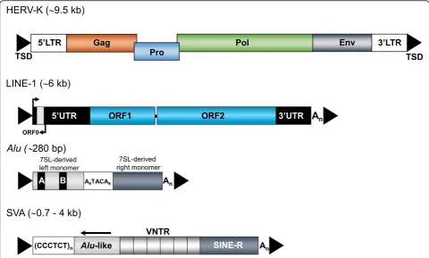
in trans can act at low frequency on any cellular polyadenylated mRNA, resulting in the generation of processed pseudogenes [56, 57]. Members of the primate-specific Alu family are approximately 300-bp long, have a dimeric structure, and are transcribed by RNA polymerase III (reviewed in [58, 59]) (Fig.2). The Fig. 2Retrotransposons in the human genome. Currently, only LINE-1 (L1),Aluand SVA elements are verifiably still mobilized in humans.A full length HERV-K provirus is ~ 9.5 kb long, codes for group-specific antigen (Gag), protease (Pro), polymerase (Pol) and envelope (Env) proteins and is flanked by ~ 1-kb long terminal repeats (LTRs) with the 5’LTR including the HERV-K promoter. A functional full length L1 element is ~ 6 kb in length, harbours three open reading frames (ORF0, ORF1 and ORF2) at which ORF1 and ORF2 are separated by a 63-bp noncoding spacer region. The 5′untranslated region (5’UTR) harbours both sense and antisense promoter.Aluelements comprise ~ 280-300 bp, are composed of two 7SL-RNA derived monomers separated by an A-rich connector (A5TACA6), contain an internal A and B box promoter, and end in a poly A tail (An).
family of Alu elements is, with > 1 million copies, the most abundant TE in our genome [35], and represents the currently most active group of retrotransposons in humans with ~ one thousand mobilization-competent copies per genome. The SVAs are hominid-specific com-posite non-coding retroelements, with an average length of ~ 2 kb, and represent the youngest family of active hu-man TEs (Fig. 2). There are roughly 2700 SVA copies per human genome, most of which are full-length and about 20–50 of which may be active [60–67].
In sum, all above listed mobilization-competent non-LTR retrotransposons may collectively account for thousands of somatic insertions [68]. It was calculated that up to 5% of newborns harbour a new retrotransposition event, and > 120 known human disease-causing insertions of L1 s,Alus and SVAs have been identified to date [69–73]. L1-mediated insertions have sporadically resulted in a wide number of human genetic disorders, including hemophilia A (L1), cystic fibrosis (Alu) [74] and X-linked dystonia-par-kinsonism (SVA) to cite a few reviewed in [71]. Altered ex-pression of TEs and animation of genomic L1 sequences also appear to be hallmarks of cancer, and can be respon-sible for driving mutations in tumorigenesis [75, 76]. In-deed, the random mobilization of L1 in our genome implies that virtually any disorder can be generated by the mutagenic activity of non-LTR retrotransposons, and L1-mediated retrotransposition events occur approximately once in every 250 pathogenic human mutations [77].
TE insertions can affect host gene expression by various mechanisms and impact stem cell development
When expressed, TEs can affect developmental processes either by their encoded gene products, which could in-fluence phenotype and/or function of the host cell, or through their genomic de novo insertion, that could re-sult in genetic changes in the host genome. L1-mediated retrotransposition events can be associated with the in-duction of local genomic instability, including DNA de-letions at the insertion site, 3′- or 5′-transductions, and non-allelic homologous recombination (reviewed in [32,
78]). Additionally, new retrotransposon insertions into genes can act as insertional mutagens, alter gene expres-sion and interfere with host gene function [79].
Although fewer than 1 of 10,000 TEs is still capable of mobilization [71], a far greater proportion can influence gene expression in cis and in trans (reviewed in [80]). This is because TEs, such as ERVs, L1 and SVA ele-ments, contain transcription factor binding sites that promote transcription by RNA polymerase II or, in the case ofAluelements, by RNA polymerase III. Therefore, the promoter(s) of resident and new TE(s) that inte-grated into or close to host genes, can alter gene expres-sion, and even lead to the appearance of novel transcripts that encompass part of the coding region.
Activity of non-LTR retrotransposons in human pluripotent stem cells
Primary milieu for the evolutionary struggle between TEs and the host genome are pluripotent cells of the early mammalian embryo and germ cells
To ensure their evolutionary success, TEs need to mobilize in germ cells and/ or during early development, before germline partitioning, to guarantee transmission of new retrotransposition events to the next generation. A genome-wide epigenetic switch in the early mammalian embryo, characterized by global DNA demethylation, is necessary to activate the programme of embryonic devel-opment. This ‘epigenomic reset’ is thought to provide a window of opportunity for retrotransposons to mobilize and create heritable insertions [92], as L1 expression is also controlled by DNA methylation [93]. Consistently, previous studies have shown that i) L1 mRNAs are present in human immature diploid oocytes from in vitro fertilization donors [94], ii) both L1-encoded proteins, ORF1p and ORF2p, are expressed in prespermatogonia of fetal testes and in germ cells of adult testes [95], and iii) hESCs overexpress a combination of potentially functional full-length L1 and core Alu elements as well as non-functional L1 and Alu RNAs (Fig. 3) [79, 96–100]. The observed L1 expression levels in these pluripotent
stem cells inversely correlate with both their low global DNA methylation levels, and their levels of L1 promoter methylation [97,101,102]. It has been reported that only a specific subset of L1 retrotransposons, likely under the combined influence of activators or repressors and local chromatin constraints, is active at any given period of early embryogenic development type [67, 96, 103, 104]. While expression of members of the youngest L1 lineages, L1Hs and L1PA2, is highest in hESCs, expression of a sub-set of L1 lineages, primarily L1PA3, L1PA4, L1PA5 and L1PA6, which were predicted to have entered the ances-tral genome between 26.8 and 7.6 million years ago, is re-duced [98]. Thus, these data demonstrated that RNAs from active and “hot” L1 s are expressed in hESCs and hiPSCs. Using an engineered L1 retrotransposition re-porter assay in cultured cells [40], it was demonstrated that the cellular environment of human and murine ESCs support L1 retrotransposition (Fig.3), and that L1 de novo insertions in cultivated hESCs can occur into genes and lead to small deletions of genomic DNA at the target site [97, 105, 106]. In agreement with these data demonstrat-ing that host factors essential for retrotransposition of functional L1 mRNAs are present in hESCs, it was verified that endogenousAluYa5 elements are trans-mobilized in cultured hiPSCs [79].
Somatic and heritable L1-mediated retrotransposition in pluripotent stem cells during early development
Studies of transgenic mouse models established to inves-tigate engineered L1 retrotransposition in vivo, and, more recently, the sequencing of mouse pedigrees, con-firmed that the majority of heritable de novo retrotran-sposition events in mice occur in preimplantation embryos, and indicate that embryonic cells are likely to be the natural habitat for L1-mediated retrotransposition [107–113]). Notably, such L1 mobilization in pluripotent cells of pre-implantation embryos could result in the new insertion being a mosaic within somatic and germ-line cells in adult mice. Although it was reported that L1 transcript levels peak after fertilization at the 2-cell-stage and decrease from the 2- to the 8-cell stages in mice [114], it remains unclear at which time point after fertilization L1-retrotransposition is initiated. Endogen-ous L1 elements were shown to be active in mEndogen-ouse early primordial germ cells, before the PIWI/piRNA retro-transposon defense pathway is activated in male embry-onic gonads [113,115].
By contrast, we currently know relatively little about TE expression and timing of retrotransposition in humans. An example that unambiguously proves that endogenous L1 mobilization can occur early in human embryogenesis was reported by van den Hurk and colleagues: they char-acterized a mutagenic L1 insertion resulting in choroider-emia, a rare recessive X-linked eye disorder, in an affected male patient whose mother was a somatic and germline mosaic for the pathogenic L1 insertion [116]. Due to a 3′ transduction carried by the mutagenic L1 insertion, the authors were able to identify the specific donor L1 elem-ent and verify that it was retrotransposition-competelem-ent [116, 117]. Taken together, data strongly suggest that, in humans, new heritable L1 andAluinsertions can likely ac-cumulate during early embryogenesis, and to some extent in germ cells [97,107,110]. Retrotransposition events me-diated by L1 were estimated to occur, at a minimum, in 1 of 20 meioses forAlu, 1 of 20 to 200 meioses for L1, and 1 of 900 meioses for SVA [118]. Clearly, future studies should determine the timing of L1 retrotransposition dur-ing embryogenesis and a rate of retrotransposition in modern humans.
Very recently, studies in mice provided evidence that the observed transcriptional L1 activation may have a role in mouse ESCs and pre-implantation embryos. Activation of L1 transcription after fertilization regulates global chro-matin accessibility at the beginning of development, and therefore could be important for normal embryonic devel-opment [119]. Also, the L1 RNA seems to act as a nuclear scaffold to repress the transcriptional program specific to the 2-cell embryo and, consequently, the L1 transcript could be required to exit from the 2-cell stage [120]. These data suggest that L1-encoded RNAs and proteins
have additional roles in embryonic development. These findings seem paradoxical, as despite the mutagenic po-tential of L1 during embryogenesis, they suggest that L1 activity could be required for proper embryonic develop-ment and even to maintain ESC identity.
TE activity as parameter to define the naïve pluripotent state in hESCs
Mouse ESCs and iPSCs were proposed to represent a naïve state of pluripotency corresponding to the inner cell mass, whereas hESCs/iPSCs correspond to a more ad-vanced, or ‘primed’, state of pluripotency found in the postimplantation epiblast [121]. Indeed, it has been chal-lenging to define the naïve state of pluripotency in hESCs, particularly in view of the expanding number of protocols for deriving putative naïve human cells [122,123]. Putative naïve human cultured cells were examined according to their resemblance to human pluripotent cells in vivo. Three comparative parameters for evaluating the naïve state of human pluripotent stem cells were established: (1) the expression profile of TEs based on single-cell RNA-seq data [124], (2) the DNA methylation landscape of human preimplantation development [125], and (3) the chromosome X inactivation status of female human ESCs [126, 127]. Comprehensive examination of the “ transpo-scriptome”was found to offer a more sensitive measure of the correspondence between pluripotent stem cells and the early human embryo than gene expression profiling [128]. Constituting a unique model system to dissect mechanisms of early human development, naïve hESCs provide an excellent cellular model to interrogate the function of transposable elements that become activated during early human development [128].
Cultured human pluripotent stem cells support expression and mobilization of endogenous TEs
their functional consequences, is of paramount signifi-cance. A direct consequence of reprogramming, due to genomewide epigenomic changes [130], is the activation of full-length L1 mRNA expression; indeed, L1-ORF1p and L1 RNA transcript levels are upregulated by up to four orders of magnitude compared to their respective parental cells [79,100,132,133]. To investigate if hiPSCs provide the environment for L1 retrotransposition, Wis-sing and colleagues exploited an in vitro L1 retrotranspo-sition assay, and detected similar levels of L1-engineered retrotransposition as those previously reported in hESCs [97,100]. These data demonstrated that the cellular milieu of both hESCs and hiPSCs contains the host factors re-quired for L1 retrotransposition. In order to clarify if the transcriptional activation of endogenous L1 elements in hiPSCs leads to L1-mediated mobilization, targeted se-quencing was performed on cultured hESC and hiPSC lines, followed by PCR validation of candidate de novo L1,
Aluand SVA insertions in multiple laboratories [79]. Not-ably, the sequencing of a small number of lines revealed ongoing L1, Alu and SVA retrotransposition in hiPSCs; Similarly, oneAlude novo insertion was found to have oc-curred during the cultivation of the hESC line H9, which was the only hESC line analyzed in this study [79]. Thus, these studies confirmed that hESCs and hiPSCs are a nat-ural habitat for L1-mediated retrotransposition, consistent with heritable retrotransposition during early human em-bryogenesis. However, the currently available datasets on L1 mobilization in hiPSCs vs hESCs are not sufficient to allow conclusions on the frequency of L1-mediated retro-transposition events in individual hESCs or hiPSCs. Remarkably, an earlier whole-genome sequencing (WGS)-based study in mouse ESCs failed to detect endogenous L1 retrotransposition [134]. Lack of retrotransposition in mESCs when compared to hESCs/hiPSCs might reflect distinctly different properties of human primed [79] and mouse naïve [134] pluripotency states; or the limitations of the method employed for detecting de novo retrotran-sposition events, namely genome-wide DNA sequencing with [79] or without [134] a previous retrotransposon en-richment step. Overall, these data might suggest that the genome of mESCs would be more stable than hESCs/ hiPSCs, although several studies have presented compel-ling evidence demonstrating ongoing (and heritable) L1 retrotransposition during early mouse embryogenesis [107, 113]. Thus, it is also possible that cultured mESCs fail to recapitulate this aspect of mouse early embryonic biology, and additional studies are required to solve this contradiction.
Occurrence and structures of de novo retrotransposition events in cultured hESCs and hiPSCs
An intriguing aspect of L1 retrotransposition studies in hESCs/hiPSCs is that most validated insertions are
full-length. In hiPSCs, 57–66% of all validated endogen-ous and engineered L1 de novo retrotransposition events were full-length [79, 100]. Another report found seven potential de novo L1 insertions in two hiPSC lines using targeted L1 sequencing, but could not PCR-validate or fully characterize the genomic integration sites of these events [133]. The success in the identification of L1 mobilization events depends on multiple factors such as heterogeneity of the investigated cell population, and methodological factors such as stem cell culture condi-tions, population bottlenecks in cultured cells, high-throughput sequencing method used, bioinformatic parameters, and how candidate L1 insertions are vali-dated, which can affect the results decisively. For ex-ample, a recent study applied WGS to nine hiPSC lines [135] and did not identify any de novo retrotransposon insertions, and far fewer mutations overall when com-pared to earlier studies [25,26]. While an early report in hESCs validated a small number of engineered but 5′-truncated L1 insertions [97], additional insertion characterization in hESCs has recently revealed that > 70% of them are full-length (Cano and Garcia-Perez, personal communication). The finding that 66–75% of engineered L1 retrotranposition events in hESCs and hiPSCs are full-length is in stark contrast to the situation in cancer cell lines, where only ~ 5% of engineered L1 de novo insertions represent full-length elements [136]. While this discrepancy warrants additional research, it is tempting to speculate that due to L1 overexpression, host factors involved in 5′truncation might be titrated/ sequestered in hESCs/hiPSCs allowing a higher rate of full-length insertions in these cells. Notably, other fea-tures of de novo L1 insertions events do not differ sig-nificantly between cancer cells and hESCs/hiPSCs; For example, ~ 50% of all validated L1 retrotransposition events in both cell types occurred into introns of host genes [79, 100, 136]. Similar to transformed cells, in-tronic L1 insertions into hiPSCs can interfere with the transcription of the affected host gene [79].
Epigenomic reprogramming causes a constant derepression of endogenous TEs
retrotransposons, but also of currently inactive HERV-K, HERV-H and HERV-W elements [132]. A sharp increase of L1 and SVA transcription was observed several days after transduction with reprogramming vectors, and both RNAs remained highly expressed upon culturing hiPSCs [132]. While there was only little heterogeneity in L1 ex-pression levels between tested hESC lines, considerable heterogeneity between hiPSC clones was observed [132]. Comparison of expression levels of individual endogenous retroelements between hiPSC clones derived from a single donor and issued from the same reprogramming experi-ment uncovered striking differences, notably for HERV-H, HERV-K and L1Hs. Thus, these data suggest that the gen-ome wide epigenomic reprogramming might not occur to the same extent in all reprogrammed cells, and that hESCs might represent a more realistic model to study human early embryogenesis processes. It was hypothesized that transcriptionally activated functional L1Hs elements, whose antisense promoters can affect the expression of neighbouring genes [139], have a similar effect during or after reprogramming of hiPSCs [132]. The interference of
cis- and trans-acting transcriptional regulatory elements of preexisting or de novo TE insertions with the transcrip-tion of TE-close genes may result in phenotypic anomalies difficult to detect through conventional assays, such as blockade of differentiation to particular lineages, predis-position to oncogenic changes, aberrant release of bio-active molecules, altered immunogenicity or ectopic activation of disease-related genes in iPSCs or their pro-geny [67, 132]. In hiPSCs, unsteady production of tran-scripts from TE-integrants normally silenced in ESCs and secondary activation of neighbouring genes was noted [67]. This phenomenon could contribute to the ineffi-ciency of reprogramming by stochastically activated genes that affect the path to pluripotency [140]. Transcriptional perturbation of TE-close genes may also plague iPSCs and their progeny with phenotypic anomalies [141]. These findings argue for an in-depth survey of the genomic, transcriptional, and epigenetic state of the repetitive gen-ome of iPSC clones, that are to be used for basic research or clinical applications. In these studies, bona fide estab-lished hESC lines could offer an excellent reference to de-tect harmful changes in hiPSCs that might limit their applications. Indeed, and while the use of hESCs on re-generative medicine is clearly limited, hESCs have become an excellent benchmark to establish clinically safer human pluripotent embryonic stem cell-like cells.
Activity of endogenous non-LTR retrotransposons in adult stem cells
Breaking the dogma–TE activity in adult stem cells of the brain
TEs are the prototype of“selfish DNA”, whose only pur-pose is to generate more copies of themselves. Thus, it
was assumed for a long time that most TE activity in mammals might occur only in cellular niches that trans-mit genetic information to the next generation, as this would ensure the evolutionary success of TEs. Although pioneer work by Barbara McClintock discovered TEs in somatic tissues of corn [142], the lack of TE activity in the mammalian soma has been a long-standing dogma. How-ever, this “dogma” was recently challenged with the de-scription of TE activity in mammalian healthy somatic cells, specifically in adult stem cells of the brain. The first evidence for ongoing mobility of L1 s in adult stem cells was obtained from neural precursor cells (NPCs) derived from rat hippocampus neural stem cells [112]. It was dem-onstrated that rodent NPCs can support elevated levels of retrotransposition using engineered human L1 reporter el-ements. By applying a transgenic mouse model it was demonstrated that L1 s generate genomic somatic mosai-cism during brain development and in adult mice. Con-sistently, human NPCs isolated from fetal brain or derived from hESCs were shown to express moderate levels of L1 RNA and L1-ORF1p (Fig.3), and to significantly support elevated levels of engineered L1 retrotransposition in vitro [143]. These and previous studies in mice (i.e., [112]) rep-resented a major breakthrough in the TE field, because it was demonstrated for the first time that the endogenous L1 copy number is increased in several regions of the hu-man brain (including hippocampus) when compared to other human somatic tissues [143, 144]. More recently, the use of an adenoviral/L1 hybrid vector [145] has allowed demonstrating L1 retrotransposition in non-dividing mature neuronal cells, differentiated from hESCs [99] (Fig. 3). While L1 s are usually repressed in somatic tissues, the mechanism responsible for L1 activa-tion in neuronal progenitor cells has been studied (for re-view see [146, 147]). While the L1 promoter whose activity is known to be regulated by CpG methylation [93,
148], and is hypermethylated in mature neurons, it is less methylated than in fibroblasts [143] and these data sug-gest that additional factors beside DNA methylation might allow L1 to be expressed in neurons. The most accepted current model for the regulation of L1 expression and ret-rotransposition in neurons and NPCs involves the activity of Sox2 and WNT3A in combination with DNA methyla-tion. Briefly, Sox2, a negative regulator of neuronal differ-entiation, is suggested to interact with the L1 promoter and repress L1 expression in rodent and human neural stem cells [112, 143]. Methyl-CpG-binding protein 2 (MeCP2) has been demonstrated to associate with the L1 promoter in a methylation-dependent manner, and to repress L1 expression in neural stem cells [149,
stimulate L1 expression through the canonical Wnt pathway [151].
Somatic L1 retrotransposition events accumulate during various developmental steps
Above studies mostly used engineered L1 reporter con-structs, although more recently sequencing-based ap-proaches have been used to demonstrate ongoing endogenous L1 retrotransposition in the human brain, not only in NPCs but also in neurons [144, 152–154]. Several high-throughput sequencing methods for the de-tection of endogenous retrotransposition events have been recently developed and applied to brain-derived samples (reviewed in [155]). Retrotransposon capture se-quencing (RC-seq, [156]), and L1-seq [72] are two of the methods used to demonstrate L1 retrotransposition in neuronal cells isolated from cerebral cortex, caudate nu-cleus and hippocampus. Although these studies have re-ported significant variability in the inferred rate of L1 mobilization in the brain [144, 152–154,157], the main conclusion from these studies is that somatic L1 retro-transposition events are accumulated during a variety of neural development stages, including early progenitor cells and mature neuronal cells. In summary, and while current estimates of L1 retrotransposition rates in neu-rons range from 0.04–0.6 to 13.7 L1 insertions per cell (see [111, 146, 157]), it is clear now that our brain is composed of a mosaic of genomes.
L1 retrotransposition occurs at very low level in the majority of tested adult stem cell types
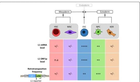
Is L1 active in other somatic tissues besides the brain, or can all adult somatic stem cell types of the human body support L1 retrotransposition? In order to address this question, Macia and colleagues compared L1 expression and engineered L1 retrotransposition among various hu-man cell types using isogenic cells [99]. To this end, the differentiation capability of hESCs was exploited to pro-duce relatively pure populations of adult stem cells, which were then used to study L1 expression and retrotransposi-tion. Notably, this approach allowed comparing different cell types that contain the same genetic background. Thus, L1 expression and retrotransposition was analyzed in kera-tinocytes (KERs, considered multipotent embryonic progenitor cells generating epidermal barrier, hair and nails), NPCs, fully differentiated neurons, MSCs and HSCs (Fig.3). This allowed comparing NPCs and neurons to an-other ectoderm-derived cell type (KERs) and to multipo-tent stem cell types derived from mesoderm (MSCs and HSCs). Remarkably, data revealed that all cell types tested express endogenous L1-mRNAs and encoded L1-ORF1p, although there were significant differences in their expres-sion levels (Fig. 3): L1-RNAs and L1-ORF1p were expressed at similar low levels in all cell types but NPCs,
which expressed moderate levels of gene products as re-vealed by RT-qPCR, immunoblot analysis and confocal mi-croscopy [99]. Consistently, the absence of detectable full-length L1 transcripts from human bone marrow-de-rived MSCs was demonstrated [158]. Analysis of the DNA-methylation status of L1 promoters in the various differentiated cell types tested uncovered an inverse cor-relation with L1 expression levels [99], which is consistent with the hypothesis that DNA methylation is a major mechanism to inhibit L1 retrotransposition in somatic cells [159].
To investigate whether KERs, HFFs, neurons and multi-potent NPCs, MSCs and HSCs support L1 retrotransposi-tion, Macia and colleagues took advantage of a hybrid adenovirus-based L1 vector that was specifically designed to conduct experiments in non-dividing cells [160]. Con-sistent with L1 expression data, L1 retrotransposition in all cell types tested, was extremely low, except for NPCs (Fig.
primordial germ cells (PGCs), at least in mice [113]. In humans, PGCs represent the primary undifferentiated stem cell type that differentiates towards gametes (sperm-atozoa and oocytes). Due to the therapeutic potential of PGCs for the treatment of infertility [165], the impact of L1 activity in those cells should be given due regard. In summary, among the adult stem cell types tested to date, somatic L1 retrotransposition is restricted to NPCs and PGCs, and does not seem to be a generic property of mul-tipotent adult stem cells.
Regulation of TE activity in pluripotent stem cells The human host is in a constant battle with TEs such as L1 to prevent their amplification and overreaching activ-ity, because functional, active L1 elements carry substan-tial mutagenic potential. Somatic L1-mediated retrotransposition in somatic cells, such as stem cells, has also been linked to disease, especially in the context of cancer [75, 76,162, 166]. Mobility restriction of these elements is therefore critical to maintain genome stabil-ity, especially during germline establishment and early embryogenesis, as new TE insertions in these cells can potentially be transmitted to the next generation [167].
During the first few days of embryogenesis, most TEs are targeted by silencing mechanisms and kept under con-trol during early embryogenesis by epigenetic silencing and post-transcriptional regulation of L1 mRNAs [99,168,
169]. Epigenetic silencing of TE expression is mediated by 1) Krüppel-associated box domain-containing zinc finger proteins (KRAB-ZFPs), mediators of heterochromatic for-mation [98] 2) cytosine methylation, a process governed by the action of DNA methyltransferase-3 like protein in germ cells [93], and 3) ten-eleven translocation (TET) family proteins [170]. TEs are recognized by sequence-specific proteins- or RNA-based repressors, with subsequent recruitment of heterochromatin-inducing complexes [171]. Histone methylation, histone deacetyla-tion, and DNA methylation control TE expression and re-press their cis-acting transcriptional components, which would otherwise activate neighboring genes via promoter or enhancer effects [172–178]. Histone repressive marks play a major role in this process, and trimethylation of his-tone 3 at lysine 9 (H3K9me3) is a modification found in a wide variety of TEs, including ERVs, LINEs, SINEs, and SVAs in hESCs [98,179–184]. Excellent reviews have been published recently on how the host controls the activity of active TEs [67,110,155]. In the following section, we will address those host-encoded mechanisms that were re-ported to interfere with the retrotransposition cycle specif-ically in pluripotent stem cells.
Restriction of TE transcription
KRAB-ZFPsare the largest family of transcriptional reg-ulators in higher vertebrates [185] and play a major role
in the early embryonic control of vertebrate TE expres-sion, including ERV, LINE, SINE and SVA elements [98,
178,184]. Harbouring an N-terminal KRAB domain and a C-terminal array of DNA-binding zinc fingers, they mediate silencing by recruiting the cofactor KRAB-associated protein 1 (KAP1, also known as TRIM28), which acts as a docking protein for proteins with heterochromatin-forming activities, including the H3K9 methyltransferase SETDB1 [178, 186]. The KRAB/KAP1 system represses transcription of endogen-ous retroelements primarily via histone deacetylation, H3K9 trimethylation, and HP1 recruitment, with DNA methylation occurring only secondarily to ensure the permanence of the silencing process [187,188].
Evolutionary old L1 subfamilies that are between ~ 7 and 25 million years of age are silenced in hESCs [98,
185, 189] through specific KRAB-ZFPs such as ZNF93/ KAP1, and there is evidence suggesting that KRAB-ZFPs also bind the younger and retrotransposition-competent L1Hs elements [185]. Beside L1s, in hESCs two-thirds of endogenous retroelements bound by KAP1 are SVAs, suggesting KAP1 is a major repressor of SVA activity. In summary, it is now well established that KRAB-ZFP/ KAP1 complexes control TE activity in hESCs, including retrotransposition-competent elements harbouring the potential to affect genomic integrity, and help maintain-ing transcriptional homeostasis and normal differenti-ation of ESCs.
TE family in these cells, tended to be more hypomethy-lated compared to non-overexpressed copies in naïve cells [128]. Thus, DNA methylation is key to control L1 expres-sion and mobilization, and is the major TE controlling mechanism during gastrulation [125].
TET proteins: While the primary mechanism under-lying DNA demethylation during the early embryonic period is replication-coupled dilution of 5mC [199,
200], an active mechanism dependent on TET enzymes has recently been uncovered to erase DNA methylation at specific loci in this period [170, 201–203]. TET en-zymes catalyze the oxidation of 5-methylcytosine to 5-hydroxymethylcytosine (5hmC), and further to 5-for-mylcytosine (5fC) and 5-carboxylcytosine (5caC), which can be replaced with unmodified cytosine by base excision repair [204, 205]. Some of the TET proteins are highly expressed in ESCs and blastocysts [206], and it was re-ported that TET binding and demethylation at particular TE classes, such as LTR retrotransposons, acts in concert with pluripotency factors Nanog, Oct4 and Sox2 to main-tain expression of a subset of genes in ESCs [170]. Deple-tion of TET1 and TET2 in mESCs has been shown to cause loss of 5hmC in the 5′region of L1 [207]. In hESCs, TET proteins were shown to preferentially bind to evolu-tionary young, functional L1 elements, and participate in their active demethylation, but do not interact with older, inactive subfamilies. Although TETs drive L1 demethyla-tion, L1s can be kept repressed through the TET-dependent recruitment of the transcriptional repres-sor SIN3A. The SIN3A co-repressive complex binds func-tional L1s in mouse and human ESCs ensuring their repression in a TET-dependent manner [92, 170]. Thus, and instead of being only positive regulators of L1 expres-sion, TET enzymes may have a dual role in TE regulation by also recruiting SIN3A to demethylated L1 elements. A recent report provided evidence that the methyl-CpG binding domain as well as the adjacent non-sequence spe-cific DNA binding domain of MeCP2 mediate repression of TET1-induced L1 mobilization [208]. Also, the KZFP/ KAP1 complex was recently reported to maintain hetero-chromatin and DNA methylation at TEs in naïve murine ESCs partly by protecting these loci from TET-mediated demethylation [209].
Small RNAs classes include PIWI-interacting RNAs (piRNAs), endogenously produced small interfering RNAs (siRNAs) or micro RNAs (miRNAs), and the importance of small RNA-based repression in the control of L1 ex-pression in human pluripotent stem cells has been re-ported and reviewed [114, 210, 211]. Double-stranded RNAs (dsRNAs) with sequence specificity to portions of the transcriptome that originate from TEs with bidirec-tional promoters, such as L1s, was suggested to serve as substrate for the production of siRNAs [212]. Moreover, dsRNA produced from the 5’UTR of L1 may trigger the
interferon-dependent restriction factor ribonuclease L in some cancer cells, leading to L1 mRNA degradation [213].
piRNAsare a complex class of small non-coding RNAs that comprise mostly 24–32 nucleotides, specifically inter-act with the PIWI protein subfamily of the ARGONAUTE family [214], and act to repress mobile genetic elements in the germline of Drosophila and mammals [169]. piRNAs are generated by transcription of long TE clusters, result-ing in the accumulation of short mature piRNAs in the cytoplasm by the ping-pong mechanism [215]. piRNAs then act as guides to destroy complementary TE tran-scripts by endonucleolytic cleavage. PIWI-mediated con-trol is indeed triggered by the recognition of L1-proximal sequences by a complex encompassing a member of the PIWI subclade of Argonaute proteins and L1-derived piR-NAs, which leads to L1 transcriptional inhibition via DNA methylation [216–218]. The piRNAs–PIWI system and DNA methyltransferases, acting downstream of piRNA action, play a crucial role in the early embryonic control of the youngest and mobile L1 lineages in human pluripo-tent cells [98,186,211,219].
A role for miRNAs and the miRNA biogenesis ma-chinery in controlling human non-LTR retrotransposons has been suggested by demonstrating that a complex termed Microprocessor, which comprises the RNase III type enzyme Drosha and its partner DGCR8, catalyzes the nuclear step of microRNA biogenesis [220,221] and binds L1, Alu and SVA-derived small RNAs in human cells [210]. The results suggest that Microprocessor rec-ognizes and processes structural regions within retrotransposition-competent L1 andAluelements lead-ing to a decrease of functional L1 andAluRNAs which could result in a reduction of L1 andAlu retrotransposi-tion frequencies [169, 210]. miR-128, a member of the class of miRNAs encompassing 20- to 24-nt-long non-coding RNAs that inhibit translational initiation and stimulate decay of mRNA targets [222, 223], was re-cently reported to decrease both amounts of full-length L1 RNA and engineered L1 retrotransposition frequen-cies in a cell culture based assay in human tumor cells and induced pluripotent stem cells [198].
Post-transcriptional control of TEs
APOBEC3 proteins The human APOBEC3
to 10-fold during reprogramming [132]. A3B is a nuclear protein that is also expressed in hiPSCs [105,231,232] and was demonstrated to restrict L1 retrotransposition in both hESCs [105] and hiPSCs in a deaminase-independent man-ner [211]. While it is likely that A3A inhibits L1 retrotran-sposition by deaminating transiently exposed L1 DNA [233], both A3C and A3DE were reported to restrict L1 retrotransposition through editing-independent mecha-nisms by interacting with ORF1p, thereby interfering with L1 RT activity [234, 235]. Additional mechanistic studies are required, because contradictory results were found among cell lines and studies.
SAMHD1, a dGTP-activated deoxynucleoside triphos-phate triphosphohydrolase that inhibits L1 retrotranspo-sition in actively dividing cells by reducing the L1 ORF2p level [236], was demonstrated to be transiently induced during the initial 10 days of reprogramming [132], and could therefore play a role in the restriction of L1 mobilization during this period. In addition to diminishing L1 reverse transcription by reducing L1 ORF2p expression [236], SAMHD1 was recently re-ported to stimulate the formation of stress granules and thus enhance the sequestration of L1 RNP complexes in these granules [237].
TEX19.1 is expressed in germ cells, pluripotent cells and the placenta of mammals [238] and was recently shown to regulate L1-ORF1p levels and mobilization of engineered L1 elements in pluripotent mouse embryonic stem cells [106]. Mouse TEX19.1, and its human
ortholog TEX19, physically interact with L1-ORF1p, and can regulate L1-ORF1p abundance at postranslational level through stimulating its polyubiquitylation and proteasome-dependent degradation [106].
For the sake of completeness, we also mention the fol-lowing host-encoded factors which also restrict L1 retro-transpositrion mostly by post-transcriptional mechanisms, but have not been reported to restrict L1 mobilization in stem cells so far: (i) MOV10, an RNA helicase that medi-ates access of the RNA-induced silencing complex to mes-senger RNAs [239, 240] (ii) ZAP, an antiviral member of the poly (ADP-ribose) polymerase (PARP) protein family [241, 242] (iii) TREX-1, a three-prime DNA exonuclease [243,244].
DNA transposons as tools for genetic engineering of stem cells
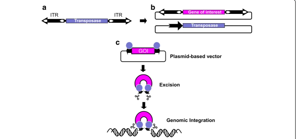
An important point of intersection between stem cells and transposable elements is genetic engineering for the purposes of deciphering disease mechanisms and for gene- and cell-based therapeutics. DNA (Class 2) trans-posons (Fig. 4a) became major tools for genome manip-ulations in a wide range of cell types, including stem cells (reviewed by [245–247]. The mobility of DNA transposons invariably depends on two functional com-ponents: a protein component called the transposase and a DNA component called inverted terminal repeats (ITRs) supporting binding of and cleavage by the trans-posase (Fig. 4a). Due to their attributes including
a
c
b
efficient gene transfer in human cell types, ease of ex-perimental manipulation, and detailed understanding of the transposition process, theSleeping Beauty, piggyBac and Tol2 transposons have become the transposons of choice for stem cell engineering. What is common to all gene transfer applications by any transposon is the setup of a conditional, two-component gene delivery system, in which a gene of interest flanked by the transposon ITRs is mobilized out of standard plasmid vectors by the trans-posase that is conditionally and transiently expressed in the relevant cells (Fig.4b and c). Stem cells are ideal tar-gets for gene therapy applications. It is of great hope to consider stem cells to achieve tissue repair or to restore and replenish cells in the background of a genetic disease. Following a transposon-based genetic manipulation step to introduce a gene of interest, such as a therapeutic gene rendering stable phenotypic correction, stem cells can be expanded in vitro and then subjected to differentiation into particular cell lineages according to the specific thera-peutic need. There is now widespread evidence for robust transposon-mediated gene transfer in several, clinically relevant stem cell types, such as ESCs, iPSCs, CD34+ hematopoietic stem cells (HSC), MSCs, or myoblasts.
Transgene expression in hESCs by means of theSleeping
Beautytransposon system
A proof of concept study established efficient and stable transgene expression in hESCs using theSleeping Beauty transposon system. In this study, transposons, carrying different transgenes coding for genetic markers, were ef-fectively delivered to undifferentiated hESCs. The source of transposase was either DNA or mRNA. Molecular analysis indicated that 98% of stable gene transfer re-sulted from transposition events. These genetically engi-neered hESCs were then differentiated into teratomas in vivo and mature hematopoietic cells in vitro and the progeny maintained stable transgene expression [248]. Similarly successful gene transfer in hESCs was obtained with the piggyBac system as well, where fluorescent re-porters were introduced into ESCs, and these remained functional following in vitro differentiaton. ThepiggyBac transposon system also allowed for seamless restoration of the insertion site following transposon removal by a second round of transient transposase expression [249]. The Sleeping Beauty transposon was also used to intro-duce a CAG promoter-driven GFP expression cassette to hESCs. During differentiation experiments the CAG promoter yielded outstanding GFP expression in cardio-myocytes allowing for specific labeling of cardiomyo-cytes in a spontaneously differentiated, mixed cell population [250]. The same transposon was also used to introduce a genetically encoded Ca2+ions indicator (GCaMP2) into hESCs allowing for real-time Ca imaging without the need for dyes. Cardiomyocytes differentiated
from these ESCs were characterized by their spontaneous contractions and Ca2+ signal oscillations, presenting a powerful tool for pharmacological screening assays [251].
Multiple applications of transposon systems in the hiPSC field
During the early developments in the iPSC field, a signifi-cant step was achieved when non-viral methods were used to achieve reprogramming. BothpiggyBac[252] and
promoter was delivered by the transposon, and genomic modification was achieved following sgRNA delivery. Fol-lowing a transient transposase expression the inducible Cas9 cassette was removed yielding a genome-edited iPSC clone with seamless transgene removal [261].
Efficient gene transfer into mobilized HSCs and human CD34+cord blood cells
A rapidly progressing area for transposon-mediated gene therapy applications is represented by HSCs. Transposon-based tools hold great promise for render-ing efficient gene correction without the potential risks inherent to viral delivery methods, which resulted in se-vere adverse reactions in clinical trials in the past [262,
263]. HSCs have the potential for self-renewal and main-tenance of the ability to differentiate into hematopoietic lineages, and are thus ideal targets for gene therapy ap-plications in hematologic diseases. HSCs can be effi-ciently modified by the Sleeping Beauty and piggyBac transposon systems [264]. Especially, developments of
Sleeping Beautytransposons were taking shape in recent years enabled by the highly efficient SB100X hyperactive transposase in CD34+ HSCs [265]. The Sleeping Beauty transposon system not only supports the efficient gene transfer into mobilized CD34+ HSCs, but also into hu-man cord blood CD34+ cells as shown in a model of sickle cell disease [266]. Further developments of the transposon-mediated delivery were also undertaken in recent years to target CD34+ HSCs. For example, an in-teresting and promising optimization step was under-taken when the transposon was delivered to the cells as a minicircle in combination with transposase supplied as mRNA. This approach led to improved cell survival and reduced cytotoxicity in the HSCs providing also several biosafety advantages over conventional delivery methods [267]. The combination of the Sleeping Beauty trans-poson system and adenoviral vectors in a hybrid vector system was also proposed as a promising method to achieve in vivo gene delivery. This approach holds broader clinical application potential for gene therapy as it may circumvent the need for myeloablation and trans-plantation. In this study HSCs were mobilized into per-ipheral blood in a transgenic, humanized mouse model, and were targeted using a hybrid adenovirus/transposon vector system injected intravenously in vivo, resulting in functional HSCs homing back to the bone marrow stably expressing the transgene [268].
MSCs are also in the focus of regenerative medicine, however, there are no breakthrough applications yet, and the field is still hampered by many controversies. Never-theless, it has been evidenced that both Sleeping Beauty andpiggyBactransposon-based gene transfer is applicable in MSCs as well. The genetically engineered MSCs are still characterized by the ability to undergo osteogenic,
myogenic, and adipogenic differentiation after modifica-tions with the transposons [257, 269]. piggyBac-mediated gene transfer of IFNγ into adipose-derived MSCs was used in a mouse model of melanoma to show that the IFNγ-expressing MSCs engrafted into tumor stroma, inhibited tumor growth and angiogenesis, and prolonged the survival of mice [270].
Myoblasts are self-renewing adult muscle progenitor cells, which differentiate into skeletal muscle cells and could potentially be harnessed for cell therapy of muscle disorders. Both Sleeping Beauty- and piggyBac-mediated gene transfer methods can be applied to efficiently modify myoblasts and were shown to be useful tools in delivering therapeutic genes into myoblasts. Proper dysferlin expres-sion as well as highly efficient engraftment of the engi-neered myoblasts was evidenced in the skeletal muscle of dysferlin-deficient mice [271], whereas transposon vectors encoding microdystrophins and delivered into myoblasts have been shown to yield proper expression levels in dif-ferentiated multinucleated myotubes [272].
Insertional mutagenesis and genetic labeling studies
steady-state blood production is maintained by the succes-sive recruitment of thousands of clones, each with a min-imal contribution to mature progeny. These results demonstrated that large numbers of long-lived progeni-tors are the main drivers of steady-state haematopoiesis during most of adulthood [277].
The risk of genotoxicity caused by DNA transposon activity
As discussed above in the context of endogenous TEs, one of the most important risk factors also associated with the use of transposon-based gene transfer tools in stem cells is genotoxicity. In the context of a transposon vector system, at least two fundamental properties can contribute to gen-otoxicity: i) interaction of the transposase with endogenous human DNA sequences or human proteins with the trans-poson vector sequences and ii) the genome-wide insertion profile of the vector. With respect to“off-target” cleavage of the transposase, the use of theSleeping BeautyandTol2 systems appears to be safe in human cells. Both of these transposons originate from fish genomes, and the mamma-lian lineage does not contain transposons sufficiently simi-lar to allow cleavage by these transposases. Vice versa, human cells do not express proteins that could re-mobilize a genomically integratedSleeping BeautyorTol2vector. In contrast, the human PGBD5 transposase-derived protein was reported to mobilize piggyBac transposon vectors in human cells [278], thereby presenting potential implica-tions for human applicaimplica-tions [279].
Characterization of the target site selection properties of different vector systems is highly useful for ranking the different vector types and designs according to their genotoxic potential [280]. The Sleeping Beauty trans-poson displays the least deviation from random with re-spect to genome-wide distribution: no apparent bias was seen for either heterochromatin marks or euchromatin marks and only a weak correlation with transcriptional status of targeted genes was detected [281]. This is in marked contrast to target site distributions of several other transposons including Tol2[264, 282], and
piggy-Bac[264,283,284] that favor integration into genes and near chromatin marks characteristic of active transcrip-tion units (e.g., H3K27 acetylatranscrip-tion and H3K4 mono-methylation). ThepiggyBactransposon, in particular, has been shown to favor open chromatin, expressed genes and TSSs (±5 kb) associated with DNaseI hypersensitive sites, H3K4Me3 marks and Pol II-bound regions in mouse and human cells [283–288]. These observations collectively suggest that Sleeping Beauty might be the safest currently available transposon for therapeutic gene delivery in clinical trials.
Conclusions
Stem cell therapies have been expected to bring substantial benefit to patients suffering a wide range of diseases and
injuries. It was expected that the benefits of bone marrow transplants for patients needing reconstruction of their hematopoietic and immune system would apply to stem cell transplants of other cell types, and optimism has been high for the utilization of embryonic and induced pluripo-tent stem cells for a variety of applications. However, be-fore these promising stem cells or their differentiated derivatives are administered to patients, genomic integrity of these cells has to be ensured to guarantee that these cells remain therapeutically functional and are not tumori-genic. In recent years, a remarkable amount of data accu-mulated showing that the activity of endogenous TEs can be one source of genomic destabilization in stem cells, and constitutes a risk for the biosafety of stem cell-based ther-apies. By giving an overview of the potentially mutagenic activity of TEs in human multipotent and pluripotent stem cells, the consequences of their activity for the genomic in-tegrity and host gene expression, we provide arguments for a thorough characterization of TE activity and its conse-quences in the individual stem cell lines before their thera-peutic utilization in patients in order to ensure biosafety of these stem cells cells and/or their applied derivatives.
Acknowledgments
We thank Dr. B. Völker (Paul-Ehrlich-Institut) for his assistance in generating Fig.1, Dr. J. Garcia-Perez (Univ. Edinburgh, UK) for ongoing discussions and critical reading of the manuscript, and two anonymous reviewers for constructive comments.
Funding
S.R.H. and P.T.R. are funded by the Government of Spain (MINECO, RYC-2016-21395 and SAF2015–71589-P [S.R.H.]; PEJ-2014-A-31985 and SAF2015– 71589-P [71589-P.T.R.]). GGS is supported by a grant from the Ministry of Health of the Federal Republic of Germany (FKZ2518FSB403).
Availability of data and materials
The dataset used to generate Fig.1is available in the repository of the US Food and Drug Administration (FDA),www.clinicaltrials.gov.
Authors’contributions
SRH, AS, ZI and GGS wrote the manuscript. PTR and NVHF generated the figures. All authors read and approved the submitted manuscript.
Ethics approval and consent to participate Not applicable.
Consent for publication Not applicable.
Competing interests
The authors declare that they have no competing interests.
Publisher’s Note
Springer Nature remains neutral with regard to jurisdictional claims in published maps and institutional affiliations.
Author details
1Division of Medical Biotechnology, Paul-Ehrlich-Institut,
Paul-Ehrlich-Str.51-59, 63225 Langen, Germany.2Host-Pathogen Interactions,
Biology II, Faculty of Pharmacy, University of Granada, Campus Universitario de Cartuja, 18071 Granada, Spain.
Received: 5 November 2018 Accepted: 27 February 2019
References
1. Mason C, Dunnill P. A brief definition of regenerative medicine. Regen Med. 2008;3:1–5.https://doi.org/10.2217/17460751.3.1.1.
2. Thomson JA, Itskovitz-Eldor J, Shapiro SS, Waknitz MA, Swiergiel JJ, Marshall VS, Jones JM. Embryonic stem cell lines derived from human blastocysts // Embryonic Stem Cell Lines Derived from Human Blastocysts. Science. 1998; 282:1145–7.https://doi.org/10.1126/science.282.5391.1145.
3. Takahashi K, Yamanaka S. Induction of pluripotent stem cells from mouse embryonic and adult fibroblast cultures by defined factors. Cell. 2006;126: 663–76.https://doi.org/10.1016/j.cell.2006.07.024.
4. Yu J, Vodyanik MA, Smuga-Otto K, Antosiewicz-Bourget J, Frane JL, Tian S, et al. Induced pluripotent stem cell lines derived from human somatic cells. Science. 2007;318:1917–20.https://doi.org/10.1126/science.1151526. 5. Lodi D, Iannitti T, Palmieri B. Stem cells in clinical practice: Applications and
warnings. J Exp Clin Cancer Res. 2011;30:9. https://doi.org/10.1186/1756-9966-30-9.
6. O'Donoghue K, Fisk NM. Fetal stem cells. Best Pract Res Clin Obstet Gynaecol. 2004;18:853–75.https://doi.org/10.1016/j.bpobgyn.2004.06.010. 7. Avots A, Harder F, Schmittwolf C, Petrovic S, Muller AM. Plasticity of
hematopoietic stem cells and cellular memory. Immunol Rev. 2002;187:9– 21.https://doi.org/10.1034/j.1600-065X.2002.18702.x.
8. Barriga F, Ramírez P, Wietstruck A, Rojas N. Hematopoietic stem cell transplantation: Clinical use and perspectives. Biol Res. 2012;45:307–16. https://doi.org/10.4067/S0716-97602012000300012.
9. Ullah I, Subbarao RB, Rho GJ. Human mesenchymal stem cells - current trends and future prospective. Biosci Rep. 2015.https://doi.org/10.1042/ BSR20150025.
10. Chaudhary D, Trivedi RN, Kathuria A, Goswami TK, Khandia R, Munjal A. In vitro And In vivo Immunomodulating Properties of Mesenchymal Stem Cells. Recent Patents Inflamm Allergy Drug Discov. 2018;12:59–68.https:// doi.org/10.2174/1872213X12666180227105924.
11. Squillaro T, Peluso G, Galderisi U. Clinical Trials With Mesenchymal Stem Cells: An Update. Cell Transplant. 2016;25:829–48.https://doi.org/10.3727/ 096368915X689622.
12. Trounson A, McDonald C. Stem Cell Therapies in Clinical Trials: Progress and Challenges. Cell Stem Cell. 2015;17:11–22.https://doi.org/10.1016/j.stem. 2015.06.007.
13. Ratcliffe E, Glen KE, Naing MW, Williams DJ. Current status and perspectives on stem cell-based therapies undergoing clinical trials for regenerative medicine: Case studies. Br Med Bull. 2013;108:73–94.https://doi.org/10.1093/bmb/ldt034. 14. Kimbrel EA, Lanza R. Current status of pluripotent stem cells: Moving the
first therapies to the clinic. Nat Rev Drug Discov. 2015;14:681–92.https://doi. org/10.1038/nrd4738.
15. Reubinoff BE, Pera MF, Fong CY, Trounson A, Bongso A. Embryonic stem cell lines from human blastocysts: Somatic differentiation in vitro. Nat Biotechnol. 2000;18:399–404.https://doi.org/10.1038/74447.
16. Trounson A, DeWitt ND. Pluripotent stem cells progressing to the clinic. Nat Rev Mol Cell Biol. 2016;17:194–200.https://doi.org/10.1038/nrm.2016.10. 17. Mandai M, Kurimoto Y, Takahashi M. Autologous Induced Stem-Cell-Derived
Retinal Cells for Macular Degeneration. N Engl J Med. 2017;377:792–3. https://doi.org/10.1056/NEJMc1706274.
18. Cyranoski D. Japanese man is first to receive‘reprogrammed' stem cells from another person. Nature. 2017.https://doi.org/10.1038/nature.2017.21730. 19. Ben-David U, Benvenisty N. Analyzing the genomic integrity of stem cells.
Cambridge: StemBook; 2008.https://doi.org/10.3824/stembook.1.150.1. 20. Lefort N, Perrier AL, Laâbi Y, Varela C, Peschanski M. Human embryonic stem
cells and genomic instability. Regen Med. 2009;4:899–909.https://doi.org/10. 2217/rme.09.63.
21. Maitra A, Arking DE, Shivapurkar N, Ikeda M, Stastny V, Kassauei K, et al. Genomic alterations in cultured human embryonic stem cells. Nat Genet. 2005;37:1099–103.https://doi.org/10.1038/ng1631.
22. Martins-Taylor K, Xu R-H. Concise review: Genomic stability of human induced pluripotent stem cells. Stem Cells. 2012;30:22–7.https://doi.org/10. 1002/stem.705.
23. Baker DEC, Harrison NJ, Maltby E, Smith K, Moore HD, Shaw PJ, et al. Adaptation to culture of human embryonic stem cells and oncogenesis in vivo. Nat Biotechnol. 2007;25:207–15.https://doi.org/10.1038/nbt1285. 24. Ben-David U, Mayshar Y, Benvenisty N. Large-scale analysis reveals acquisition
of lineage-specific chromosomal aberrations in human adult stem cells. Cell Stem Cell. 2011;9:97–102.https://doi.org/10.1016/j.stem.2011.06.013. 25. Gore A, Li Z, Fung H-L, Young JE, Agarwal S, Antosiewicz-Bourget J, et al.
Somatic coding mutations in human induced pluripotent stem cells. Nature. 2011;471:63–7.https://doi.org/10.1038/nature09805.
26. Hussein SM, Batada NN, Vuoristo S, Ching RW, Autio R, Närvä E, et al. Copy number variation and selection during reprogramming to pluripotency. Nature. 2011;471:58–62.https://doi.org/10.1038/nature09871. 27. Laurent LC, Ulitsky I, Slavin I, Tran H, Schork A, Morey R, et al. Dynamic
changes in the copy number of pluripotency and cell proliferation genes in human ESCs and iPSCs during reprogramming and time in culture. Cell Stem Cell. 2011;8:106–18.https://doi.org/10.1016/j.stem.2010.12.003. 28. Mayshar Y, Ben-David U, Lavon N, Biancotti J-C, Yakir B, Clark AT, et al.
Identification and classification of chromosomal aberrations in human induced pluripotent stem cells. Cell Stem Cell. 2010;7:521–31.https://doi. org/10.1016/j.stem.2010.07.017.
29. Liang G, Zhang Y. Genetic and epigenetic variations in iPSCs: Potential causes and implications for application. Cell Stem Cell. 2013;13:149–59. https://doi.org/10.1016/j.stem.2013.07.001.
30. Tapia N, Schöler HR. Molecular Obstacles to Clinical Translation of iPSCs. Cell Stem Cell. 2016;19:298–309.https://doi.org/10.1016/j.stem.2016.06.017. 31. Kazazian HH, Wong C, Youssoufian H, Scott AF, Phillips DG, Antonarakis SE.
Haemophilia A resulting from de novo insertion of L1 sequences represents a novel mechanism for mutation in man. Nature. 1988;332:164–6.https:// doi.org/10.1038/332164a0.
32. Beck CR, Garcia-Perez JL, Badge RM, Moran JV. LINE-1 elements in structural variation and disease. Annu Rev Genomics Hum Genet. 2011;12:187–215. https://doi.org/10.1146/annurev-genom-082509-141802.
33. Marchi E, Kanapin A, Magiorkinis G, Belshaw R. Unfixed endogenous retroviral insertions in the human population. J Virol. 2014;88:9529–37. https://doi.org/10.1128/JVI.00919-14.
34. Naveira H, Bello X, Abal-Fabeiro JL, Maside X. Evidence for the persistence of an active endogenous retrovirus (ERVE) in humans. Genetica. 2014;142: 451–60.https://doi.org/10.1007/s10709-014-9789-y.
35. Lander ES, Linton LM, Birren B, Nusbaum C, Zody MC, Baldwin J, et al. Initial sequencing and analysis of the human genome. Nature. 2001;409:860–921. https://doi.org/10.1038/35057062.
36. Speek M. Antisense promoter of human L1 retrotransposon drives transcription of adjacent cellular genes. Mol Cell Biol. 2001;21:1973–85. https://doi.org/10.1128/MCB.21.6.1973-1985.2001.
37. Swergold GD. Identification, characterization, and cell specificity of a human LINE-1 promoter. Mol Cell Biol. 1990;10:6718–29.https://doi.org/10.1128/MCB.10.12.6718. 38. Athanikar JN, Badge RM, Moran JV. A YY1-binding site is required for
accurate human LINE-1 transcription initiation. Nucleic Acids Res. 2004;32: 3846–55.https://doi.org/10.1093/nar/gkh698.
39. Wei W, Gilbert N, Ooi SL, Lawler JF, Ostertag EM, Kazazian HH, et al. Human L1 retrotransposition: Cis preference versus trans complementation. Mol Cell Biol. 2001;21:1429–39.https://doi.org/10.1128/MCB.21.4.1429-1439.2001. 40. Moran JV, Holmes SE, Naas TP, DeBerardinis RJ, Boeke JD, Kazazian HH. High
Frequency Retrotransposition in Cultured Mammalian Cells. Cell. 1996;87: 917–27.https://doi.org/10.1016/S0092-8674(00)81998-4.
41. Cost GJ. Human L1 element target-primed reverse transcription in vitro. EMBO J. 2002;21:5899–910.https://doi.org/10.1093/emboj/cdf592. 42. Luan DD, Korman MH, Jakubczak JL, Eickbush TH, Luan DD, Korman MH,
et al. Reverse transcription of R2Bm RNA is primed by a nick at the chromosomal target site: A mechanism for non-LTR retrotransposition. Cell. 1993;72:595–605.https://doi.org/10.1016/0092-8674(93)90078-5.
43. Denli AM, Narvaiza I, Kerman BE, Pena M, Benner C, Marchetto MCN, et al. Primate-specific ORF0 contributes to retrotransposon-mediated diversity. Cell. 2015;163:583–93.https://doi.org/10.1016/j.cell.2015.09.025. 44. Piskareva O, Schmatchenko V. DNA polymerization by the reverse
transcriptase of the human L1 retrotransposon on its own template in vitro. FEBS Lett. 2006;580:661–8.https://doi.org/10.1016/j.febslet.2005.12.077. 45. Brouha B, Schustak J, Badge RM, Lutz-Prigge S, Farley AH, Moran JV,