Sorrentino
et al.
R E S E A R C H
Open Access
Conservation and evolutionary divergence in the
activity of receptor-regulated smads
Gina M Sorrentino
1,2, William Q Gillis
2, Jamina Oomen-Hajagos
2,3and Gerald H Thomsen
2*Abstract
Background:Activity of the Transforming growth factor-β(TGFβ) pathway is essential to the establishment of body axes and tissue differentiation in bilaterians. Orthologs for core pathway members have been found in all
metazoans, but uncertain homology of the body axes and tissues patterned by these signals raises questions about the activities of these molecules across the metazoan tree. We focus on the principal canonical transduction proteins (R-Smads) of the TGFβpathway, which instruct both axial patterning and tissue differentiation in the developing embryo. We compare the activity of R-Smads from a cnidarian (Nematostella vectensis), an arthropod (Drosophila melanogaster), and a vertebrate (Xenopus laevis) inXenopusembryonic assays.
Results:Overexpressing NvSmad1/5 ventralizedXenopusembryos when expressed in dorsal blastomeres, similar to
the effects ofXenopusSmad1. However, NvSmad1/5 was less potent than XSmad1 in its ability to activate
downstream target genes inXenopusanimal cap assays. NvSmad2/3 strongly induced general mesendodermal
marker genes, but weakly induced ones involved in specifying the Spemann organizer. NvSmad2/3 was unable to
induce a secondary trunk axis inXenopusembryos, whereas the orthologs fromXenopus(XSmad2 and XSmad3)
andDrosophila(dSmad2) were capable of doing so. Replacement of the NvSmad2/3 MH2 domain with theXenopus XSmad2 MH2 slightly increased its inductive capability, but did not confer an ability to generate a secondary body axis.
Conclusions:Vertebrate and cnidarian Smad1/5 have similar axial patterning and induction activities, although NvSmad1/5 is less efficient than the vertebrate gene. We conclude that the activities of Smad1/5 orthologs have been largely conserved across Metazoa. NvSmad2/3 efficiently activates general mesendoderm markers, but is unable to induce vertebrate organizer-specific genes or to produce a secondary body axis inXenopus. Orthologs dSmad2 and XSmad3 generate a secondary body axis, but activate only low expression of organizer-specific genes that are strongly induced by XSmad2. We suggest that in the vertebrate lineage, Smad2 has evolved a specialized role in the induction of the embryonic organizer. Given the high level of sequence identity between Smad orthologs, this work underscores the functional importance of the emergence and fixation of a few divergent amino acids among orthologs during evolution.
Keywords:TGFβ, BMP, Smad, Animal body patterning, Evolution of signal transduction
Background
In developing animal embryos the Transforming Growth Factor-β (TGFβ) superfamily of ligands and signaling pathways regulate cell fate decisions, pattern formation, growth and organogenesis. Canonical TGFβ signals are transduced by Smad proteins operating in either of two major signaling branches, the bone morphogenetic
protein (BMP) and Activin/Nodal pathways. The unique receptor-regulated Smad (R-Smad) protein sequences determine the specificity of each R-Smad for upstream receptors and downstream cofactors and target genes. Recently, orthologs of the core members of the TGFβ pathway have been identified outside of Bilateria, in ani-mals that lack the degree of complexity seen in bilaterian symmetry and tissue-types [1]. These animals possess TGFβgenes even though none have a true dorsoventral axis or mesoderm, and the sponge lacks definitive germ layers altogether. TGFβ superfamily ligands and their
* Correspondence:[email protected]
2
Department of Biochemistry and Cell Biology, Stony Brook University, Life Sciences Building room 450, Stony Brook, NY 11794-5215, USA
Full list of author information is available at the end of the article
signal transduction components are not found in the choanoflagellate Monosiga brevicollis (the eukaryotic outgroup to Metazoa), which indicates that this growth factor system is restricted to Metazoa [1-3].
Discovery of key conserved developmental gene path-ways has led to the paradigm of a shared‘genetic toolkit’: a gene network that generates the variety of animal body forms by differential deployment. Work has been done to reveal the evolutionary history of many gene networks by mapping their presence or absence onto phylogenetic trees. It has been tempting to reconstruct the presence of morphological features along with the presence of a gene network in animal ancestors at key nodes, such as the ancestors of Bilateria and Eumetazoa [4]. However, some authors reject these reconstructions on the grounds that conservation of genes involved in core gen-etic regulatory networks does not necessitate the pres-ence of the particular morphologies known to be regulated by these networks [5]. These disagreements highlight the need for functional testing when studying the meaning of these orthologous gene networks.
We approached the question of functional conserva-tion by testing the ability of non-bilaterian gene pro-ducts to function in a developing vertebratein vivo. We focus on the Smad proteins, which operate both as intra-cellular transducers of TGFβfamily receptor signals and as transcription factors. Failure of Smad signaling and abnormal downstream gene regulation causes funda-mental disruption of body axes and cell fate determi-nation. Three subtypes of Smads are involved in TGFβ signaling [6-8], the receptor-regulated (R), the common (Co) and the inhibitory Smads (I). R-Smads are phos-phorylated at a C-terminal pair of serine residues when an extracellular ligand binds to Type I and II receptors, forming a signaling complex. Phosphorylated R-Smads then bind to a Co-Smad to form a trimeric complex that facilitates additional interactions with transcription fac-tors on promoter elements of target genes. Smad signal-ing is regulated at the level of receptors and R-Smad/ Co-Smad complexes by I-Smads [6]. With a few excep-tions, most non-vertebrate taxa have four Smad genes, an R-Smad in the Activin/Nodal pathway (AR-Smad), an R-Smad in the BMP pathway (BR-Smad), a Co-Smad, and an I-Smad. Vertebrates typically have multiple co-pies of each due to gene duplication events [3], which raise major questions about whether duplicated Smads have retained ancestral activities and/or evolved diver-gent functions.
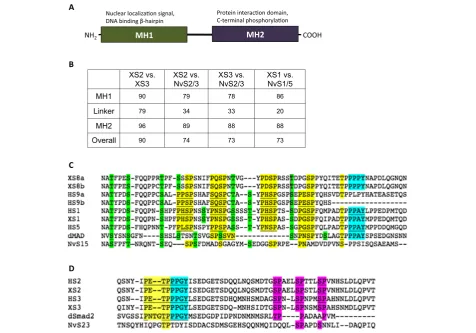
Smads are considered highly conserved in their bio-chemical and biological functions, and they are structu-rally defined by the presence of two characteristic‘MAD homology’ (MH) domains that determine functionality. Generally speaking, the N-terminal MH1 domain binds directly to DNA and contains a nuclear localization signal,
and the C-terminal MH2 domain contains binding sites for the numerous potential protein co-factors that make up the transcriptional complexes (Figure 1A) [6,8]. R-Smad proteins terminate at a consensus SSXS polypeptide, of which the last two serines become phosphorylated in response to receptor activation [6] (see Additional file 1). Co- and I-Smads lack this consensus. The MH1 and MH2 domains are separated by a linker region that can be highly variable among species and even Smad subtypes, but serves important regulatory functions by providing sites for non-TGFβ receptor-driven phosphorylation and targeting by E3 ubiquitin ligases [8].
Vertebrates have three BR-Smads that transduce BMP signals - Smad1, Smad5, and Smad8/9 (see Additional file 1). In Xenopus, XSmad1 is the major embryonic intracellular transducer of BMP signals, and its ectopic expression in dorsal embryonic regions mimics the effects of BMP overexpression such as loss of dorsal cell identity resulting in tadpoles that are almost entirely composed of ventral tissues, lacking heads and neural tissues as a consequence of respecification [9]. Func-tional conservation of BR-Smad orthologs across taxa has been shown by the ectopic expression of dMad, the XSmad1 ortholog from Drosophila, that when injected dorsally into Xenopus embryos causes the same cata-strophic loss of head and neural tissues as overexpres-sion of the native XSmad1 [9].
In contrast to vertebrates, most non-vertebrate ani-mals have just two R-Smads. With respect to the Activin-like pathway in Drosophila, an AR-Smad called
dSmad2 has been described but its activity and signifi-cance appears to be quite different than Smad2/3 in ver-tebrates [19,20]. The protein dSmad2 is activated by the Activin-type receptor Baboon, and loss of Baboon func-tion (and thus dSmad2 funcfunc-tion) causes minor problems with cell proliferation and growth, but does not affect body patterning [20]. In fact, dSmad2 overexpression in prospective ectoderm of Xenopus animal caps causes Activin-like induction of mesoderm [19], but the level to which dSmad2 shares functional homology with verte-brate Smad2 or Smad3 was not tested.
Smad family members have been identified in all meta-zoan clades, but the extent to which there is (or is not) functional conservation among the Smads, particularly
across highly divergent taxa such as non-bilaterians and chordates, is an important question to answer that will inform the evolution of this protein family. In the present study, we used qualitative and quantitative methods to examine whether the functions of the R-Smads have been conserved sufficiently during metazoan evolution to allow R-Smads from a cnidarian to participate in the TGFβ signal transduction network during early verte-brate embryogenesis. We have chosen two exemplar taxa for this study,Xenopus laevis(the African clawed frog, a model organism for functional studies) and the model cnidarian Nematostella vectensis (the starlet sea ane-mone). TheNematostellaBR-Smad ortholog, NvSmad1/5, has been identified, and aNematostellaAR-Smad ortholog (NvSmad2/3) was found previously and evaluated in a phylogenetic analysis of the NvSmad family, but it has not been experimentally tested for function [2].
Experiments presented here test the abilities of Nema-tostella and DrosophilaR-Smad orthologs to induce ex-pression of downstream pathway genes and pattern tissues in the Xenopusembryo. We also probe the acti-vities of individual Smad domains using chimeric con-structs from Xenopus Smad2 and Nematostella Smad2/ 3. We find that cnidarian R-Smad proteins activate BMP and Activin/Nodal responses, but not at the efficiency of the nativeXenopusproteins. However, we reveal qualita-tive differences in the ability of NvSmad2/3 to function in the developing vertebrate. Notably, vertebrate Smad2 and Smad3 have different signaling abilities, and only the bilaterian orthologs of Smad2/3 are capable of indu-cing ectopic axial structures in Xenopus embryos. Our findings show a deep conservation of fundamental Smad activities across 650 million years of animal evolution, but divergence in the smaller scale fine-tuning of gene activation, reflecting different evolutionary histories of the two major Smad TGFβsignaling pathways.
Methods
Xenopus, Nematostella, and Drosophilaclones
TheXenopus Smad1,Smad2, andSmad3andNvSmad1/ 5 clones were already available in the Thomsen Lab (Stony Brook University). NvSmad2/3 was cloned di-rectly out of cDNA prepared from total RNA of Nema-tostella planulae. The primers were designed from a predicted protein sequence [NCBI: XP_001631607], which was identified using a Basic Local Alignment Search Tool (BLAST) search with XSmad2 sequence (forward primer: 50 ATGACTTCCCTGTTGCCT 30, re-verse primer: 50 CTACGATACCGAGGAGAT 30). The PCR amplification was carried out with Platinum
™
Taq DNA Polymerase High Fidelity ( Life Technologies, Invi-trogen, Grand Island, NY). The PCR conditions were as follows: 94°C for 2 minutes (1 cycle); 94°C for 30 se-conds, 56°C for 30 sese-conds, 68°C for 1.5 minutes (40 cycles); and 68°C for 2 minutes. The Drosophila dSmad2 (or Smox) clone was a gift from the lab of Dr. Spyros Artavanis-Tsakonas (Harvard University) and the Drosophila Protein Interaction Map group. All clones were subcloned into the plasmid pCS2 containing three HA tags 50 of the gene start site. TheXSmad2-ΔExon3 clone was a gift from the laboratory of Malcolm Whitman at Harvard University.
Sequence analysis
Once subcloned, all clones were sequenced and checked against the correct protein sequence from GenBank. To create the alignments and pairwise comparisons used for Figure 1 and Additional file 1, we aligned the amino acid sequences by hand in MacVector (MacVector, Inc., Cary, NC), saved them as subdomain alignments, and opened them in ClustalW (European Bioinformatics Institute,
Cambridge, UK, http://www.clustal.org) to calculate pair-wise percent identity scores [see Additional file 2 for accession numbers].
Chimera assembly
Amino acid boundaries for MAD Homology domains in XSmad2 and NvSmad2/3 are given in their entries at NCBI. MH1 chimera: [XSmad2: 1 to 172] + [NvSmad2/ 3: 131 to 423]. Linker chimera: [NvSmad2/3: 1 to 130] + [XSmad2: 173 to 267] + [NvSmad2/3: 224 to 423]. MH2 chimera: [NvSmad2/3: 1 to 223] + [XSmad2: 268 to 467]. In order to create the chimeric constructs, fragments were generated by PCR from XSmad2 and NvSmad2/3
clones [see Additional file 3 for primer locations and sequences]. The PCR amplification was carried out with Platinum
™
Pfx DNA Polymerase from (Life Technolo-gies). The PCR conditions were as follows: 94°C for 4 minutes (1 cycle), 94°C for 30 seconds, 55°C for 30 seconds, 68°C for 1 minute (35 cycles) and 68°C for 30 minutes. Primers were designed to amplify the desired region from one species and add approximately 10 nucleotides of the intended adjacent region of the other species, to generate fragments that would partially over-lap within the chimeric product. Chimeric sequences were then generated by placing the appropriate frag-ments together in a PCR reaction and adding the primers corresponding to the ends of the desired chimeras. The fragments were ligated into pGEM-T vector and sub-cloned into an HA-tagged pCS2 vector. Chimeras were verified by sequencing.Messenger RNA synthesis
Clones were linearized and messenger RNA (mRNA) for microinjection was made from each clone using the Amplicap
™
SP6 High Yield Message Maker kit (Epicentre Biotechnologies, Madison, WI). The mRNA was purified using a Qiagen RNeasy kit (Qiagen Inc., Valencia, CA), tailed using the Poly(A) Polymerase Tailing Kit (Epicentre Biotechnologies), and purified again before use.Xenopusembryo injections
Care and Use Committee Guidelines and was approved by the Stony Brook University Internal Review Board.
Translation assessment
Western blotting was performed to check for expression of the Heamaglutinin Antigen (HA) peptide tags and equalize translation levels. Embryos were lysed with a pipet tip in PBS 1% Triton at stage 11, at the same time as the animal caps from the same experiment were ready for harvesting. Lysates were spun at 4°, and soluble pro-tein was mixed 1:1 with loading buffer and loaded in a 5% polyacrylamide gel. An Anti-HA primary antibody from Santa Cruz (sc-805) used at 1:500; the loading con-trol was Abcam anti-β-Actin (ab 8229), used at 1:750. The secondary antibody was Alexa Fluor 680 goat anti-rabbit IgG from Life Technologies (A-21109), used at 1:10,000 [see Additional file 4 for full western blots and loading controls].
Xenopusanimal cap assay
Messenger RNA was injected into the animal pole of both blastomeres at 2-cell stage; animal caps were har-vested at stage 8 and cultured in 0.5× Marc’s Modified Ringers (MMR) buffer until stage 11. Cells were lysed with Proteinase K and total RNA was extracted from the animal caps and whole embryo controls using phenol: chloroform extraction, followed by ethanol precipitation. Next, cDNA was synthesized using 1 μg of total RNA and SuperScript II Reverse Transcriptase enzyme from Invitrogen (Life Technologies). Then, cDNA samples were analyzed on a Roche Diagnostics LightCycler 480 System using SYBR
™
Green Mastermix I from Roche Diagnostics (Indianapolis, IN). Animal cap cDNA was compared to cDNA from a whole embryo, representing the endogenous expression levels. For each primer pair in each experiment, serial dilutions of whole embryo cDNA were used to create the standard curve to which all samples were compared in order to calculate concen-tration of PCR product. Once concenconcen-trations were acquired and imported into Excel, raw values were nor-malized to the level of Ornithine Decarboxylase (ODC), a housekeeping gene. See Additional file 5 for a table of LightCycler primer sequences and quantitative RT-PCR (qPCR) conditions, and their references.Results and discussion
NematostellaSmads contain the highly conserved MAD-homology domains that define bilaterian Smads
First, we revisited the presence and identities of R-Smads in
Nematostella. Previous work identified one AR-Smad (NvSmad2/3) and one BR-Smad (NvSmad1/5) [2,3], and our re-examination of genomic and cDNA sequences con-firmed those earlier identifications, but since the NvSmad2/
3 ortholog was only reported as a predicted protein [NCBI: XP_001631657], we isolated a full-length copy of this cDNA (see Methods). We then performed pairwise align-ments of all R-Smad orthologs fromXenopusand Nematos-tella to validate their relationships and highlight their unique features [see Additional file 1 and Additional file 2 for detailed alignments and accession numbers].
We found that the amino acid sequences of the MAD homology domains are highly conserved betweenXenopus
and Nematostella (Figure 1B). The N-terminal MH1 DNA-binding domain is more conserved in the Smad1/5 category (86%) than in the Smad2/3 category (78 to 79%). The C-terminal MH2 protein-interacting domain is the most conserved in each R-Smad category, and is equally conserved between Smad1/5 and Smad2/3 (88 to 89%). The linker region is less conserved than the MAD ho-mology domains, 20% in Smad1/5 and 33 to 34% in Smad2/3. Since the linker region is more variable yet con-tains important sites for post-translational regulation, we performed a second, more inclusive alignment of linker domains in order to investigate the status of several im-portant sites. We included R-Smad orthologs from the human and fromDrosophila melanogasterin this part of this analysis [see Additional file 2 for accession numbers]. Figure 1C and D show alignments of the important resi-dues of the linker regions.
The human Smad1/5/9 linker contains four conserved proline-X-serine-proline (PXSP) consensus sites for MAPK phosphorylation [22], which are putatively present inXenopus Smad8a and 8b (Figure 1C, yellow). The Drosophila dMad linker contains two conserved MAPK sites (Figure 1C, underline and yellow) [23], and the NvSmad1/5 linker shows one potential site (Figure 1C). With the exception of human Smad9b, vertebrate and Drosophila Smad1/5/8 orthologs share the PPXY motif that binds Smurf1, an E3 ubiquitin ligase that, once bound, will bring about ubiquitin-mediated degradation of these Smads [24] (Figure 1C, cyan). The linker of NvSmad1/5, however, lacks this site (Figure 1C). The dMAD linker also contains eight serine/threonine phosphorylation sites for GSK3 [23], which show variable conservation in the other orthologs (Figure 1C, green). The vertebrate orthologs contain seven of these predicted sites, and the linker of NvSmad1/5 con-tains potentially five of them.
The human Smad2 and Smad3 orthologs contain a MAPK consensus site [25] that is also found inXenopus
are present in theXenopusandDrosophilaorthologs, and two of which appear in NvSmad2/3 (Figure 1D, magenta). These analyses illustrate that cnidarian R-Smad linker regions may have fewer points of regulation compared to bilaterian R-Smads, suggesting that NvSmad1/5 could be regulated in a different manner from bilaterian orthologs.
Overexpression ofNvSmad1/5causes ventralization phenotypes inXenopusembryos
Bilaterian BR-Smad orthologs can ventralize Xenopus
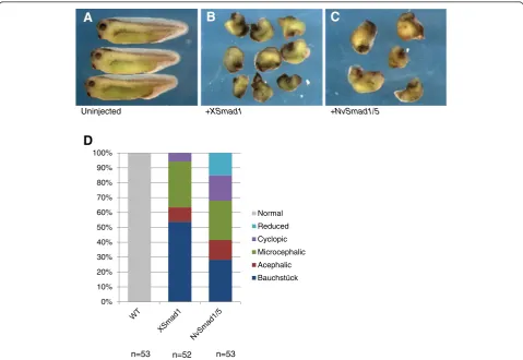
embryos when ectopically expressed in dorsal tissues. We tested whether NvSmad1/5 could function similarly when ectopically expressed in vivoin Xenopusembryos. We compared the phenotype from ectopic expression of NvSmad1/5 to that of XSmad1 (the Smad5 gene is not present in Xenopus laevis, and XSmad8 transcripts are scarce during gastrulation [29]). We found that ectopic dorsal expression of NvSmad1/5 generated the hallmarks of BMP overexpression: ventralization and obliteration of head structures. By stage 34, uninjected wild type
tadpoles had obvious head and neural structures (Figure 2A), whereas tadpoles that had been injected with XSmad1 mRNA showed a range of ventralization phenotypes, the most severe of which are shown in Figure 2B. Injection of NvSmad1/5mRNA also showed a range of ventralization effects, the most severe of which are shown in Figure 2C.
To quantify the range of effects, we used Kao and Eli-son’s DorsoAnterior Index (DAI) to score the severity of the ventralization phenotypes on a scale of 0 (most se-verely ventralized) to 5 (normal) [30]. Overall, the XSmad1 phenotypes scored as more severe than the NvSmad1/5 phenotypes (Figure 2D). The weighted means of the XSmad1 and NvSmad1/5 phenotypes were 0.89 and 1.77, respectively. The standard deviation of the XSmad1 scores was less than that of the NvSmad1/5 scores, 1.0 and 1.4 respectively. The XSmad1 overex-pression phenotype is overall more severe and has less range, whereas the NvSmad1/5 phenotype is less severe and shows more variation. These results indicate that
A
B
C
Uninjected +XSmad1 +NvSmad1/5
0% 10% 20% 30% 40% 50% 60% 70% 80% 90% 100%
Normal
Reduced
Cyclopic
Microcephalic
Acephalic
Bauchstück
D
n=53 n=52 n=53
the NvSmad1/5 protein functions in the Xenopus
embryo and successfully generates the expected ventrali-zation effects of BMP activity, but it is less potent than the native XSmad1 protein under the same conditions.
NvSmad1/5 induces downstreamBMPmarker gene expression in Xenopus
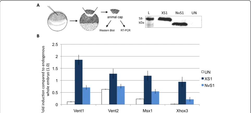
The observation that ectopic expression of NvSmad1/5 and XSmad1 results in similar ventralization phenotypes led us to compare their inductive activity more precisely, and determine whether NvSmad1/5 has the ability to initiate similar downstream gene expression inXenopus. To do this, we used Xenopusanimal cap assays to com-pare the expression levels of ventral marker genes known to be downstream of BMP signaling. We used tagged expression vectors and western blotting to con-firm equal protein translation levels before performing RT-PCR analysis (Figure 3A) [see Additional file 4 for western blot loading controls].
In three out of four cases, NvSmad1/5 induced expres-sion at a level significantly higher than that of the unin-jected animal caps (Figure 3B). NvSmad1/5 was able to induce downstream BMP pathway membersVent1, Msx1,
andXhox3at levels higher than in uninjected animal caps, yet at roughly half the levels induced by the native
XSmad1 protein. However, in all cases, NvSmad1/5 failed to induce expression equal to endogenous levels in the whole embryo (set as 1.0 on the Y-axis for all RT-PCR ana-lyses). We were not able to see a clear induction response byVent2, which may be due to high levels of endogenous
Vent2expression. Thus, despite the absolute differences in activity between NvSmad1/5 and XSmad1, NvSmad1/5 can initiate transcription ofXenopusBMP target genes.
NvSmad2/3induces expression of a subset of markers of the Activin/Nodal pathway
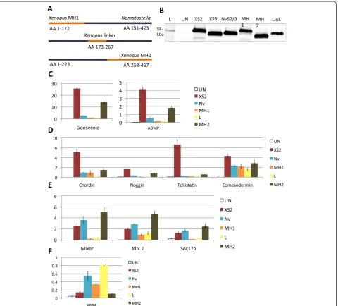
In order to test the functional conservation of verte-brate and cnidarian AR-Smad orthologs, we examined the ability of NvSmad2/3 to initiate Activin/Nodal sig-naling in the Xenopus animal cap. Equal protein trans-lation levels were confirmed using western blotting before RT-PCR analysis (Figure 4A) [see Additional file 4 for western blot loading controls]. Unlike the uni-formity of marker induction by NvSmad1/5, the induc-tion response to XSmad2 and NvSmad2/3 showed two clear patterns: for some markers NvSmad2/3 showed only a fraction of the inductive power of the native XSmad2, whereas for other markers, NvSmad2/3 was equal to or greater than XSmad2 in its inductive abili-ties (see Figure 4B-E red and teal bars).
To investigate these patterns, we included additional AR-Smad orthologs. We chose the DrosophilaAR-Smad dSmad2 as a protostome representative and XSmad3 as the second vertebrate AR-Smad ortholog. Upon repeat-ing these experiments with all four treatments, further trends became evident. We were able to split Activin/ Nodal markers into four classes based upon their in-ductive response. Class I included goosecoidand ADMP, two genes expressed strictly in the Spemann organizer
of the developing amphibian. Both of these were strongly induced by XSmad2 and less so by the other orthologs (Figure 4B). Class II markers were induced strongly by XSmad2 and dSmad2, and responded poorly to XSmad3 and NvSmad2/3 (Figure 4C). Class II included three BMP-inhibitors -chordin, noggin,and follistatin, as well aseomesodermin, another gene associated with dorsaliza-tion. In contrast, Class III markers were induced strongly by XSmad3, while XSmad2, NvSmad2/3, and dSmad2
showed relatively less response (Figure 4D). Class III markers are more general mesendoderm-related Activin/ Nodal markersmix2, mixer,andsox17α.
Xbrachyury was in a class by itself, Class IV (Figure 4E). Xbra induction by Smad2/3 orthologs was generally low. The highest induction was by NvSmad2/3 and reached almost 60% of endogenous level in the
Xenopus embryo (1.0 on the Y-axis in all RT-PCR ana-lyses). To test whether we were experimenting at the appropriate dosage (4 ng), we compared three different dosages of NvSmad2/3 and XSmad2 - 2 ng, 5 ng, and 10 ng. Results were similar; NvSmad2/3 induced more strongly, while XSmad2 induced very weakly (Figure 4F).
Xbraresponse to the lower doses of NvSmad2/3 remained consistent with previous results, while Xbraresponse to the highest dose of NvSmad2/3 dropped to the low level ofXbraresponse to XSmad2.
Substituting the NvSmad2/3 MH2 with the XSmad2 MH2 increases inductive capability
The Smad2/3 orthologs showed very particular induc-tion patterns in our Xenopus animal cap assays. We wished to determine whether the differences in activity between XSmad2 and NvSmad2/3 might reflect evolu-tionary specialization of specific regions of XSmad2, par-ticularly whether any single domain from XSmad2 could increase the capability of NvSmad2/3 to induce orga-nizer markers inXenopus.To this end, we created three chimeras that replaced the domains in NvSmad2/3 one at a time with XSmad2 domains (Figure 5A), and tested their inductive abilities in animal cap assays with the same set of markers as above. We confirmed equal translation levels with western blotting before RT-PCR (Figure 5B) [see Additional file 4 for western blot load-ing controls]. The linker chimera (‘Link’ in Figure 5B) showed a slightly lower amount of protein than the others at 4 ng mRNA injection. It remained at a lower level even at 8x the injection concentration of the other treatments (data not shown), so we kept the injection concentrations equal.
Interestingly, the four classes of markers from our pre-vious experiment were largely consistent in this experi-ment as well. In Class I markers goosecoid and ADMP, substitution of the XSmad2 MH2 domain (“MH2 chimera”) led to a gain in inductive ability over the wild type NvSmad2/3, to about 50% of the level of XSmad2 induction (Figure 5C). For Class II markers chordin,
follistatin, andeomesodermin, the MH2 chimera showed very slight enhancement in inductive ability, but that was still only a fraction of the level of induction observed with XSmad2 (Figure 5D). For Class III markers, NvSmad2/3 inductive ability was already slightly higher than that of XSmad2, and the MH2 chimera showed a modest increase (Figure 5E). For Xbra, the Class IV
marker, the MH2 chimera had significantly less in-ductive activity than NvSmad2/3 (Figure 5F).
In all cases, substitution of the XSmad2 MH1 domain (‘MH1 chimera’) had a negative effect on the inductive capacity of NvSmad2/3 (Figure 5C-F). Likewise, swap-ping in the XSmad2 linker region for the NvSmad2/3 linker region (‘linker chimera’) resulted in a drop in in-ductive ability of nearly every marker tested. Again,
Xbra showed its own unique response pattern; it was the only marker to respond more strongly to the linker chimera than to the wild type NvSmad2/3 (Figure 5F). The Xbra response levels to wild type XSmad2 and NvSmad2/3 correspond to our previous dosage observa-tions (Figure 4E).
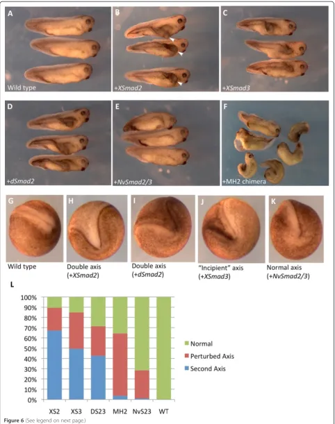
NvSmad2/3 does not induce the formation of a second body axis when ectopically expressed inXenopus embryos
NvSmad2/3 shows a complicated activity pattern in re-gard to its induction of dorsal mesoderm markers and Activin/Nodal targets. This calls into question the level of Smad2/3 functional conservation within Metazoa. It has been shown previously that Smad2 from the mouse can induce a second body axis inXenopusembryos [31], one with trunk and tail characteristics but lacking a head. This is nearly identical to axial structures induced by ectopically-expressed Xenopus activin [32] and indi-cates that Smad2 function is conserved among vertebrates. We performed ectopic expression experiments to deter-mine whether the ability to induce a second body axis is unique to the vertebrate Smad2 ortholog. Alternatively, that ability could be inherent to both of these vertebrate Smad2/3 paralogs, to all bilaterian Smad2/3 orthologs, or more generally to all metazoan Smad2/3 orthologs.
We observed a very strong secondary axis phenotype caused by bilaterian Smad2/3 orthologs (Figure 6A-D). The secondary axis was evident as a second set of neural folds at neurula stage (Figure 6G-K) and developed into an unmistakable secondary trunk by tadpole stage (Figure 6B, white arrowheads). XSmad2 produced a se-condary axis in 65% of embryos, whereas XSmad3 did so in about 50% of embryos, and dSmad2 in 45% (Figure 6L). In another 25 to 35% of cases, both proteins did not generate a distinct secondary axis, but did create a small“incipient” second axis at the neurula stage (for example, Figure 6J) that was subsumed into the primary axis during development and eventually manifested as the‘perturbed’axis of the tadpole [see Additional file 6].
of NvSmad2/3. The MH2 chimera did not improve upon the ability of NvSmad2/3 to produce a secondary body axis, but it perturbed the natural axis in upwards of 50% of embryos (Figure 6F, L).
These data agree with other data we present here that suggest that bilaterian Smad2/3 orthologs have developed functions that non-bilaterian orthologs are un-able to perform in vivo. These data also support our
results indicating that swapping XSmad2 domains onto NvSmad2/3 cannot bestow full functional abilities.
NvSmad1/5, but not NvSmad2/3, can recapitulate activity of bilaterian orthologs
NvSmad1/5 engaged the Xenopus pathway well enough to cause very severe ventralized phenotypes (Figure 2) and activate transcriptional targets (Figure 3), although
at a lower level than XSmad1. We found that ectopic ex-pression of NvSmad2/3 was unable to induce a second-ary axis inXenopus embryos, and showed differences in downstream induction of Activin/Nodal markers when compared to XSmad2, including the BMP inhibitors nog-gin, chordin, and follistatin, and the organizer-specific genes goosecoid and ADMP. All of these except ADMP
are known to have cnidarian orthologs [33]. Interest-ingly, NvSmad2/3 induced the general mesendoderm markers at the same level as some of the bilaterian orthologs (Class III, Figure 4D). There is no ortholog of
nodal known in Nematostella, but NvActivin is expressed in the endoderm during gastrulation [33]. Likewise, the Sox17 ortholog NvSoxF1 is expressed broadly in the endoderm following gastrulation (there are no definitive orthologs of mix2 or mixer yet known to be expressed in developing Nematostella endoderm) [34]. Our data are further evidence thatActivinsignaling via AR-Smads to pattern endoderm is an ancient and conserved mechanism in metazoan development.
One alternative explanation for the differential activation of gene targets by NvSmad2/3 in our experiments could be a dose-dependence. Experiments incubating Xenopus ani-mal caps with Activin ligand have revealed striking dose-dependent induction of mesodermal markers including
Xbra and goosecoid by Activin, which are activated at low and high doses of Activin respectively [35,36]. We observed a concordant Xbra dose-dependent response to ligand-independent overexpression of either Xenopus or
Nematostella Smad2/3 (Figure 4F and results not shown). We reasoned that if the particular dose of Smad2/3 was responsible for these differences in gene induction, then programming the animal cap system with graded concen-trations of NvSmad2/3 (up to 10 ng) might yield sufficient activity to replicate the inductive patterns observed with XSmad2 (for example, induction of Xbra and Xgsc at re-spectively low and high levels of NvSmad2/3). To the con-trary, however, the response patterns of most markers remained consistent for all three doses tested (Additional file 7). Increasing the level of NvSmad2/3 to 10 ng did not activate the goosecoidgene even to a level induced by the lowest amount of XSmad2 (see Additional file 7).
We propose that the differences in cnidarian versus bilaterian Smad2/3 activity reflect evolutionary diver-gence, which has rendered NvSmad2/3 unable to engage the necessary signaling, transcriptional, or other neces-sary cofactors in the Xenopus system. This may be due to lack of key microdomains or amino acid residues that are present in Xenopus and other bilaterian Smad2/3 orthologs which facilitate more efficient or complete en-gagement and activation of target genes. For instance, Smad2 and Smad3 proteins make complexes with Smad4, FAST-1, p53 and other co-factors in order to enter the nucleus, bind DNA, and transcribe target genes [13,35,37]. The low inductive activity of NvSmad2/3 in Xenopus could be due to NvSmad2/3 forming transcriptional complexes that are weak, un-stable, and/or inactive. Smads are also a common target of TGFβ signal regulation by other pathways, such as FGF (via MAPK) and Wnt (via GSK3) [36], thus there are various ways in which the subtle protein sequence differences between NvSmad2/3 and vertebrate Smad2 and 3, especially those in the linker domain, could lead to differences in activity.
Despite the low inductive ability of NvSmad1/5 rela-tive to XSmad1, it could still re-pattern the Xenopus
embryo to cause severe significant ventralization of dor-sal tissues. This was not the case with NvSmad2/3, which could not induce the secondary body axis observed with overexpression of XSmad2, XSmad3, or dSmad2 (Figure 6E, G). Mouse Smad2 can also generate a very pronounced second axis inXenopusembryos [31], which builds the case that bilaterian Smad2/3 orthologs have a function that the non-bilaterian NvSmad2/3 is not able to perform. This suggests fine-scale divergence in the case of Smad1/5 and larger-scale divergence in the evolutionary history of Smad2/3.
Vertebrate Smad2 and Smad3 have different activity
There are numerous indicators that vertebrate Smad2 and Smad3 have different activities. There is evidence of exclusive co-factors for each in zebrafish [38], and verte-brate Smad2 and Smad3 differ in their mechanisms of nuclear import and their regulation by ubiquitination
(See figure on previous page.)
Figure 6Ectopic overexpression ofNvSmad2/3does not induce a secondary axis inXenopusembryos.Injection of 0.5 ngXSmad2mRNA into the marginal zone of one ventral vegetal blastomere at the 8-cell stage can produce a secondary body axis inXenopusembryos.
(A) Uninjected, wild type tadpoles. (B) Tadpoles that were injected withXSmad2show a classic secondary body axis phenotype (marked with white arrowheads in this photo only). (C) Injection ofXSmad3shows a clear secondary axis. (D)dSmad2is able to induce the formation of a second body axis. (E)NvSmad2/3is not able to generate a second body axis, but can perturb the original axis. (F) The MH1 chimera acutely perturbs the original axis, but generates a complete second axis in only a few cases. Embryos were scored for axial phenotypes at neurula stage. Examples: (G) wild type, (H) double axis that would result in a second body axis at tadpole stage (result ofXSmad2mRNA, in this case), (I) another double axis (caused bydSmad2mRNA, in this case), (J)‘incipient’axis that will eventually get subsumed into the primary axis and result in the‘perturbed axis’phenotype (result ofXSmad3mRNA, in this case, though it could be caused by any of the treatments),
[8,13,26,39]. Their divergent gene induction activities in our animal cap assays also suggest a division of labor. Most significantly, XSmad2 shows greater transactiva-tion of markers associated with the Spemann organizer, particularly genes encoding dorsalizers such as the BMP inhibitors chordin, noggin, and follistatin. XSmad3, on the other hand, is more efficient in the activation of ge-neral mesendodermal genes such as mix2 and mixer,
and the endoderm-specific gene sox17α (Figure 4C). This division of labor agrees with the observations that Smad3 might be more involved in TGFβ-mediated cell cycle control in some cell lines, reflected by the findings that mutations in Smad3 are more prevalent in some types of cancer [13]. Mouse gene knockout phenotypes also indicate that Smad2 may have a greater role than Smad3 during embryonic development, with Smad3 contributing more to the regulation of cell stasis [15].
NvSmad2/3 has comparable inductive ability to XSmad3 (stronger with mesendodermal genes, weaker with organizer-related genes), whereas XSmad2 and dSmad2 show similar inductive ability (stronger with organizer-related genes, weaker with mesendodermal genes). This makes it tempting to propose that XSmad3 retains deep ancestral function similar to NvSmad2/3; however, functional testing showed that XSmad3 pro-duces a secondary body axis in the same manner as XSmad2 and dSmad2, while NvSmad2/3 does not (Figure 6L). This creates a very complicated picture of Smad3; it has the ability to control the embryonic orga-nizing center and induce dorsal tissue fates as well as Smad2, butin vitroit shows more affinities for induction of mesendoderm-related genes. We infer that the Smad2/3 progenitor may have acquired its ability to con-trol the evolving vertebrate organizer before the duplica-tion event, and that the ‘division of labor’ after the duplication event appears to be superficial, affecting the protein’s activity rather than its actual function.
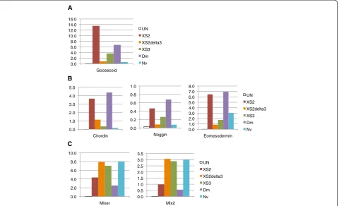
One important contributor to this division of labor be-tween vertebrate Smad2 and Smad3 may have been the evolution of exon 3 in vertebrate Smad2. This exon encodes a 30-amino acid insertion positioned within the MH1 domain immediately adjacent to the predicted DNA-binding hairpin [see Additional file 1]. This inser-tion prevents proper DNA binding by Smad2, but Smad3, lacking this insert, binds DNA. Interestingly, an alternatively spliced version of Smad2 mRNA encodes a protein that does not include exon 3 (known as
Smad2-ΔExon3) and this variant of Smad2 has been shown to bind to DNA [40]. Smad2ΔExon3 splice variant tran-scripts and protein have been found in gastrula stage
Xenopus embryos [41], and various mammalian cell lines. We have tested the ability of Xenopus
Smad2-ΔExon3 to activate Activin/Nodal signaling markers, and our results indicate that the activity of XSmad2ΔExon3
is, more similar to that of XSmad3 and NvSmad2/3 than it is to XSmad2 (Figure 7). The functional importance of exon 3 in Smad2 signaling, and its origin during verte-brate evolution merits further analysis in the future.
The MH2 domain has the largest influence on R-Smad induction capability
The results of our chimeric R-Smad analysis underscore the importance of the MH2 domain as a determinant of gene activation, and illustrate an interesting aspect of se-quence conservation versus signaling activity. The MH2 domain is the most conserved protein domain between R-Smad orthologs from various species (Figure 1B) [see Additional file 1], yet despite this high degree of se-quence conservation, replacement of the MH2 domain in NvSmad2/3 with the XSmad2 MH2 shows the great-est enhancement of NvSmad2/3 activity (Figure 5C, D). This points to the importance of the few amino acid residues that vary between the MH2 domains of Xen-opus and Nematostella proteins, which may not be revealed by natural mutagenesis (for example, cancer mutations) or directed changes. These types of substitu-tions are most frequently reported in the MH2 when they have a significant effect on Smad signaling, such as those of the loop-strand pocket that are involved in re-ceptor docking and specificity [42], those in the co-factor binding hydrophobic pocket [43,44], or those essential to Smad trimerization [45,46]. Our observed patterns of dif-ferential downstream gene induction between species are more subtle than these large effects, and indeed, in the great majority of cases, residues that are reported to be functionally important are conserved across species (Additional file 1). To reveal which residues contribute to the induction patterns reported here, we suggest fur-ther experimentation with chimeric constructs, especially single amino acid replacements of positions known for greater variability.
onto XSmad2 in order to restore DNA binding abi-lity and test whether there is a difference in down-stream gene expression or ability to induce a second axis by XSmad2.
In general, replacing the NvSmad2/3 linker region with that of XSmad2 decreased its inductive ability. Given the low protein level of the linker chimera relative to the other Smad2/3 proteins we assayed (Figure 5B, last column), the XSmad2 linker domain may destabilize the NvSmad2/3 protein structurally or by introduction of additional sequences that direct post-translational modifications. The NvSmad2/3 linker lacks motifs that are essential for these regulatory processes (Figure 1D), including a proline-proline-X-tyrosine (PPXY) consensus motif targeted by Smad ubiquitin-ligases such as Smurf2 [26,48]. Interestingly, we were unable to identify clear Smurf1 or Smurf2 orthologs in the Nematostella ge-nome or ESTs, which appears to correspond to the ab-sence PPXY motifs in either Nematostella Smad. Addition of the Xenopus linker is predicted to cause NvSmad2/3 to undergo a more complex level of regula-tion in vivo in Xenopus embryos than wild type
NvSmad2/3 might in the sea anemone, likely making the chimera sensitive to Smurf2 or NEDD4-L mediated ubi-quitylation and degradation.
Despite its apparent lack of activity on many endoge-nous Xenopus genes, the linker chimera induced down-stream Activin/Nodal target geneseomesodermin, mix.2, and Xbra at levels that approach or exceed those observed in the uninjected whole embryo (Figure 5D, E). This indicates that the linker chimera is not simply non-functional, but instead that its unique combination of se-quence features renders it suited to induce only a subset of Activin/Nodal response genes. To address this possi-bility, it would be interesting to point-mutate some of the specific kinase target residues in the NvSmad2/3 linker to create sites that confer vertebrate-like linker regulation, and test the activities of such mutants. This would help distinguish the effects of linker-driven post-translational regulation from transcriptional activity of the Nematostella nd Xenopus proteins. Conversely, it would be interesting to replace the XSmad2 linker with that of NvSmad2/3 and test whether the decrease in linker regulation sites has any effect on the ability of
0.0 2.0 4.0 6.0 8.0 10.0 12.0 14.0 16.0 Goosecoid UN XS2 XS2delta3 XS3 Dm Nv 0.0 1.0 2.0 3.0 4.0 5.0 Chordin 0.0 0.2 0.4 0.6 0.8 1.0 Noggin 0.0 1.0 2.0 3.0 4.0 5.0 6.0 7.0 8.0 Eomesodermin UN XS2 XS2delta3 XS3 Dm Nv 0.0 2.0 4.0 6.0 8.0 10.0 Mixer 0.0 0.5 1.0 1.5 2.0 2.5 3.0 3.5 Mix2 UN XS2 XS2delta3 XS3 Dm Nv A B C
XSmad2 to activate target marker genes. Our results raise interesting questions about the evolution of R-Smad functions during metazoan diversification. For ex-ample, we would like to understand how differences in R-Smad protein sequences correlate with the acquisition or loss of target genes (and protein cofactors) among testable species in major taxonomic clades, particularly at nodes where Smad gene duplications have occurred or where Smad signaling pathway complexities have been streamlined by genome reduction. This would re-quire a greater breadth ofin vivofunctional tests, assay-ing activities of orthologous Smads between species. A desirable next extension of the present study would be to test wild-type orthologs and chimeric R-Smads in
Nematostella embryonic assays (and ideally Drosophila
embryos as well). Such tests would provide additional in-formation about the evolution of Smad structure and function as well as provide important information about the biological actions of Smad signals in cnidarian germ layer specification and cell fate determination.
Conclusions
In this study we compared and contrasted the signaling activities of the two R-Smads ofNematostella with their bilaterian orthologs, in the context of a developing verte-brate. We find that the BMP-specific R-Smad, NvSmad1/ 5, can pattern (ventralize) the mesoderm ofXenopus laevis
embryos and activate downstream genes in a similar, albeit less efficient, manner than a vertebrate ortholog,Xenopus
Smad1. This speaks to a deep conservation of function within the BMP pathway of bilaterians and earlier-diverging metazoan groups. Further, we find that the Activin R-Smad, NvSmad2/3, is a strong inducer of mesendodermal and definitive endoderm genes, suggest-ing that the development of endoderm via Smad2/3 sig-naling is also an ancient and conserved system. However, the cnidarian NvSmad2/3 fails to induce a secondary body axis in Xenopusembryos and is inconsistent in its ability to activate downstream target genes compared to its bila-terian counterparts XSmad2, XSmad3, and the sole Dro-sophilaAR-Smad, dSmad2.
Based on our results and previous reports, we propose that the bilaterian ancestor solidified a novel role for the Smad2/3 ortholog in controlling body patterning that the NvSmad2/3 is unable to perform. Furthermore, our ani-mal cap assays are the first to test the inductive activities of Smad2 and Smad3 side by side, and indicate different target gene affinities for the two, with XSmad2 having sub-stantially greater effects on organizer-specific genes than general mesendodermal genes, whereas XSmad3 displays converse actions. This demonstrates an intriguing division of labor that leads us to suggest that vertebrate Smad2 has evolved novel activities that govern the vertebrate orga-nizer. Compellingly, the division of labor between these
duplicates is relatively“superficial,” being that both verte-brate AR-Smads and theDrosophilaortholog dSmad2 are capable of patterning dorsal tissues and inducing a se-condary axis inXenopusembryos.
The MH2 domain has a major influence on AR-Smad in-ductive capability, yet this domain is 96% identical in XSmad2 and XSmad3, highlighting the importance of par-ticular residues whose random mutation is not lethal to the organism, but may instead bring about slight functional changes that can be selected on and affect evolutionary di-vergence. Activity tests on a more comprehensive set of R-Smad orthologs gathered from major taxonomic groups should be very informative about the evolution of R-Smad structure/function and target gene regulation.
Additional files
Additional file 1:Protein sequence alignments of R-Smad orthologs.The alignment highlights functionally and structurally important residues and regions present in R-Smads. In the MH1 domain (orange), there is a nuclear localization signal (brick red), a DNA bindingβ hairpin (teal), and residues that make up some or most of the
hydrophobic core of the molecule (purple) [46]. Four residues coordinate a zinc atom at the center of the molecule (green triangles) [49]. In the MH2 domain (pink), there are sites of trimer stabilization (lilac), residues that are critical for trimerization contacts (green stars), residues that contribute to a hydrophobic pocket (blue stars) to bind a cysteine from an adjacent Smad molecule (open blue star), a‘loop strand pocket’ involved with macromolecular interactions (moss green), and two C-terminal serines which are phosphorylated to activate the R-Smad (yellow diamonds) [45]. The loop strand pocket of the MH2 region also contains several residues that bestow receptor specificity [42]. Blue and red boxes indicate residues that are sub-type specific between R-Smads [50]. Important residues in the linker region have already been discussed in detail in Figure 1. Note that this is not meant to be a comprehensive list of R-Smad proteins across phyla or of all residues contributing to R-Smad structure or function; please consult references for studies of these and other proteins and functional sites.
Additional file 2:Table of accession numbers and details about proteins used in the alignments.Details of the orthologs of R-Smads from human,Xenopus laevis,Drosophila melanogaster, andNematostella vectensisused in this analysis.
Additional file 3:Primer sequences and experimental PCR design to create the chimeric constructs.The table contains all primers to create all sections of each of the three chimeric constructs. The diagram shows the primers used to amplify particular sections of the constructs. Full constructs were amplified from combined sections by PCR with end-point primers. Relative lengths of the constructs are depicted. See Methods sections for a full explanation of design and method.
Additional file 4:Loading controls for western blots.Protein translation levels were detected with an antibody to the HA tags of the HA-RSmads expressed from mRNA made in vitro from the pCS2 expression vector. (A) From left to right: protein ladder, XSmad1, NvSmad1, and uninjected control embryos. The non-specific band signals indicate equal protein loading on the gel (blue arrow). (B) Left to right: protein ladder, XSmad2, XSmad3, dSmad2, NvSmad2/3, and uninjected control. 40 kDaβ-Actin loading control band can be seen where indicated (blue arrow). (C) Left to right: protein ladder, water injection (control), uninjected embryo, XSmad2, XSmad3, NvSmad2/3, MH1 chimera, MH2 chimera, and linker chimera. Non-specific bands indicate equal loading across the gel (blue arrow).
Additional file 6:Further examples of Smad2/3 overexpression ‘perturbed axis’phenotypes.Examples of the‘perturbed axis’ phenotype in tadpoles at stages 33 to 34. This phenotype was observed at some level by any of the treatments in our experiments.
Additional file 7:Dosage experiments with three concentrations of XSmad2 and NvSmad2/3.Dosage experiments showed that increasing or decreasing the mRNA concentration does not significantly change the gene induction patterns produced by XSmad2 and NvSmad2/3 in animal cap assays.
Abbreviations
ADMP: Anti-dorsalizing morphogenetic protein; AR-Smad: Activin-Nodal Pathway smad; BMP: Bone morphogenetic protein; BR-Smad: an R-Smad in the BMP pathway; Co-Smad: Common smad; DAI: DorsoAnterior Index; EST: Expressed sequence tag cDNA; I-Smads: Inhibitory smads; MAPK: Map kinase; MH: MAD homology domains in Smad proteins; PXSP: Proline-any-serine-proline peptide consensus; qPCR: Real-time quantitative RT-PCR; R-Smads: Receptor-regulated smads; RT-PCR: Reverse-transcriptase polymerase chain reaction; TGFβ: Transforming growth factor-β.
Competing interests
The authors declare that they have no competing interests pertaining to the work and conclusions submitted herein.
Authors’contributions
GMS carried out the molecular cloning and sequence alignment, performed the microinjections, western blots, and RT-PCR analyses, photographed the specimens, prepared figures, and drafted the manuscript. WQG guided the sequence alignment and RT-PCR analyses, performed initial microinjections, provided essential experimental and intellectual guidance, and edited the manuscript. JOH designed and cloned the chimera constructs, prepared additional figures, and edited the manuscript. GHT provided fundamental experimental and intellectual guidance, provided materials and clones, and edited the manuscript. All authors read and approved the final manuscript.
Acknowledgements
The authors wish to thank M.A. O’Leary for indispensable manuscript assistance and intellectual support, Dr. Spyros Artavanis-Tsakonas for the Drosophila dSmad2clone, and Dr. Malcolm Whitman for the XSmad2ΔExon3 clone. GMS wishes to thank the Gabor Inke Foundation of the Anatomical Sciences Department of Stony Brook University for funding. WQG was supported by a National Institute of Diabetes and Digestive and Kidney Diseases postdoctoral training grant (5T32DK007521) from the National Institutes of Health. JOH is supported by National Institutes of Health Stony Brook Genetics Training Grant 5T32GM007964. GHT is funded by National Institutes of health award number 5R01GM080462.
Author details
1Department of Anatomical Sciences, Stony Brook University, Health Science Center T-8, Stony Brook, NY 11794-8018, USA.2Department of Biochemistry and Cell Biology, Stony Brook University, Life Sciences Building room 450, Stony Brook, NY 11794-5215, USA.3Graduate Program in Genetics, Stony Book University, Life Sciences Building room 120, Stony Brook, NY, USA.
Received: 3 April 2012 Accepted: 9 August 2012 Published: 1 October 2012
References
1. Richards GS, Degnan BM:The dawn of developmental signaling in the metazoa.Cold Spring Harb Symp Quant Biol2009,74:81–90.
2. Matus DQ, Thomsen GH, Martindale MQ:Dorso/ventral genes are asymmetrically expressed and involved in germ-layer demarcation during cnidarian gastrulation.Curr Biol2006,16:499–505.
3. Huminiecki L, Goldovsky L, Freilich S, Moustakas A, Ouzounis C, Heldin CH: Emergence, development and diversification of the TGF-beta signalling pathway within the animal kingdom.BMC Evol Biol2009,9:28. 4. De Robertis EM:Evo-devo: variations on ancestral themes.Cell2008,
132:185–195.
5. Erwin DH:Early origin of the bilaterian developmental toolkit.Philos Trans R Soc Lond B Biol Sci2009,364:2253–2261.
6. Attisano L, Wrana JL:Smads as transcriptional co-modulators.Curr Opin Cell Biol2000,12:235–243.
7. Schmierer B, Hill CS: TGFbeta-SMAD signal transduction: molecular specificity and functional flexibility. Nat Rev Mol Cell Biol 2007, 8:970–982.
8. Massague J, Seoane J, Wotton D:Smad transcription factors.Genes Dev 2005,19:2783–2810.
9. Thomsen GH:Xenopusmothers against decapentaplegic is an embryonic ventralizing agent that acts downstream of the BMP-2/4 receptor.
Development1996,122:2359–2366.
10. Graff JM, Bansal A, Melton DA:XenopusMad Proteins Transduce Distinct Subsets of Signals for the TGFb Superfamily.Cell1996,85:479–487. 11. Callery EM, Smith JC, Thomsen GH:The ARID domain protein dril1 is
necessary for TGF(beta) signaling inXenopusembryos.Dev Biol2005, 278:542–559.
12. Hoodless PA, Tsukazaki T, Nishimatsu S, Attisano L, Wrana JL, Thomsen GH: Dominant-negative Smad2 mutants inhibit activin/Vg1 signaling and disrupt axis formation inXenopus.Dev Biol1999,207:364–379.
13. Brown KA, Pietenpol JA, Moses HL:A tale of two proteins: differential roles and regulation of Smad2 and Smad3 in TGF-beta signaling.J Cell Biochem2007,101:9–33.
14. Howell M, Mohun TJ, Hill CS:XenopusSmad3 is specifically expressed in the chordoneural hinge, notochord, and in the endocardium of the developing heart.Mech Dev2001,104:147–150.
15. Weinstein M, Yang X, Deng C-X:Functions of mammalianSmadgenes as revealed by targeted gene disruption in mice.Cytokine & Growth Factor Rev2000,11:49–58.
16. Jia S, Ren Z, Li X, Zheng Y, Meng A:Smad2and Smad3are required for mesendodermal induction by transforming growth factor-B/nodal signals in zebrafish.J Biol Chem2008,283:2418–2426.
17. Dick A, Mayr T, Bauer H, Meier A, Hammerschmidt M:Cloning and characterization of zebrafishsmad2, smad3,andsmad4.Genes Dev2000, 246:69–80.
18. Jia S, Wu D, Xing C, Meng A:Smad2/3 activities are required for induction and patterning of the neurectoderm in zebrafish.Dev Biol2009,333:273–284. 19. Das P, Inoue H, Baker JC, Beppu H, Kawabata M, Harland RM, Miyazono K,
Padgett RW:Drosophila dSmad2 and Atr-I transmit activin/TGFbeta signals.Genes Cells1999,4:123–134.
20. Brummel T, Absollah S, Haerry TE, Shimell MJ, Merriam H, Raferty L, Wrana JL, O’Connor MB:TheDrosophilaActivin receptor Baboon signals through dSmad2 and controls cell proliferation but not patterning during larval development.Genes Dev1999,13:98–111.
21. Yukita A, Michiue T, Danno H, Asashima M:XSUMO-1 is required for normal mesoderm induction and axis elongation during earlyXenopus development.Dev Dyn2007,236:2757–2766.
22. Kretzschmar M, Doody J, Massague J:Opposing BMP and EGF signalling pathways converge on the TGF-beta family mediator Smad1.Nature 1997,389:618–622.
23. Eivers E, Demagny H, Choi RH, De Robertis EM:Phosphorylation of Mad controls competition between wingless and BMP signaling.Sci Signal 2011,4:ra68.
24. Zhu H, Kavsak P, Abdollah S, Wrana JL, Thomsen GH:A SMAD ubiquitin ligase targets the BMP pathway and affects embryonic pattern formation.Nature1999,400:687–693.
25. Sapkota G, Alarcon C, Spagnoli FM, Brivanlou AH, Massague J:Balancing BMP signaling through integrated inputs into the Smad1 linker.Mol Cell 2007,25:441–454.
26. Lin X, Liang M, Feng X-H:Smurf2 Is a Ubiquitin E3 ligase mediating proteosome-dependent degradation of Smad2 in transforming growth factor-B signaling.J Biol Chem2000,275:36818–36822.
27. Kretzschmar M, Doody J, Timokhina I, Massague J:A mechanism of repression of TGFbeta/ Smad signaling by oncogenic Ras.Genes Dev 1999,13:804–816.
28. Grimm OH, Gurdon JB:Nuclear exclusion of Smad2 is a mechanism leading to loss of competence.Nat Cell Biol2002,4:519–522.
29. Yanai I, Peshkin L, Jorgensen P, Kirschner MW:Mapping gene expression in twoXenopusspecies: evolutionary constraints and developmental flexibility.Dev Cell2011,20:483–496.
31. Baker JC, Harland RM:A novel mesoderm inducer, Madr2, functions in the activin signal transduction pathway.Genes Dev1996,10:1880–1889. 32. Thomsen G, Woolf T, Whitman M, Sokol S, Vaughan J, Vale W, Melton DA:
Activins are expressed early inXenopusembryogenesis and can induce axial mesoderm and anterior structures.Cell1990,63:485–493. 33. Matus DQ, Pang K, Marlow H, Dunn CW, Thomsen GH, Martindale MQ:
Molecular evidence for deep evolutionary roots of bilaterality in animal development.Proc Natl Acad Sci USA2006,103:11195–11200.
34. Magie CR, Pang K, Martindale MQ:Genomic inventory and expression of Sox and Fox genes in the cnidarianNematostellavectensis.Dev Genes Evol2005,215:618–630.
35. Takebayashi-Suzuki K, Funami J, Tokumori D, Saito A, Watabe T, Miyazono K, Kanda A, Suzuki A:Interplay between the tumor suppressor p53 and TGF beta signaling shapes embryonic body axes inXenopus.Development 2003,130:3929–3939.
36. Fuentealba LC, Eivers E, Ikeda A, Hurtado C, Kuroda H, Pera EM, De Robertis EM:Integrating patterning signals: Wnt/GSK3 regulates the duration of the BMP/Smad1 signal.Cell2007,131:980–993.
37. Yeo CY, Chen X, Whitman M:The role of FAST-1 and Smads in transcriptional regulation by activin during earlyXenopus embryogenesis.J Biol Chem1999,274:26584–26590.
38. Esguerra CV, Nelles L, Vermeire L, Ibrahimi A, Crawford AD, Derua R, Janssens E, Waelkens E, Carmeliet P, Collen D,et al:Ttrap is an essential modulator of Smad3-dependent Nodal signaling during zebrafish gastrulation and left-right axis determination.Development2007, 134:4381–4393.
39. Fukuchi M, Imamura T, Chiba T, Ebisawa T, Kawabata M, Tanaka K, Miyazono K: Ligand-dependent degradation of Smad3 by a ubiquitin ligase complex of ROC1 and associated proteins.Mol Biol Cell2001,12:1431–1443.
40. Dennler S, Itoh S, Vivien D, ten Dijke P, Huet S, Gauthier JM:Direct binding of Smad3 and Smad4 to critical TGF beta-inducible elements in the promoter of human plasminogen activator inhibitor-type 1 gene.EMBO J 1998,17:3091–3100.
41. Faure S, Lee MA, Keller T, Dijke P, Whitman M:Endogenous patterns of TGFb superfamily signaling during earlyXenopusdevelopment.
Development2000,127:2917–2931.
42. Lo RS, Chen YG, Shi Y, Pavletich NP, Massague J:The L3 loop: a structural motif determining specific interactions between SMAD proteins and TGF-beta receptors.EMBO J1998,17:996–1005.
43. Randall RA, Germain S, Inman GJ, Bates PA, Hill CS:Different Smad2 partners bind a common hydrophobic pocket in Smad2 via a defined proline-rich motif.EMBO J2002,21:145–156.
44. Schiro MM, Stauber SE, Peterson TL, Krueger C, Darnell SJ, Satyshur KA, Drinkwater NR, Newton MA, Hoffmann FM:Mutations in protein-binding hot-spots on the hub protein Smad3 differentially affect its protein interactions and Smad3-regulated gene expression.PLoS One2011, 6:e25021.
45. Wu JW, Hu M, Chai J, Seoane J, Huse M, Li C, Rigotti DJ, Kyin S, Muir TW, Fairman R, Massagué J, Shi Y:Crystal structure of a phosphorylated Smad2. Recognition of phosphoserine by the MH2 domain and insights on Smad function in TGF-beta signaling.Mol Cell2001,8:1277–1289. 46. Shi Y, Wang YF, Jayaraman L, Yang H, Massague J, Pavletich NP:Crystal
structure of a Smad MH1 domain bound to DNA: insights on DNA binding in TGF-beta signaling.Cell1998,94:585–594.
47. Jones JB, Kern SE:Functional mapping of the MH1 DNA-binding domain of DPC4/SMAD4.Nucleic Acids Res2000,28:2363–2368.
48. Chen C, Matesic LE:The Nedd4-like family of E3 ubiquitin ligases and cancer.Cancer Metastasis Rev2007,26:587–604.
49. Chai J, Wu JW, Yan N, Massague J, Pavletich NP, Shi Y:Features of a Smad3 MH1-DNA complex. Roles of water and zinc in DNA binding.J Biol Chem 2003,278:20327–20331.
50. Hao R, Chen L, Wu JW, Wang ZX:Structure of Drosophila Mad MH2 domain.Acta Crystallogr Sect F Struct Biol Cryst Commun2008,64:986–990.
doi:10.1186/2041-9139-3-22
Cite this article as:Sorrentinoet al.:Conservation and evolutionary divergence in the activity of receptor-regulated smads.EvoDevo2012 3:22.
Submit your next manuscript to BioMed Central and take full advantage of:
• Convenient online submission
• Thorough peer review
• No space constraints or color figure charges
• Immediate publication on acceptance
• Inclusion in PubMed, CAS, Scopus and Google Scholar
• Research which is freely available for redistribution