R E V I E W
Open Access
What is a segment?
Roberta L Hannibal
1,2and Nipam H Patel
1*Abstract
Animals have been described as segmented for more than 2,000 years, yet a precise definition of segmentation remains elusive. Here we give the history of the definition of segmentation, followed by a discussion on current controversies in defining a segment. While there is a general consensus that segmentation involves the repetition of units along the anterior-posterior (a-p) axis, long-running debates exist over whether a segment can be composed of only one tissue layer, whether the most anterior region of the arthropod head is considered segmented, and whether and how the vertebrate head is segmented. Additionally, we discuss whether a segment can be composed of a single cell in a column of cells, or a single row of cells within a grid of cells. We suggest that‘segmentation’be used in its more general sense, the repetition of units with a-p polarity along the a-p axis, to prevent artificial classification of animals. We further suggest that this general definition be combined with an exact description of what is being studied, as well as a clearly stated hypothesis concerning the specific nature of the potential homology of structures. These suggestions should facilitate dialogue among scientists who study vastly differing segmental structures.
Keywords:Evolution, Metamere, Pseudosegment, Segmentation
Why is the definition of segmentation important?
‘The only dogmatic statement we are justified in
making is, that when a region exhibits during development a sufficient number of the essential structures of a typical segment, it may be assumed to
be at true metamere. What is“sufficient”has to be
decided in each case.’ES Goodrich, 1897 [1]
‘It is difficult to find a [concept] in the whole of
zoology that is so vaguely defined, but, at the same
time, so universally employed as…metamerism.’RB
Clark, 1964 [2]
Arthropods, annelids, and chordates are some of the most successful and diverse animal groups, as defined by species number and anatomical complexity. The success of these groups may be due to their segmented trunk re-gion, which may enhance locomotion and feeding [2-4]. While these groups share the trait of trunk segmenta-tion, it is controversial whether segmentation is homolo-gous, since these groups are more closely related to unsegmented phyla than to each other (Figure 1A) [5-11].
If segmentation is difficult to evolve, the most parsimonious explanation for three unrelated segmented groups would be that segmentation evolved once, but was subsequently lost in all taxa related to the arthropods, annelids, and vertebrates. In this case, we would expect to find evidence of loss of segmentation in related groups. Conversely, if segmentation is easy to gain, we would expect to find multiple unrelated segmented taxa. In fact, there may be such evidence in modern and fossil taxa, as there are a number of groups besides the arthropods, annelids, and vertebrates that display serially repeated units, and could therefore be considered segmented (Figure 1B) [7,12,13]. However, in order to use these groups to infer the evolutionary history of segmentation, we must first resolve whether these taxa are actually segmented. The main obstacle in resolving this issue is that there is no precise definition of segmentation. Instead, there is a range of definitions, depending on what animals and what parts of these animals were studied by a particular author. Here, we give an overview of the various definitions of segmentation, and we discuss current controversies in defining a segment.
The history of the definition of segmentation
The Greeks first recorded the observation that some ani-mals are made of segments, reiterated units along the
* Correspondence:[email protected] 1
Departments of Molecular and Cell Biology and Integrative Biology, University of California, 519A LSA #3200, Berkeley, CA 94720-3200, USA Full list of author information is available at the end of the article
anterior-posterior (a-p) axis. Aristotle [15] named and
classified a group of animals as‘insects’, because of their
segmental nature. Entomon (‘insect’in Greek) is derived
from the Greek word entomos, meaning ‘to cut up’, and
was used because these animals had ‘nicks’ or ‘cuts’ on
their back or bellies, corresponding to boundaries be-tween segments. Latin and related modern languages
continued this theme, as the English word ‘insect’ is
derived from the Latin insecure, which also means ‘to
cut up’.
The scientific revolution of the seventeenth century brought science to the forefront of Western society and set the stage for renewed interest in segmentation in the nineteenth century. During the 1800s, Cuvier grouped arthropods and annelids into the now defunct taxon
Articulatabecause of their similar segmental
morpholo-gies [16]. Goodrich also considered arthropod and anne-lid segments homologous. From studies on arthropods and annelids, Goodrich defined a segment as a unit, marked off from the rest of the body by transverse grooves, containing a mesodermal hollow space (coelom), a pair of nephridia (excretory glands), and a pair of ventral ganglia [1]. Goodrich also noted that, in polychaetes and arthropods, a segment also contains a pair of appendages. Besides these morphological characteristics, Goodrich used a developmental characteristic, the sequential addition of segments from anterior to posterior, to define segmentation.
Goodrich’s definition does not accurately describe all
segments. As Goodrich acknowledged, segments con-taining all of the above criteria are rarely found, al-though some features can transiently be seen during
development [1]. Also, Goodrich’s definition excludes
segmentation in the arthropod Drosophila and in the
chordates. InDrosophila, segments are formed by
simul-taneously subdividing the entire body, in contrast with
Goodrich’s developmental requirement of adding
ments from the posterior end [1,17]. In chordates, seg-ments are added progressively from the posterior, but
they do not have a number of Goodrich’s other
morpho-logical characteristics [18]. Another caveat to Goodrich’s
definition is that if the formation of reiterated structures is linked, then using all of them to define a segment would be no more informative then using one of them [19]. For example, segmentation of the coelom, neph-ridia, and ganglia might all be based on the same mo-lecular pattern. Then, since all three traits would be a read-out of the same pattern, any or all of them could be used equivocally to define that segmental pattern.
Around the same time as Goodrich, Bateson defined
segmentation as a‘more-or-less’coincident repetition of
elements from many organ systems along the a-p body axis [20]. Unlike Goodrich, however, Bateson did not de-fine what these elements had to be, and based his defin-ition on trunk segmentation in vertebrates, as well as
segmentation in arthropods and annelids. While Bateson’s
definition is applicable to trunk segmentation in the ar-thropods, annelids and chordates, many scientists prefer a more precise definition.
While segments can be thought of as the repetition of a variety of structures, Clark suggested that reiteration of coelomic sacs and accompanying muscle was the de-fining characteristic of a segment [2,3]. The coelom is a fluid-filled body cavity derived from the mesoderm. Clark suggested that segmentation involving the coelom and muscles has evolved because of the need for better locomotion, as a large, unsegmented, coelomic sac would have impeded movement. Clark proposed that the division of the coelom facilitates movement by allowing Chordates Echinoderms Hemichordates Arthropods Kinorhynhs Nematodes Nematomorphs Priapulids Annelids Brachiopods Bryozoa Gastrotrichs Molluscs Nemertea Rotifers Platyhelminthes
Segmented Phyla
A
Chordates Echinoderms Hemichordates Arthropods Kinorhynhs Nematodes Nematomorphs Priapulids Annelids Brachiopods Bryozoa Gastrotrichs Molluscs Nemertea Rotifers Platyhelminthes
Segmented and Pseudosegmented Phyla
Conflicting reports on whether Pseudosegmented
B
Figure 1Phylogenetic relationship among segmented and unsegmented phyla.Phylogeny of bilatarians based on [14]; segmented and pseudosegmented animals identified after [7,12,13].
the body to bend at the regions between compartments and that this division of the coelom is accompanied by muscle segmentation.
Clark’s theory could potentially explain the advantage
of segmentation in the arthropods, annelids, and chor-dates, since segmentation could have evolved for im-proved locomotion in all of these groups. However, segmentation does not seem to be correlated with any kind of locomotion. Clark himself acknowledged that ribbon-like animals swim in the same manner whether they are segmented or not, although he suggested that extant animals might not be good representatives of the ancestors of segmented phyla [2]. Like swimming, bur-rowing does not correlate with segmentation, since many burrowing worms are not segmented [13,21].
Another caveat to Clark’s theory is DuPorte’s [22]
ar-gument that,‘There is no real foundation for the belief
in a fundamental relation between coelomic sacs and
metamerism.’ DuPorte suggests that coelomic sacs may
be a stage in mesoderm differentiation and therefore does not have a direct relationship to segmentation. He based this on the observation that protostome meso-derm originates as a solid mass, but only differentiates into coelomic sacs after this mass has already been
di-vided into segments. Clark’s definition of segmentation
has also been used sparingly in modern times, perhaps because of the emergence of the arthropod ectoderm as a model for segmentation. Experimental evidence sug-gests that, in arthropods, the ectoderm can segment nor-mally without the mesoderm [23-27].
Analysis of modern molecular data has also failed to produce a concise definition of segmentation. Instead of finding a few genes and mechanisms that could be used to define segmentation, studies have yielded a large number [28]. Even when homologous genes or gene families are involved in segmentation, they often play different roles in different animals [8,11,29]. For ex-ample, although members of the Notch, Wnt, and fibro-blast growth factor signaling pathways oscillate to produce segmental trunk mesoderm in mouse, chick, and zebrafish, the individual genes that oscillate differ between species [29]. This may be only a minor differ-ence, in that it might not matter which component of the pathway oscillates, as long as some components os-cillate [10]. Major differences are also found, especially among different species of arthropods. Within arthro-pods, there are multiple changes in which genes appear to be involved in segmentation, and, unlike many other
arthropods and vertebrates, Drosophila does not have
any evidence of Notch or other oscillators (for example, see [30-33]). Moreover, morphologically similar seg-ments can be formed by different developmental and molecular mechanisms in the same animal. For example, although all of the somites, segmental units in vertebrate
trunk mesoderm, appear morphologically homologous in zebrafish, there is variation in how they form, depending on their position along the a-p axis [34]. The develop-ment of the anterior trunk, posterior trunk, and tail so-mites depend on different genes or have different degrees of dependence on the same genes, or both. In-stead of yielding a precise definition of a segment, mod-ern molecular studies have highlighted the complexity of segmentation.
Current controversies in the field of segmentation
While there is a general agreement that segmentation involves reiterated units along the a-p body axis, there are still a number of points of contention. The major de-bates surrounding the definition of a segment are: (1) whether a segment can be composed of only one tissue layer, (2) whether the anterior arthropod head is consid-ered segmented, and (3) whether and how the vertebrate head is segmented. An additional complicating factor for defining segmentation is whether a segment can be com-posed of a single cell in an a-p column of cells, or a sin-gle row of cells within a grid of cells. We will discuss this issue first, as it has bearing on the contentions over segmentation in the literature.
Can a segment be a single cell in a column of cells?
During development, some animals have an arrangement of cells along the a-p axis in which each single cell (or row of single cells) could be considered a segment. For
example, the notochord of the sea squirtCiona savignyi
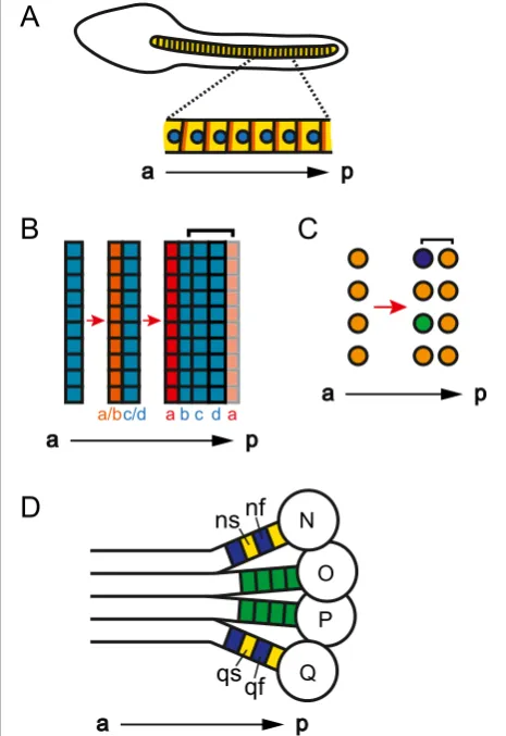
is composed of a single column of cells (Figure 2A) [35].
Similarly, the trunk of the arthropodParhyale hawaiensis,
as well as the trunks of other malacostracan crusta-ceans, is composed of columns of cells where segments arise from single-cell-wide rows within a grid of cells (Figure 2B,C) [36-38]. For a column of cells along the a-p axis to be considered a column of segments, each cell or row of cells needs a definable anterior and terior (Figure 3A,B) [39]. Having an anterior and a pos-terior distinguishes each cell from its neighbors, while making each cell a reiteration of a unit. In segments composed of two or more rows of cells, an a-p segmen-tal pattern can be accomplished by having different morphology or gene expression in anterior versus pos-terior rows of a single segment (segment polarity). In segments composed of only a single cell, or that are only a single cell wide, the anterior and posterior of the single cell must exhibit molecular or morphological a-p asymmetry (cell polarity).
The most obvious molecular mechanism to distinguish the anterior from the posterior of cells is the planar-cell polarity pathway. Indeed, expression of members of this pathway supports the hypothesis that each cell in the
(Figure 2A) [35]. By expressing taggedCionaPrickle and
Strabismus proteins, Jiang et al. [35] revealed
localiza-tion of these proteins at the anterior edge of each noto-chord cell. Additionally, they found that each notonoto-chord nucleus is asymmetrically positioned near the posterior
of each cell. These data show that eachCionanotochord
cell has a-p cell polarity, supporting the classification of
the Ciona notochord as segmented, with the segmental
units being single cells along the a-p axis. Figure 2A single cell (row) may be a segment. (A)Lateral view
of aCiona savignyilate tailbud-stage embryo where the notochord is composed of a column of single cells (yellow). The planar-cell polarity proteins Prickle and Strabismus (orange) are located at the anterior and the nucleus (blue) is located at the posterior of each cell. (Aafter [35].)(B, C)Segmentation in the trunk ofParhyale hawaiensis. Left side of the embryo is depicted, right side is mirror image. Red arrows represent progression in time.(B)Once ectodermal cells condense into rows (PSPRs), each PSPR divides to produce one parasegment of ectoderm. After the first PSPRs division,Ph-hedgehog
(Ph-hh, orange) is expressed in the anterior row (row a/b) [40]. After the second division, both En andPh-hh(red) are expressed in the anterior row (row a) [36]. While the division of one PSPR produces one parasegment of ectoderm, in general, one segment’s worth of ectoderm (bracket) forms from the Engrailed (En) negative cells of one parasegment (rows b to d), and the En positive cells from another parasegment (row a) [36].(C)One row of mesoblasts produces one segment’s worth of mesoderm (bracket). After the first mesoblasts division,Ph-twist(green) andPh-even-skipped(purple) are expressed in a subset of the anterior daughters [27,37,40].(D)Segmentation in the leech ectoderm. One side of the embryo is depicted, other side is mirror image. The ectoderm is formed from the progeny of four ectoteloblasts, N, O, P, and Q, [41]. Each progeny, or blast cell (green), of O and P gives rise to one segmental unit. However, two adjacently produced blasts cells from N and Q, ns (yellow) and nf (blue) and qs (yellow) and qf (blue), respectively, give rise to one segmental unit. a, anterior; p, posterior, PSPR, parasegment precursor row.
A
[
?
a
p
B
[
a
p
C
[
a
p
D
[
a
p
Figure 3A segment can be composed of one or more tissue layers.Bracket marks one cell or segment, except in(A), where it marks one putative cell or segment.(A)Yellow circles represent cells (putative segments) in one layer of tissue.(B)Each cell in a column of cells can be called a segment if there is a-p cell polarity in each cell. Cells (circles) now have a-p cell polarity, represented by difference in coloration from yellow (anterior) to red (posterior).
UnlikeCiona, a-p cell polarity has not yet been found in
the trunk of the malacostracan arthropod Parhyale
hawaiensisduring the stage where segments, or more
ac-curately for the ectoderm, parasegments, are destined to form from single-cell-wide rows of cells within a grid of
cells.Parhyalesegments are composed of both ectoderm
and mesoderm. In the trunk, ectodermal parasegments form via the division of parasegment precursor rows (PSPRs), while mesodermal segments form via the division of mesoblasts, which are produced via the asymmetrical division of mesodermal stem cells (mesoteloblasts;
Figure 2B,C) [36,37]. In Parhyale, each
parasegment/seg-ment has polarity after the first division of the PSPRs and mesoblasts, as there is differential gene expression in the anterior versus posterior row of each segment (Figure 2B,C) [27,37,40]. It will be interesting to determine whether a-p polarity is established before PSPR and mesoblast division, as well as what molecular pathways govern this patterning. As both cell types divide along the a-p axis, there may be intrinsic asymmetric determinants, and therefore a-p cell polarity, in the PSPRs and mesoblasts. Alternatively, the PSPRs and mesoblasts may only have intrinsic instructions for dividing in an a-p orientation, and then require subse-quent signaling from the anterior of the embryo to distin-guish the anterior daughter from the posterior daughter.
The discovery of molecular markers of a-p polarity in
Parhyale segmental precursor cells will also resolve the
question of whether segmental precursor cells are seg-ments if they do not divide. If the ectoderm is ablated in
Parhyale, mesoteloblasts still divide to form mesoblasts
[27]. However, these mesoblasts do not divide nor do they express known markers of segment polarity nor-mally seen after the first mesoblast division. If these mesoblasts have a-p cell polarity, they would be seg-ments, and segmentation of the ectoderm and meso-derm could be considered to occur independently of one another. If individual mesoblasts are not considered seg-ments because they lack a-p polarity, then the mesoderm requires either a permissive or an instructive signal from the ectoderm to gain segmental identity. An instructive signal from the ectoderm would further complicate the debate on defining a segment. If the ectoderm is re-quired to impart a segmental pattern onto the
meso-derm, then segmentation in Parhyale could be seen as
only an ectodermal characteristic, and the general defin-ition of segmentation would then include that the seg-mental pattern must be an intrinsic property of the germ layer being studied. We do not suggest using in-trinsic pattern as a criteria for segmentation, as gather-ing this level of information would make it hard, if not impossible, to define even universally agreed upon seg-mental tissue as segmented.
As with malacostracan arthropods, segments in anne-lids can form from single-cell-wide precursors. In the
leech, segments form from the asymmetrical division of teloblasts (Figure 2D) [41]. On either side of the body, progeny of four ectoteloblasts, N, O, P, and Q, and one mesoteloblast, M, come together to form a segment. Each progeny, or blast cell, of M, O, and P gives rise to one segmental unit. Although, unlike the malacostracan
Parhyale, where a single-cell-wide row of cells gives rise
to one segment/parasegment, in the leech, the progeny of each blast cell can spread over more than one seg-ment and intermix with progeny of neighboring blast cells. Moreover, for the leech teloblasts N and Q, two adjacently produced blast cells give rise to one segmental unit. N alternatively gives rise to the blast cells ns and nf, while Q alternatively gives rise to qs and qf [41,42]. These alternate blast cells have different fates, as ns gives rise mostly to anterior neurons and epidermis, while nf gives rise to mostly posterior neurons, peripheral neu-rons, and neuropil glia, and qs gives rise to both ventral and dorsal cells, while qf only gives rise to dorsal cells [42]. Experiments with the more tractable N lineage sup-port the hypothesis that ns and nf are different from birth. Ablation experiments indicate that ns and nf are not an equivalence group [42]. Additionally, molecular segment polarity is found in the progeny, as the acti-vated form of the cell cycle protein Cdc42 is expressed in higher levels in ns versus nf [43]. These data suggest that N, and by extension, Q, produce segments with in-trinsic segmental polarity. Therefore, the N and Q lineage provide a model for further studies on how telo-blasts may impose intrinsic segmental polarity on their progeny. It will be interesting to explore whether N and Q use similar mechanisms as the single progeny telo-blasts M, O and P, and also as telotelo-blasts in other systems such as the malacostracans.
The vertebrate trunk axons are an excellent example of how segmental pattern can be non-intrinsic. Verte-brate trunk axons are arranged in a reiterated pattern along the a-p axis. These axons contribute to the overall segment polarity within each trunk segment by running through the anterior half of each somite [44]. However, the segmental arrangement of axons is extrinsic, caused by axon guidance cues in the anterior half of the somite. Therefore, the segmental pattern of axons is purely dependent on the polarity of the somites. These data suggest that trunk axons must be considered with the somites in order to be segmental.
Can a segment be composed of only one tissue layer?
often called pseudosegments or metameres, depending on
the author [7,12,13]. Here, we will use ‘pseudosegments’
to refer to these structures, as‘metameres’has often been
used as a synonym for segments (for example, [1]). While the only difference between the pseudosegmen-ted and segmenpseudosegmen-ted animals may be taxonomic classifica-tion, one possible biological difference could be that the arthropods, annelids, and chordates are the only groups with segments composed of both ectodermal and meso-dermal derivatives. Segments in arthropods and annelids are derived from both the ectoderm and mesoderm and have segmental pattern in both tissue layers (Figure 3C) [11,25]. In vertebrates, although trunk segments are composed of both the ectoderm-derived nervous system and the mesoderm, there is only segmental pattern in the mesoderm (Figure 3D). There is pattern in the ectoderm-derived rhombomeres of the vertebrate head, but, as there is contention about the segmental status of rhombomeres, they will be discussed in more depth in a later section.
Some pseudosegmented animals have reiteration only in the ectoderm, but there are other so-called pseudo-segmented animals with reiterations in both the ecto-derm and the mesoecto-derm. The bdelloid rotifers, some species of nematodes, and chiton, a type of mollusk, only have reiteration in the ectoderm [12]. Bdelloid rotifers have repeated rings of intraepithelial skeletal laminae, some species of nematodes have repeated cuticular rings, and chitons have reiterated dorsal plates. However, other so-called pseudosegmented animals have reiterations in both the ectoderm and the mesoderm. The bodies of kinorhynchs are composed of 13 to 14 units with repeti-tive ganglia, muscles, and epidermal and cuticular struc-tures [12]. Therefore, the criteria of segments being composed of both ectoderm and mesoderm is not
suffi-cient to separate ‘classically’ segmented and so-called
pseudosegmented animals. Moreover, as there are many invertebrate animals whose development and anatomy are still not well characterized, there are likely to be more species that have segmental pattern in at least one tissue layer. More data on pseudosegmented animals will also help determine whether there is an actual distinc-tion between reiterated structures in segmented versus pseudosegmented phyla.
An evolutionary argument also suggests that requiring segments to be composed of both ectodermal and meso-dermal derivatives, versus one tissue layer, is an artificial distinction [45]. According to Budd [45], instead of con-sidering segmentation as a property of the entire animal, segmentation should be thought of as a property of an organ system. Budd defines segmentation as a character-istic of organ systems, because he views the evolution of segmentation as a gradual accumulation of reiterated organ structures. If one organ system becomes reiterated
first and another system becomes reiterated later in evo-lution, then the distinction between having multiple reit-erated structures, or segments, and having only one reiterated structure, or pseudosegments, is artificial. Therefore, any reiterated organ system should be consid-ered segmented, and there would be no reason to con-sider the segmented organ systems of arthropods, annelids, and chordates as distinct from the segmented organ systems of pseudosegmented animals.
Just as segmentation could evolve in one organ system at a time, segmentation could also be secondarily lost in one organ system but not another. Evidence for second-ary simplification is found in Echiura and in mollusks. Echiura is a group of marine worms that are most prob-ably highly derived annelids [46-48]. While Echiura share many developmental traits with the annelids, they lack the epidermal and muscular segmentation of bona fide segmented worms, leading to debate over their rela-tionship. However, recent phylogenies place them within the annelids [47,48]. Moreover, immunohistochemical analysis of neuronal markers shows that the nervous
sys-tem of the Echiura Bonellia viridis is arranged in an
or-ganized, serial fashion, similar to the nervous systems of segmented worms [46]. These data suggests that the Echiura evolved from a segmented ancestor and later lost most segmental characteristics. As with the case of Echiura, segmentation may have been secondarily lost in some mollusks. The cephalopods are considered to be unsegmented. However, recent phylogenies group them with the Monoplacophorans, shelled deep-sea mollusks that have the segmental characteristics of serially re-peated gills, nephridia, and muscles [49,50]. While these data could suggest that Monoplacophorans independ-ently evolved segmentation, closer examination of the chambered nautilus instead suggests that segmentation may have been lost in cephalopods [51]. The nautilus has two pairs of gills, kidneys, and atria, which can be interpreted as secondary simplification from a seg-mented ancestor. These examples of probable secondary loss of segmentation in annelids and mollusks suggest that there is no biological distinction between segments composed of many versus one organ system, and there-fore argue against the requirement that segments be composed of multiple tissue layers.
Is the tip of the arthropod head a segment?
the context of how phylogenetic assumptions can influ-ence segmental classifications.
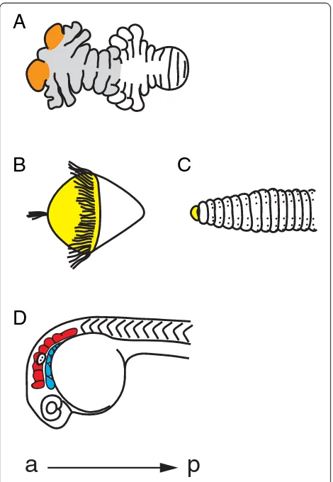
The anterior-most region of the arthropod head, the ocular lobe (protocerebrum), is different from head and trunk segments, as it contains the brain and does not bear a set of antennae or other appendages (Figure 4A) [52,53]. However, the ocular lobe also has many mor-phological similarities to the rest of the segments, mak-ing it hard to classify as either unsegmental or segmental [22,52,53]. The difficulty in using morphology to classify the ocular lobe as a segment may have lead
researchers to depend overly on phylogenetics to solve this question. Before the new molecular phylogeny, the
Articulata hypothesis placed the annelids as close
rela-tives to the arthropods. Therefore, it was assumed that the arthropods had an unsegmented anterior region, homologous to the unsegmented anterior region, or pro-stomium, of annelids (Figure 4B,C) [1,52]. The annelid prostomium lies in front of the mouth and contains the brain and sense organs. The prostomium is considered unsegmented because its embryonic origin is different from the segmented body and because it does not have characteristics of other segments, such as coelomic sacs and nephridia [52]. Additionally, in annelid species that have a trochophore larvae, the prostomium (episphere), is located anterior to the first ciliary ring (prototroch). If the annelid prostomium and the arthropod ocular lobe were homologous, then, based on annelid data, the ocu-lar lobe would not be considered a segment. However, since the new molecular phylogeny places the arthro-pods and annelids in two separate megagroups of the bilatarians, the homology of the annelid prostomium and arthropod ocular lobe, and thus the unsegmented nature of the ocular lobe, has come into dispute [5].
While molecular data likely resolved the relationship between arthropods and annelids, molecular studies have not yet solved the question of whether the ocular
lobe is a segment. In Drosophila, analysis of mutations
in genes with roles in head development suggests that it is a segment [55]. Also, segmental and appendage genes are expressed in the ocular lobes of many arthropod spe-cies [36,55-61]. However, expression of segment polarity or appendage genes is limited to one, often transient, re-gion per ocular lobe, unlike other segments, where there is a persistent domain of strong expression. This may be due to the highly derived nature of the head, or, since these genes are all pleiotropic, this may indicate a funda-mental difference in the ocular lobe versus the undis-puted segments of the rest of the body. While this problem may never be fully solved, studying the arthro-pod head will undoubtedly yield interesting insights that would never be uncovered if the assumption about head homology had not been questioned.
What parts of the vertebrate head are segmented?
Vertebrates have structures in their heads that could be considered segmental, but that are distinct from their trunk segments. Controversy exists on whether these head structures are segmented as they do not easily fit into the already tenuous definition of segmentation formed from studies on trunk segments. Moreover, re-searchers have divided the vertebrate head it into seg-ments in numerous different ways, some of which may be artificial. To clarify what parts of the head are most likely to be segmented, here we give an overview of how
A
B
C
D
a
p
Figure 4Annelid, arthropod, and vertebrate heads. (A)The segmental nature of the anterior region, or ocular lobe, of the arthropod head is disputed (orange). Ventral view of a 96-hour
some head structures are probably misconstrued as seg-mented, followed by a brief discussion of the head struc-tures that have the most evidence for being considered segmented, the rhombomeres and pharyngeal arches.
The vertebrate head has been divided into segments in many different ways [62-70]. While the vertebrate head probably contains a number of independently segmented structures, there is little or no reliable morphological or molecular data to support some of these claims. Two ex-amples are the somitomeres and prosomeres. Somito-meres are defined as segmental structures of the paraxial mesoderm that resemble somites, the trunk mesodermal segments [66,67]. Somitomeres are an attractive theory, as their existence would suggest that the head and trunk share a unified segmental developmental program. How-ever, there is only disputed scanning electron micro-scope data to support their existence, and no data to support a shared segmental program between the verte-brate head and trunk. Prosomeres, or forebrain seg-ments, would also provide a framework to organize the vertebrate head [70,71]. However, while the forebrain may be partitioned, there is neither repeated morpho-logical pattern nor molecular segmental polarity to sup-port segmentation.
Hox gene expression has often been used to
sup-port the claim that the vertebrate head is segmented
(for example, see [63,67]). Hox gene expression often
correlates with anterior segmental or parasegmental boundaries and Hox proteins determine what type of structure will form from each segment [72,73]. However,
this does not imply that Hox genes are a marker of
segmentation and, therefore, Hox expression should
not be used to define a body region, such as the head, as
segmented. In support of not equating nested Hox gene
expression to segmentation, knock-out, and overexpression
of Hox genes alters segment identity, but do not prevent
the formation of segments [72,73]. Moreover, many
un-segmented animals expressHoxgenes along their a-p axis
but are not segmented, no matter what definition is used (for example, see [74,75]). Although clearly important
for patterning segmental structures,Hoxgene expression
by itself should not be used as molecular evidence of segmentation.
While more evidence is needed to support many of the claims for segmentation in the vertebrate head, there is morphological and molecular evidence to support two structures, the rhombomeres and pharyngeal arches, as segmented (Figure 4D) [66]. The rhombomeres are seven transient compartments in the chordate hindbrain that control neural organization and architecture [76]. If each rhombomere were a segment, we would expect a repeated pattern of segment polarity, such as the expres-sion of a gene in only the anterior or posterior part of each rhombomere. Instead, there is a two-rhombomere
periodicity of gene expression, where the ephrin ligands
are expressed in even-numbered rhombomeres, and their
receptors, theEphs, are expressed in odd-numbered
rhom-bomeres [77]. While this is often compared to the
two-segment periodicity of pair-rule genes inDrosophila, each
segment inDrosophilaultimately has its own segment
po-larity [17]. There is still popo-larity in each rhombomere, however, since motor neurons and their axon trajectories have a repeated pattern in each rhombomere [68].
Pharyngeal arches also have segmental characteristics. The pharyngeal arches are bulges on the lateral surface of the embryonic head that give rise to skeletal and mus-cular derivatives, sensory ganglia, and motor innerva-tions [78]. The pharyngeal arches are composed from ectoderm, mesoderm, endoderm, and neural crest cells. Together, these tissues are arranged so that there is a re-iterated pattern within each arch. Morphologically, car-tilage is in the anterior region while a blood vessel is in the posterior region. Molecularly, the ETS-type
tran-scription factor polyomavirus enhancer activator 3 is
expressed in the anterior mesenchyme and in the posterior epithelium [79]. These data suggest that pharyngeal arches are segmented.
Despite their segmental characteristics, rhombomeres and pharyngeal arches are often not considered in com-parisons of segmentation among the arthropods, annelids, and chordates. Perhaps this is because rhombomeres and the neural crest component of pharyngeal arches are vertebrate innovations, and therefore do not have homologous counterparts in the arthropods and annelids. Although, as some authors suggest, these seemingly verte-brate specific segmental head structures could have been superimposed upon an ancestral segmental body plan, similar to that of the arthropods and annelids [63] (see [67] for a persuasive counterpoint). Most importantly, the a-p pattern within each rhombomere and pharyngeal arch suggest that they are segmental and therefore should be considered in further studies and discussions of segmenta-tion. As vertebrate innovations, they are especially interest-ing as models of segmental evolution in novel structures.
Conclusions
To bring the focus back to developmental and
evolu-tionary biology, we suggest that ‘segmentation’ be used
in its most general meaning: the repetition of units along the a-p axis, where each unit has a-p polarity [20,45]. This inclusive definition should then be combined with an exact description of the segmented structures in the animals or systems being discussed, as well as a clearly stated hypothesis concerning the specific nature of the po-tential homology of structures. While we support a gen-eral definition of segmentation, it is also crucial that authors be explicit in what they are implying about ances-tors and shared traits versus convergence, to facilitate the advancement of new ideas, versus circular discussions.
A broader definition of segmentation could also im-pact molecular studies. New studies on segmentation should examine multiple genes, since there is a wide range of molecular mechanisms involved in segmenta-tion, even within groups with segmental homology. Ad-vancements in sequencing technologies have made it possible to find and analyze many genes involved in the segmentation process simultaneously. Moreover, these studies can yield an unbiased list of genes involved in segmentation in a particular organism or structure, as opposed to the candidate-gene approach used in the past. It will be exciting to see how broader knowledge about segmental molecular mechanisms impacts our thoughts on the core features of segmentation and the shared homology among segmented animals.
Abbreviations
a-p:anterior-posterior; En: Engrailed;Ph-hh:Parhyale-hedgehog; PSPR: Parasegment precursor row.
Competing interests
The authors declare that they have no competing interests. No funding was received for this review.
Authors’contributions
RLH drafted the manuscript. NHP critically revised the manuscript. RLH and NHP read and approved the final manuscript.
Acknowledgements
We thank Craig Miller and David Weisblat for discussions on the nature of segmentation. We thank David Stafford and Craig Miller for helpful comments on the manuscript.
Author details
1Departments of Molecular and Cell Biology and Integrative Biology, University of California, 519A LSA #3200, Berkeley, CA 94720-3200, USA. 2Present Address: Department of Genetics, Stanford University School of Medicine, Stanford, CA 94305, USA.
Received: 15 July 2013 Accepted: 19 November 2013 Published: 17 December 2013
References
1. Goodrich ES:On the relation of the arthropod head to the annelid prostomium.Q J Microsc Sci1897,40:247–268.
2. Clark RB:Dynamics in Metazoan Evolution: The Origin of the Coelom and Segments.Oxford: Clarendon Press; 1964.
3. Clark RB:The evolution of the celom and metameric segmentation. In
The Lower Metazoa: Comparative Biology and Phylogeny.Edited by Dougherty EC. Berkeley: University of California Press; 1963:91–107. 4. Tautz D:Segmentation.Dev Cell2004,7:301–312.
5. Aguinaldo AMA, Turbeville JM, Linford LS, Rivera MC, Garey JR, Raff RA, Lake JA:Evidence for a clade of nematodes, arthropods and other moulting animals.Nature1997,387:489–493.
6. Chipman AD:Parallel evolution of segmentation by co-option of ancestral gene regulatory networks.Bioessays2010,32:60–70.
7. Couso JP:Segmentation, metamerism and the Cambrian explosion.Int J Dev Biol2009,53:1305–1316.
8. Davis GK, Patel NH:The origin and evolution of segmentation.Trends Genet1999,15:M68–M72.
9. Peel A, Akam M:Evolution of segmentation: rolling back the clock.
Curr Biol2003,13:R708–R710.
10. Richmond DL, Oates AC:The segmentation clock: inherited trait or universal design principle?Curr Opin Genet Dev2012,22:600–606. 11. Seaver EC:Segmentation: mono-or polyphyletic?Int J Dev Biol2003,
47:583–596.
12. Minelli A, Fusco G:Evo-devo perspectives on segmentation: model organisms, and beyond.Trends Ecol Evol2004,19:423–429.
13. Willmer P:Invertebrate Relationships: Patterns in Animal Evolution.Cambridge: Cambridge University Press; 1990.
14. Paps J, Baguñà J, Riutort M:Bilaterian phylogeny: a broad sampling of 13 nuclear genes provides a newLophotrochozoaphylogeny and supports a paraphyletic basalAcoelomorpha.Mol Biol Evol2009,26:2397–2406. 15. Aristotle:The History of Animals. Book IV.[http://classics.mit.edu/Aristotle/
history_anim.4.iv.html]
16. Cuvier G, Latreille P:Le règne animal distribué d’après son organisation, pour servir de base a l’histoire naturelle des animaux et d’introduction a l’anatomie comparée.Paris: Chez Déterville; 1817.
17. Davis GK, Patel NH:Short, long, and beyond: molecular and embryological approaches to insect segmentation.Annu Rev Entomol
2002,47:669–699.
18. Dequéant M-L, Pourquié O:Segmental patterning of the vertebrate embryonic axis.Nat Rev Genet2008,9:370–382.
19. Scholtz G:TheArticulatahypothesis - or what is a segment?Org Divers Evol2002,2:197–215.
20. Bateson W:Materials for the Study of Variation Treated With Especial Regard to Discontinuity in the Origin of Species.London: Macmillan; 1894. 21. Giangrande A, Gambi MC:Metamerism and life-style within polychaetes:
morpho-functional aspects and evolutionary implications.Ital J Zool1998,
65:39–50.
22. DuPorte EM:The comparative morphology of the insect head.Annu Rev Entomol1957,2:55–70.
23. Azpiazu N, Lawrence PA, Vincent J-P, Frasch M:Segmentation and specification of theDrosophilamesoderm.Genes Dev1996,10:3183–3194. 24. Bock E:Wechselbeziehungen zwischen den Keimblättern bei der
Organbildung vonChrysopa perla(L.).Rouxs Arch Dev Biol1942,
141:159–247.
25. Frasch M:Intersecting signalling and transcriptional pathways in Drosophilaheart specification.Semin Cell Dev Biol1999,10(1):61–71. 26. Haget A:Analyse expérimentale des facteurs de la morphogenèse
embryonnaire chez le coléoptèreLeptinotarsa.Bullettin Biologique de la France et de la Belgique1953,87:123–217.
27. Hannibal RL, Price AL, Patel NH:The functional relationship between ectodermal and mesodermal segmentation in the crustacean,Parhyale hawaiensis.Dev Biol2012,361:427–438.
28. Peel AD, Chipman AD, Akam M:Arthropod segmentation: beyond the Drosophilaparadigm.Nat Rev Genet2005,6:905–916.
29. Krol AJ, Roellig D, Dequéant M-L, Tassy O, Glynn E, Hattem G, Mushegian A, Oates AC, Pourquié O:Evolutionary plasticity of segmentation clock networks.Development2011,138:2783–2792.
30. Chesebro JE, Pueyo JI, Couso JP:Interplay between a Wnt-dependent organiser and the Notch segmentation clock regulates posterior development inPeriplaneta americana.Biol Open2013,2:227–237. 31. Damen WGM:Evolutionary conservation and divergence of the
segmentation process in arthropods.Dev Dyn2007,236:1379–1391. 32. El-Sherif E, Averof M, Brown SJ:A segmentation clock operating in
blastoderm and germband stages ofTriboliumdevelopment.
33. Sarrazin AF, Peel AD, Averof M:A segmentation clock with two-segment periodicity in insects.Science2012,336:338–341.
34. Holley SA:The genetics and embryology of zebrafish metamerism.
Dev Dyn2007,236:1422–1449.
35. Jiang D, Munro EM, Smith WC:Ascidian prickle regulates both mediolateral and anterior-posterior cell polarity of notochord cells.
Curr Biol2005,15:79–85.
36. Browne WE, Price AL, Gerberding M, Patel NH:Stages of embryonic development in the amphipod crustacean,Parhyale hawaiensis.Genesis
2005,42:124–149.
37. Price AL, Patel NH:Investigating divergent mechanisms of mesoderm development in arthropods: the expression ofPh-twistandPh-mef2in Parhyale hawaiensis.J Exp Zool Part B2008,310:24–40.
38. Scholtz G, Dohle W:Cell lineage and cell fate in crustacean embryos - a comparative approach.Int J Dev Biol1996,40:211–220.
39. Lawrence P:The cellular basis of segmentation in insects.Cell1981,
26:3–10.
40. Vargas-Vila MA, Hannibal RL, Parchem RJ, Liu PZ, Patel NH:A prominent requirement for single-minded and the ventral midline in patterning the dorsoventral axis of the crustaceanParhyale hawaiensis.Development
2010,137:3469–3476.
41. Weisblat D, Shankland M:Cell lineage and segmentation in the leech.
Philos Trans R Soc B Biol Sci1985,312:39–56.
42. Bissen ST, Weisblat DA:Early differences between alternate n blast cells in leech embryo.J Neurobiol1987,18:251–269.
43. Zhang SO, Kuo D-H, Weisblat DA:Grandparental stem cells in leech segmentation: differences in CDC42 expression are correlated with an alternating pattern of blast cell fates.Dev Biol2009,336:112–121. 44. Keynes RJ, Stern CD:Segmentation in the vertebrate nervous system.
Nature1984,310:786–789.
45. Budd GE:Why are arthropods segmented?Evol Dev2001,3:332–342. 46. Hessling R, Westheide W:AreEchiuraderived from a segmented
ancestor? Immunohistochemical analysis of the nervous system in developmental stages ofBonellia viridis.J Morphol2002,252:100–113. 47. Struck T, Schult N, Kusen T, Hickman E, Bleidorn C, Mchugh D, Halanych K:
Annelid phylogeny and the status ofSipunculaandEchiura.BMC Evol Biol
2007,7:57.
48. Struck TH, Paul C, Hill N, Hartmann S, Hösel C, Kube M, Lieb B, Meyer A, Tiedemann R, Purschke G:Phylogenomic analyses unravel annelid evolution.Nature2011,471:95–98.
49. Smith SA, Wilson NG, Goetz FE, Feehery C, Andrade SCS, Rouse GW, Giribet G, Dunn CW:Resolving the evolutionary relationships of molluscs with phylogenomic tools.Nature2011,480:364–367.
50. Wilson NG, Rouse GW, Giribet G:Assessing the molluscan hypothesis Serialia (Monoplacophora + Polyplacophora) using novel molecular data.
Mol Phylogenet Evol2010,54:187–193.
51. Taylor JD:Origin and Evolutionary Radiation of the Mollusca.Oxford: Oxford University Press; 1996.
52. Scholtz G, Edgecombe GD:The evolution of arthropod heads: reconciling morphological, developmental and palaeontological evidence.Dev Genes Evol2006,216:395–415.
53. Budd GE, Telford MJ:The origin and evolution of arthropods.Nature2009,
457:812–817.
54. Stephenson J:The Oligochaeta.Oxford: Clarendon Press; 1930. 55. Schmidt-Ott U, González-Gaitán M, Technau GM:Analysis of neural
elements in head-mutantDrosophilaembryos suggests segmental origin of the optic lobes.Rouxs Arch Dev Biol1995,205:31–44.
56. Janssen R:Segment polarity gene expression in a myriapod reveals conserved and diverged aspects of early head patterning in arthropods.
Dev Genes Evol2012,222:299–309.
57. Liubicich DM, Serano JM, Pavlopoulos A, Kontarakis Z, Protas ME, Kwan E, Chatterjee S, Tran KD, Averof M, Patel NH:Knockdown ofParhyale Ultrabithoraxrecapitulates evolutionary changes in crustacean appendage morphology.Proc Natl Acad Sci2009,106:13892–13896. 58. Prpic N-M, Wigand B, Damen WG, Klingler M:Expression of dachshund in
wild-type and Distal-less mutantTriboliumcorroborates serial homologies in insect appendages.Dev Genes Evol2001,211:467–477.
59. Prpic N-M, Tautz D:The expression of the proximodistal axis patterning genes Distal-less and dachshund in the appendages ofGlomeris marginata (Myriapoda:Diplopoda) suggests a special role of these genes in patterning the head appendages.Dev Biol2003,260:97.
60. Rogers BT, Kaufman TC:Structure of the insect head as revealed by the EN protein pattern in developing embryos.Development1996,
122:3419–3432.
61. Schmidt-Ott U, Technau GM:Expression of en and wg in the embryonic head and brain ofDrosophilaindicates a refolded band of seven segment remnants.Development1992,116:111–125.
62. Golden JA, Cepko CL:Clones in the chick diencephalon contain multiple cell types and siblings are widely dispersed.Development1996,
122:65–78.
63. Horder T, Presley R, Slípka J:The head problem. The organizational significance of segmentation in head development.Acta Universitatis Carolinae2010,158:1–165.
64. Jeffs P, Keynes R:A brief history of segmentation.Sem Dev Biol1990,
1:77–87.
65. Kimmel CB:Patterning the brain of the zebrafish embryo.Annu Rev Neurosci1993,16:707–732.
66. Kuratani S:Evolutionary developmental biology and vertebrate head segmentation: a perspective from developmental constraint.Theor Biosci
2003,122:230–251.
67. Kuratani S, Schilling T:Head segmentation in vertebrates.Integr Comp Biol
2008,48:604–610.
68. Lumsden A, Krumlauf R:Patterning the vertebrate neuraxis.Science1996,
274:1109–1115.
69. Noden DM, Schneider RA:Neural crest cells and the community of plan for craniofacial development: historical debates and current perspectives. InNeural Crest Induction and Differentiation.Edited by Saint-Jeannet J-P. New York: Landes Bioscience and Springer Science + Business Media, LLC; 2006:1–23.
70. Rubenstein J, Martinez S, Shimamura K, Puelles L:The embryonic vertebrate forebrain: the prosomeric model.Science1994,266:578–580. 71. Puelles L, Rubenstein JLR:Forebrain gene expression domains and the
evolving prosomeric model.Trends Neurosci2003,26:469–476.
72. Gehring WJ, Kloter U, Suga H:Evolution of theHoxgene complex from an evolutionary ground state.Curr Top Dev Biol2009,88:35–61.
73. Mallo M, Wellik DM, Deschamps J:Hoxgenes and regional patterning of the vertebrate body plan.Dev Biol2010,344:7.
74. Hejnol A, Martindale M:Coordinated spatial and temporal expression of Hoxgenes during embryogenesis in the acoelConvolutriloba longifissura.
BMC Biology2009,7:65.
75. Ryan JF, Mazza ME, Pang K, Matus DQ, Baxevanis AD, Martindale MQ, Finnerty JR:Pre-bilaterian origins of theHoxcluster and theHoxcode: evidence from the sea anemone,Nematostella vectensis.PLoS One2007,
2:e153.
76. Alexander T, Nolte C, Krumlauf R:Hoxgenes and segmentation of the hindbrain and axial skeleton.Annu Rev Cell Dev2009,25:431–456. 77. Mellitzer G, Xu Q, Wilkinson DG:Control of cell behaviour by signalling
throughEphreceptors andephrins.Curr Opin Neurobiol2000,10:400–408. 78. Graham A:Deconstructing the pharyngeal metamere.J Exp Zool Part B
2008,310:336–344.
79. Miller CT, Maves L, Kimmel CB:mozregulatesHoxexpression and pharyngeal segmental identity in zebrafish.Development2004,
131:2443–2461.
doi:10.1186/2041-9139-4-35
Cite this article as:Hannibal and Patel:What is a segment?EvoDevo