www.fm.viamedica.pl
Prenatal development of coronary arteries
in the rat: morphometric patterns
Anna Ratajska
1, Elżbieta Fiejka
1, Jolanta Siemińska
21Department of Pathological Anatomy
2Department of Experimental and Clinical Physiology, Medical University of Warsaw, Poland
[Received 4 September 2000; Revised 6 October 2000; Accepted 6 October 2000]
The aim of this work was to address morphometric patterns of coronary artery (c.a.) development in the rat based on serial section analysis of hearts at differ-ent stages of prenatal developmdiffer-ent. Studies were performed on foetal hearts 15–21 days (ED) post-conception. Paraffin sections were stained with haema-toxylin-eosin (H&E) and frozen sections were labelled with Griffonia simplicifolia I (GSI) lectin (endothelial cell marker). Coronary arteries’ luminal diameters were measured at different distances from the aortic roots and the main c.a. branch lengths were calculated from serial sections. All measured values were com-pared to heart length and to foetal stages. On ED15 precursors of c.a. were distinguished as tubes running on both sides of the outflow tract. Below the aortic valves the tubes had the largest diameter. Formation and development of c.a. proceeded by elongation of vascular tubes distally, ramification and forma-tion of the media and the adventitia. During the prenatal period the c.a. length increased approximately 14-fold, while heart length increased about 4-fold, and crown-rump length about 2.5-fold. The lumen of the proximal part of c.a. in-creased 4-fold during ED18–21. An increase in c.a. length is the fastest com-pared to the heart growth, and crown-rump growth during the foetal life.
key words: coronary artery development, angiogenesis, coronary ar-tery, rat, embryonic heart, morphometry
Address for correspondence: Anna Ratajska, PhD, Department of Pathological Anatomy, Medical University of Warsaw, ul. Chałubińskiego 5, 02–004 Warszawa, Poland, tel/fax: + 48 22 629 98 92, e-mail: [email protected]
INTRODUCTION
The earliest morphological signs of coronary artery formation can be distinguished as vascular tubes running along the ED15 rat hearts. These tubes con-sist of endothelial cells lining their lumen and mes-enchymal cells attached to them. The blood is not flowing within the lumen yet, because the vascular tubes do not form a patent connection with the aor-ta at this saor-tage [18]. It seems that the development of larger vessels which are predestined to contain the media and the adventitia starts at preferred ar-eas of the myocardium [16]. The vasculogenesis, which leads to the larger vessel development, in-cludes endothelial cell assembly and a recruitment of other cell types followed by differentiation to
smooth muscle cell phenotype of the media, fibro-blasts of the adventitia and connective tissue dep-osition [12,14,23]. The formation of the coronary ostia occurs as a consequence of the ingrowth of vascular tubes into the aorta. Although many of these activities have been addressed recently by nu-merous studies [2–5,7,8,13,17,20–23,26] some questions regarding heart vascularisation remain unresolved.
Folia Morphol., 2000, Vol. 59, No. 4
The present work was designed to: 1) address the spatio-temporal and selected morphometrical pa-rameters of coronary artery development in rats; 2) characterise growth rates of coronary arteries in relation to the foetal age, crown-rump and heart length.
METHODS
Animals and tissues
The investigation conforms to the requirements for the care and use of Laboratory Animals of the Med-ical University of Warsaw, Poland and of the Europe-an Communities Council Directive of November 24, 1986 (National Institutes of Health Publications No. 80–23, Revised 1978).
Hearts were obtained from pregnant rat dams (Wistar WAG) at various times of gestation, between ED14 and ED21 (ED21 is the last day of gestation). Foetuses were removed from uteri, under ether and chloral hydrate anaesthesia and decapitated. Then their hearts were excised and placed in Tris-Tyrode solution (pH 7.4). The foetal crown-rump and heart lengths were obtained under a dissecting microscope by the use of a calibrated micrometer scale. The hearts from the same litter were either: 1) mounted onto a chuck with OCT compound and frozen for immunohistochemical staining or 2) paraffin embed-ded after formalin fixation. While embedding all hearts were oriented with the longitudinal axis per-pendicular to the plane of section so that cross-sec-tions of coronary arteries could be obtained.
Immunohistochemical staining
Frozen serial sections were placed on sialinised slides, air-dried and stained with Griffonia simplicifolia I (GSI) lectin conjugated with rhodamine (Sig-ma)(1:100 dilution in PBS containing 1% BSA). This lectin has an affinity to alpha-methyl-D-galactopyr-anosyl group specific for endothelial cells of mi-crovasculature in rodents [1,9]. It also has an affini-ty for some basement membrane components, but to a lesser extent. The following numbers of hearts were used for serial sectioning: ED14 — four, ED15 — six, ED16 — six, ED17, ED18, ED20 and ED21 — three of each. Larger hearts (ED18–21) were cut serially at the base, but towards the apex sections were cut every 50 or 100 mm.
Light microscopy and morphometry
Paraffin sections (6 mm thick) were serially cut, and stained with haematoxylin-eosin. These sections were
used for the morphometric analysis using Multiscan computer system connected with a light microscope. The hearts from paraffin blocks of ED15, ED16, ED17, ED18 and ED21 specimens (three of each) were tak-en for morphometry. A vessel having fully developed aortic orifice and covered with the primitive media (and the adventitia) was considered a nascent coro-nary artery. A structure having an endothelial cell lining without a lumenised connection with the aor-ta was considered a coronary artery precursor. This structure was recognised by its spatial position within the embryonic heart. From each heart the longest branches of both coronary arteries were analysed, provided they were recognisable on sections and had a proper orientation (perpendicular to the plane of sections); otherwise they were not taken for the measurement. Lumen diameters of the left and the right c.a. or their precursors were measured at vari-ous section levels. The cross section of the lumen was in the shape of a circle or an ellipse. The short-est axes of ellipses were taken as the vessel’s diameter, as explained in Figure 1. The large hearts (ED18–21) were measured serially at the base but towards the apex the distal sections were measured every 50 or 100 mm. This was because the course and dimen-sions of distal c.a. did not change at these stages of development (based on the pilot study results).
Heart cranio-caudal dimension was measured and expressed as heart length.
Statistical analysis
Three types of measurement for each foetal heart were obtained. The length of the c.a. was estimated based on the thickness of sections and the number of sections in which the vessel was visible. The mean luminal diameter of the proximal part of c.a. was calculated from measurements performed on the first 15 sections nearest to the aortic roots. Finally the hearts’ lengths were obtained. Mean values were estimated from normal distribution and expressed as mean ± SEM.
The method of the least squares was used to fit the straight line and/or power function between mean luminal diameter and the foetal age. Based on the course of these lines a growth coefficient of the mean luminal diameter was estimated.
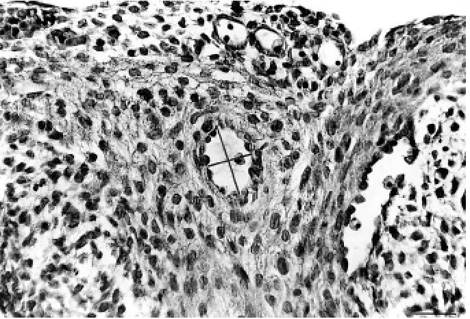
Figure 1. H&E stained section of ED15.5 heart. C.a. wall consists of the endothelial cells and the primitive tunica media visible on the cross section. The short and the long lines represent the lumen dimensions; the short line from each measurement has been taken as
lu-men diameter. Bar = 50 mm.
radii of identical length and was shown on both sides of “0 level” on the ordinate. The long axis of the vessel was represented as a straight line (abscissa). This ap-proximation neglected curvatures resulting from the natural course of the vessel within the myocardium.
In order to exclude the influence of possible arte-facts caused by the heart shrinkage upon the mea-surements, some hearts were immersed in 1M KCl solution inducing heart relaxation before fixation. Then, c.a. morphometry was performed on those hearts, as described above.
RESULTS
Immunohistochemical and histological identification of coronary artery constituents and precursor(s)
The labelling of tissue sections with Griffonia sim-plicifolia I lectin made it possible to identify all vascu-lar structures within the myocardium. Among these were structures that had an appearance of typical blood islands (Fig. 2). These structures were c.a. pre-cursors. On ED14 GSI-positive cells were seen form-ing clusters within the outflow tract (= conotruncus and truncus arteriosus) and the proximal ventricles of the heart on ED14 (Fig. 2B). On the following day these structures transformed into vascular tubes that were forerunners of the proximal coronary arteries. The vascular tubes were distinguished as coronary
artery precursors based on their localisation within the myocardium and their lumen diameters that were the largest compared to the lumen of other vascular structures (capillary plexuses).
The main tubes were running along both sides of the outflow tract and the ventricles with their dis-tal end still not far away from the base of the heart. The tube corresponding to the main branch of the left coronary artery occurred earlier in development than the tube corresponding to the future right cor-onary artery.
Folia Morphol., 2000, Vol. 59, No. 4
In ED16 hearts the endothelium-lined tubes were seen to be continuous in every section, i.e. two tubes (on each side of the aorta) were continuous with the peritruncal plexus, which at this time developed a lumenised connection with the aortic sinus. On ED17 two predominant vascular tubes with a patent lumen and arborisations towards the apex were found in the serial sections. These tubes became major branches of the coronary arteries and con-tained the multi-layered media and the adventitia. The distal end of the artery appeared on H&E stained sections as a cellular clusters with no recognisable lumen.
In older hearts (ED18–21) distal ends of coronary arteries extended further towards the apex of the heart and their branches developed the media and the adventitia.
Morphometry and graphical models
Figure 3 represents the crown-rump dimensions of the rat foetuses used for the experiment. Between ED15 and ED21 the foetuses grew 2.5 times in length.
Figure 2. ED14 heart. A) H&E stained section with blood island (arrow). Blood island is localised within the myocardium in the area of future coronary artery; ou — outflow tract; v — primitive ventricle; B) GSI — stained sections of the same stage heart. Blood island
(arrow) represents the structure that on ED15 would form vascular tube (the c.a. precursor). Bar = 50 mm.
The growth of the foetus was slow during the peri-od of our interest and had a linear character.
The heart length increased 4 times during the period studied. The growth was approximately lin-ear, with a tendency to be faster by the end of the prenatal life (Fig. 4).
On ED15 the vascular tube corresponding to the main branch of a coronary artery was aligned ap-proximately with the long axis of the heart; the total length of its main branch was about 150 mm, which is 1/8 to 1/10 of the heart length at this stage. One day later the length of the coronary artery on ED16 doubled, and equalled 1/4 to 1/6 of the heart’s length (Fig. 5). On the last day of the prenatal life the length of the c.a. was estimated to be about 14 times great-er compared to the length of the c.a. on ED15. The c.a. growth in length proceeded in a linear fashion and its rate was greater than the growth rate of both the foetus itself and its heart.
Figure 3. Development of foetuses during the prenatal period of c.a. formation represented as linear growth of the body length. The ab-scissa: embryonic day (ED) post-conception; the ordinate: crown-rump length (in mm).
Figure 4. Growth of the heart during the foetal life represented as linear dimension of the heart between ED15 and ED21.
proximal c.a. had changed during the prenatal peri-od studied. We found that the internal diameter of the proximal c.a. did not change significantly between E15 and ED18 and grew fast after ED18 (Fig. 6).
Figure 7 presents graphical models of coronary artery luminal reconstruction at various distances from the orifice at different foetal stages. Since the lumen diameter of the large vessels increased during devel-opment, we wanted to study how the c.a. lumen di-ameter had changed depending on foetal stage and
Folia Morphol., 2000, Vol. 59, No. 4
Figure 5. The c.a. growth during the foetal life (between ED15 and 21) represented as linear growth of c.a. length. The line forms a large angle with the abscissa, which is indicative of a rapid growth.
Figure 6. Graphical presentation of growth of the proximal c.a. luminal diameter during the foetal life (ED15–21).
widened and narrowed alternatively. By the end of the prenatal life (ED21) the length of the c.a. was equal to the length of the ventricles (Fig. 7C and Table 1).
We wanted to find out whether in areas of the largest lumen diameter the c.a. ramified to form two sister vessels, which would become separate branches of the c.a. The analysis of serially cut sec-tions allowed us to ascertain that there was no re-lationship between the lumen diameter and the sites of c.a. ramification. The same pattern of c.a. lumi-nal shape was also found in KCl-dilated hearts.
Table 1. Foetal dimensions and heart dimensions repre-sented as crown-rump lenght and heart lenght, respec-tively, between ED15 and ED21
ED Crown rump length [mm] Heart length [mm] (mean ± SEM) (mean ± SEM)
15 11.8 ± 0.5 1.17 ± 0.16
16 14.3 ± 0.25 1.52 ± 0.04
17 17.4 ± 0.6 2.72 ± 0.23
18 20.0 ± 0.2 03.0 ± 0.16
19 26.3 ± 1.03 3.25 ± 0.27
Figure 7. Graphical model of coronary artery luminal shape is expressed as a diameter of its lumen (Y-axis) measured at different distanc-es from the orifice (X-axis). Point “0” on the ordinate reprdistanc-esents the level of aortic orifice. “Length” is the length of the c.a. (X-axis). Three stages of prenatal life are detailed: ED15, ED16, and ED21. On ED15 the coronary orifice is usually not seen, thus the c.a. lumen is marked with XXX at point “0”. Note, that on ED15, and 16 the lumen diameter of c.a. is largest at a certain distance from the orifice.
A
B
Folia Morphol., 2000, Vol. 59, No. 4
DISCUSSION
Recently numerous studies have addressed the em-bryonic c.a. formation from vascular precursors in different species [3–6,24,26]. Our paper further de-fines the precursor of c.a. development and is the first documentation of the histomorphometric char-acteristics of coronary artery formation. Since the GSI-positive cells were recognised in ED14 hearts as separate clusters at areas corresponding to the fu-ture c.a. development, we could suggest that the first steps of the c.a. formation proceed by vasculo-genesis. Vasculogenesis and angiogenesis have been documented to occur during the embryonic coro-nary vessel development [19], however, it has not been certain whether coronary arteries developed by vasculogenesis [22]. Our data indicate further that the forerunners of the c.a. in rats are recognised on ED15 as the vascular tubes aligned with the long axis of the heart. These tubes are very short com-pared to the heart’s length: they run along the out-flow tract and reach only the proximal part of ven-tricles. Between ED16 and 21 the tubes reorganise to form the main branch of the coronary artery, whose length increases markedly in this period of time. Compared to the heart growth and crown-rump dimensions the c.a. increase in length is much faster (14-fold compared to 2.5-fold foetal growth increase). In one of the previous studies which uti-lised india ink injections on staged quail hearts it has been found that the distal part of the coronary artery is situated initially not far away from the base of the heart and later (as development proceeds) it “moves” towards the heart’s apex. This may indi-cate that with time the c.a. increases in length and its distal part is approaching the heart apex [22].
The mechanism of c.a. increase in length is un-known, but based on our graphical patterns at dif-ferent developmental stages we can speculate that the elongation of the coronary artery seems to pro-ceed by adding the tube-like or blood-island-like structures to the distal part of the forming vessel. Considering our statistical results of a rapid increase in the length of the vessel we can exclude the notion that the growth of the c.a. proceeds by a simple elonga-tion of the distal end. We suggest the mechanism of end-to-end tube coalescing for coronary artery forma-tion in embryogenesis. This mechanism based on stud-ies utilising retroviral tagging was also proposed by Mikawa & Fischman [15], and Mikawa & Gourdie [16]. On ED15 a coronary artery precursor consists of a single layer of GSI-positive cells, while the proxi-mal part is not yet connected to the aorta. At some
point along the course of the outflow tract the lu-men of the c.a. attains its maximal diameter and in some cases at this stage the vessel wall consists of two cell layers. The largest lumen diameter is found below the level of the aortic valves. Observations made by Waldo et al. [26] using india ink injections into the chicken heart have suggested that the prox-imal part of the c.a. close to the aortic sinus has a narrow lumen. These authors, however, have not presented any measurement data on this topic.
According to our previous data [18], the second layer of cells (the embryonic media) appears at the time when primordial endothelial cells are approach-ing the aorta. These results would indicate a discon-tinuous way of embryonic media formation during the first stages of the c.a. development. Once a wide lumenised connection with the aortic sinus has been established, the decreasing proximo-distal gradient of smooth muscle media coating is clearly distin-guished [11,18,22]. Thus, the largest lumen diame-ter and the start of the media formation occur at a certain distance from the aortic orifice.
Based on the mathematical criteria and morpho-histochemical analysis we can state that the increase in luminal diameter of the proximal c.a. is initially slow (ED15–18), and subsequently becomes rapid (ED18–21), in other words it proceeds according to a power function. The time course for the differenti-ation of the proximal part occurs from ED15 to ED18. Our observations and literature reports indicate that this period is characterised by a marked remodelling of the proximal c.a. in order to form a single lumen and develop an aortic orifice. The process of remod-elling also involves the peritruncal connections: some branches (slit-like channels) from the peritruncal plex-us disappear and eventually only two develop into the coronary artery stems [17,23]. Thus, while the remodelling takes place, the diameter of the proxi-mal c.a. does not increase.
At the time of the c.a. formation the crown-rump length, the heart length, and the c.a. length increase linearly. The body weight of the embryos during the prenatal life proceeds according to a power func-tion [25], whereas during the stages of coronary ar-tery formation (the second half of the embryonic life) the crown-rump length increases linearly.
as well as the addition of a second layer of cells be-gin at some distance form the aortic sinuses. After this time (ED17) maturation proceeds distally (to-wards the apex). Further elongation (after ED16) appears to result from the addition of vascular struc-tures to the distal end. We cannot exclude the possi-bility that there are also other patterns of ramifica-tion within the coronary system. The c.a. starting from one vessel ramifies to sister vessels towards the apex, which then coalesce into one tube again. This pattern of arborisation may persist to the end of the prenatal life [20].
Our data indicate that the shape of the c.a. lu-men is not changed at areas where ramifications occur and is not the result of a fixation artefact. Thus, the presented shape of the c.a. lumen may reflect the pattern of its embryonic development. Since the c.a. consists of bulgy tubes, we can speculate that these tubes are added to the distal end of c.a. while development proceeds.
ACKNOWLEDGEMENTS
The paper was partially supported by KBN grant (#401/P05/97/13) and by funds of the Medical Uni-versity of Warsaw.
REFERENCES
1. Alroy J, Goyal V, Skutelsky E (1987) Lectin histochemistry of mammalian endothelium. Histochem, 86: 603–607. 2. Conte G, Pelligrini A (1984) On the development
of the coronary arteries in human embryos, stages 14–19. Anat Embryol, 169: 209–218.
3. Bogers AJJC, Gittenberger-de Groot AC, Dubbeldam JA, Huysmans HA (1988) The inadequacy of existing theories on development of the proximal coronary ar-teries. Int J Cardiol, 20: 117–123.
4. Bogers AJJC, Gittenberger-dr Groot AC, Dubbeldam JA, Huysmans HA (1988/89) Scanning electron micros-copy substantiates histology in showing the inade-quacy of the existing theories on the development of the proximal coronary arteries and their connections with the arterial trunks. Acta Morphol Neerl-Scand, 26: 225–237.
5. Bogers AJJC, Gittenberger-dr Groot AC, Poelman RE, Péault BM, Huysmans HA (1989) Development of the origin of the coronary arteries, a matter of ingrowth or outgrowth? Anat Embryol, 180: 437–441. 6. Dettman RW, Denetclaw W, Ordahl CP, Bristow J (1998)
Common epicardial origin of coronary vascular smooth muscle, perivascular fibroblasts, and intermyocardial fibroblasts in the avian heart. Dev Biol, 193: 169–181. 7. Dbalý J, Oštádal B, Rychter Z (1968) Development of the coronary arteries in rat embryos. Acta Anat, 71: 209–222.
8. Gittenberger-de Groot AC, Bogers AJJC, Bartelings MM (1987) Aspects of normal and abnormal development of the main coronary arteries. In: Spaan JAE, Bruschke
AVG, Gittenberger-de Groot AC (eds.) Coronary Circu-lation. Dordrecht, Nyhoff, 32–42.
9. Hansen-Smith FM, Watson L. Lu DY, Goldstein I (1988) Griffonia simplicifolia I: fluorescent tracer for micro-circulatory vessels in nonperfused thin muscles and sectioned muscle. Microvasc Res, 36: 199–215. 10. Heintzberger CFM (1983) Development of myocardial
vascularization in the rat. Acta Morphol Neerl-Scand, 21: 267–284.
11. Hood LC, Rosenquist TH (1992) Coronary artery devel-opment in the chick: origin and deployment of smooth muscle cells, and the effects of neural crest ablation. Anat Rec, 234: 291–300.
12. Hungerford JE, Little CD (1999) Developmental biolo-gy of the vascular smooth muscle cell: building a mul-tilayered vessel wall. J Vasc Res, 36: 2–27.
13. Hutchins GM, Kessler-Hanna A, Moore GW (1988) De-velopment of the coronary arteries in the embryonic human heart. Circulation, 77: 1250–1257.
14. Manasek FJ (1971) The ultrastructure of embryonic myocardial blood vessels. Dev Biol, 26: 42–54. 15. Mikawa T, Fischman D (1992) Retroviral analysis of
cardiac morphogenesis: discontinuous formation of coronary vessels. Proc Natl Acad Sci USA, 89: 9504– –9508.
16. Mikawa T, Gourdie RG (1996) Pericardial mesoderm generates a population of coronary smooth muscle cells migrating into the heart along with ingrowth of the epicardial organ. Dev Biol, 174: 221–232. 17. Poelmann RE, Gittenberger-de Groot AC, Metink MMT,
Bökenkamp R, Hogers B (1993) Development of the cardiac vascular endothelium, studied with antiendot-helial antibodies in chicken-quail chimeras. Circ Res, 73: 559–568.
18. Ratajska A, Fiejka E (1999) Prenatal development of coronary arteries in the rat: morphologic patterns. Anat Embryol, 200: 533–540.
19. Rongish BJ, Torry RJ, Tucker DC, Tomanek RJ (1994) Neovascularization of embryonic rat hearts cultured in oculo closely mimics in utero coronary vessel devel-opment. J Vasc Res, 31: 205–215.
20. Tomanek RJ, Haung L, Suvarna PR, O’Brien LC, Ratajska A, Sandra A (1996) Coronary vascularization during de-velopment in the rat and its relationship to basic fibro-blast growth factor. Cardiovasc Res, 31: E116-E126. 21. Voboril A, Schiebler TH (1969) Über die Entwicklung
des Gefässversorgung des Rattenherzen. Z Anat Entw Gesch, 129: 24–40.
22. Vrancken Peeters MPFM, Gittenberger-de Groot AC, Mentink MMT, Hungerford JE, Little CD, Poelmann RE (1997) The development of the coronary vessels and their differentiation into arteries and veins in the em-bryonic quail heart. Dev Dyn, 208: 338–348.
23. Vranken Peeters MP, Gittenberger-de Groot AC, Mentink MM, Hungerford JE, Little CD, Poelmann RE (1997) Differences in development of coronary arter-ies and veins. Cardiovasc Res, 36: 101–110.
Folia Morphol., 2000, Vol. 59, No. 4
25. Vuillemin M, Pexieder T (1989) Normal stages of car-diac organogenesis in the mouse: I. Development of the external shape of the heart. Am J Anat, 184: 101–113.