Immature Teratoma of Head and Neck: Two case
reports
Hitesh Verma
1*, Arjun Dass
2, Surinder K Singhal
3, Nitin Gupta
4and Amrinder Kaur
51*,2,3,4
Department of Otorhinolaryngology and Head and Neck Surgery, Government Medical College and Hospital Chandigarh, India
5
Senior Resident, Department of Pathology, Government Medical College and Hospital Chandigarh, India
Teratoma refers to a neoplasm that recapitulates all three germ layers. Teratoma is usually developmental and sometimes congenital neoplasm which displays both solid and cystic components with gross and microscopic differentiation into a wide variety of tissues representative of all three germ layers i.e. ectoderm, mesoderm and endoderm. Teratomas of the head and neck are extremely rare and usually seen during the neonatal period. Teratomas of the head and neck due to their obscure origin, bizarre microscopic appearance, unpredictable behavior and often dramatic clinical presentation are a clinical surprise. We are presenting two case reports, where immature teratoma was present in hypopharynx and in nose.
Key words: Immature teratoma, nose, Hypopharynx, totipotent, germ layers
INTRODUCTION
Teratomas are embryonal neoplasms that arise when totipotential germ cells recapitulate normal organogenesis and give rise to more or less organoid masses in which tissues derived from all three blastodermic layers (ectoderm, endoderm, and mesoderm) can be identified ( Chakravarti, 2011;, Rothschild, 1994). Broadly, these are classified as mature, immature and malignant. Age range at presentation varies from birth to 6th decade of life(Ling, 1993). The immature form of teratoma was first described in 1960 by Thürlbeck and Scully (Trabelsi, 2002). It is found either in pure form or as a component of a mixed germ cell tumor. According to WHO, immature teratoma (IT) is defined as a teratoma containing a variable amount of immature embryonal type (generally) neuroectodermal tissue.
They occur in genital systems and other organs along the midline of the body, with similar morphology and classification (Azizkhan, 1996). The incidence of teratomas is 1 per 4000 live births, with the sacrococcygeal area being the most common site (Coppit 2000). Teratomas of the head and neck are extremely rare and teratomas occur in these locations in nearly 1-3.5% of all cases(Dadmehr, 2006). The common sites of
involvement are the neck, oropharynx, nasopharynx, orbit, and paranasal sinuses. Most neck teratomas are reported as mature, and malignant immature teratomas have been said to occur in approximately five percent of teratomas of the neck. The current treatment for tumor is surgical excision with adjuvant chemotherapy. Cisplatin, etoposide, and bleomycin (BEP) are commonly using chemotherapeutic agents for immature teratoma.
We are presenting here two case reports. In case one it involved hypopharynx and in second case it involved nose.
Case report 1
Fifty five years male patient was presented with history of difficulty in swallowing for solid more then liquid for the last 6 weeks.
*Corresponding author: Dr. Hitesh Verma, Department of Otorhinolaryngology and Head and Neck Surgery Third Floor, D block Government Medical College and Hospital, sector 32, Chandigarh, India, E-mail: hitesh_verma72@yahoo.com, Tel.: +9101722665253-2309, Fax: +9101722608488
position the text box anywhere in the document. Use the Text Box Tools tab to change the formatting of the pull quote text box.]
Case Report
He also had history of change in voice and difficulty in breathing for the last two weeks. He presented with stridor in our OPD and indirect laryngoscopic examination showed single globular mass present in right side of hypopharynx and covering endolarynx completely. Neck examination was unremarkable. The rest of local examination was normal. In view of stridor, emergency tracheostomy was performed. The X ray soft tissue neck lateral view showed mass in hypopharynx covering larynx (Figure 1).
Figure 1. X Ray soft tissue neck lateral view showed
well defined mass in hypopharynx covering
endolarynx with tracheostomy tube in situ.
Direct laryngoscopy revealed a single globular pinkish white mass with irregular surface (4×4 cm in size) which was found attached with right lateral wall of pyriform sinus by narrow stalk (Figure 2).
Figure 2. Direct Laryngoscopy revealed single globular mass in hypopharynx with narrow attachment on right lateral wall.
Endolarynx and left half of hypopharynx was normal. The base of the mass was cauterized and excised completely. The patient was kept on nasogestric tube for five days.
Tracheostomy tube was removed on 6th postoperative day. The histopathological examination showed mature pilosebaceous units along with immature mesenchymal elements revealed and diagnosis of immature teratoma was made (Figure 3). After second cycle of chemotherapy, the patient expired.
Figure 3. Photomicrograph showing immature teratoma showing mature pilosebaceous units along with immature mesenchymal elements (HE x 100)
Case report 2
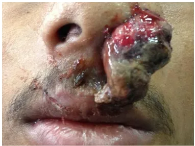
Figure 4. Radish fleshy mass coming out from left nasal cavity extending till upper lip with widening of nasal ala.
Figure 5. Contrast enhancing CT scan of Nose and PNS (coronal cut) showing homogenous soft tissue density in left side of nasal cavity and paranasal sinuses with erosion of septum and cribriform plate.
Figure 6. Photomicrograph showing focally hyperplastic respiratory epithelium lined tissue showing immature mesenchymal elements in the subepithelium (HEX200)
showing immature mesenchymal elements in the subepithelium, which was suggestive of immature teratoma (Figure 6). In post operative period, the patient
DISCUSSION
Teratomas are common tumors originating from totipotential cells and are composed of heterogenous tissues reminiscent of derivatives from any or all three germ layers in varying ratio (Duwe, 2005). The presence of tissues within the lesion that are foreign to the affected sites is a distinctive feature of this rare tumor. Most of the cases occur in neonates and older infants, contrary to our patient who were adult age-group. It has been established that there is no sex predilection in head and neck teratomas(Abemayor, 1984). A mature teratoma is typically benign and is found more commonly in females, but immature teratomas are typically malignant and are found more often in males as in our cases. As a general rule, while pediatric teratomas of the head and neck tend to be oncologically benign, adult teratomas tend to be histologically and oncologically malignant. The duration of symptoms is short in malignant form as compared to benign form of immature teratoma was seen in our cases (Ibrahim, 2012; Swetha, 2011).
Benign teratomas are solid and cystic tumors composed of a variety of both immature and mature tissues derived from all three germ layers. The epithelial component of a benign teratoma usually consists of mature squamous epithelium and immature intestinal or respiratory epithelium. Primitive neuroepithelium with rosettes, pseudorosettes or neurofibrillary matrix predominates in some tumors. Pigmented retinal epithelium can also be seen. The mesodermal component consists of fibroblasts and embryonic, immature spindle cells embedded in a myxoid matrix. Islands of cartilage, smooth muscle cells, and skeletal muscle cells exhibiting varying degrees of maturity may also be present. To make the diagnosis of teratoma, it is mandatory to find at least two of three germ layers (Smirniotopoulos, 1995). Teratomas may also be histologically immature while being oncologically benign, or they may harbor malignant components and have the potential to exhibit an aggressive biological behavior. The amount of neuroectodermal immature tissue present permits the classification of immature teratomas into three grades of increasing malignancy. Immature teratoma may matastasise to brain, liver and lung. Metastasis to brain has also been reported. The imaging appearance is typically of a large, heterogeneous mass with a prominent solid component. However, the spectrum of appearances ranges from a predominatly cystic to a predominantly solid mass. . The presence of a prominent solid component containing calcifications and small foci of fat . Cystic components may contain serous, mucinous, or
fatty sebaceous material.
The recommended management for head and neck teratomas is complete surgical excision. Chemotherapy has been successful in those with recurrent disease as
this tumor is highly chemosensitive (Ernest E. Lack ,1985). In both cases we followed the same treatment protocol as per literature. The prognosis with tumor remains guarded, it depends on early detection and aggressive management. We learn from these two cases that immature teratoma can involve any part in head and neck, it require early diagnosis and management.
REFERENCES
Chakravarti A, Shashidhar TB, Naglot S, Sahni JK (2011). Head and Neck Teratomas in Children: A Case Series. Indian J Otolaryngol Head Neck Surg, 63(2): 193–197.
Rothschild MA, Catalano P, Urken M, Brandwein M, Som P, Norton K, Biller HF (1994). Evaluation and management of congenital cervical teratoma. Case report and review. Arch Otolaryngol Head Neck Surg, 120(4): 444–448.
Ling YJ, Lai DM, Tsu HC, Lin SM (1993). Primary intracranial teratocarcinoma with extraneural metastasis- Case report and review of literature. Acta neurol Sin, 2: 332-7.
Trabelsi A, Conan-Charlet V, Lhomme C, Morice P, Duvillard P, Sabourin JC (2002). Peritoneal glioblastoma: Recurrence of ovarian immature teratoma. Ann Pathol, 22: 130-3.
Azizkhan RG, Caty MG (1996). Teratomas in childhood. Curr Opin Pediatr, 8: 287-92
Coppit GL, Perkins JA, Manning SC (2000). Nasopharyngeal teratomas and dermoids: A review of the literature and case series. Int J Pediatr Otorhinolaryngol, 52: 219-27.
Dadmehr M, Nejat F, Ansari S, Habibi Z (2006). Ruptured occipitocervical teratoma mimicking an upper cervical myelomeningocele. Case report. J Neurosurg, 104: 360-1.
Duwe BV, Sterman DH, Musani AI (2005). Tumors of the mediastinum. Chest, 128: 2893-909
Abemayor EA, Newman MDL, Bergstrom J, Dudley JG, Magidson MD, Ljung BM (1984). Teratomas of the head and neck in childhood. The Laryngoscope, 94(11): 1489-92.
Ibrahim C, Murat G, Aytekin Y, Umit B, Orhan GY (2012). A benign teratoma presenting as an obstruction of the nasal cavity: a case report.Journal of Medical Case Reports, 6:147
Swetha A, Amsavardani T, Sahara A, Krishan K (2011). Oral teratoma with a primitive neuroectodermal tumor component: a case report. International journal of Clinical Pediatric Dentistry, 4 (3): 240-244.
Ernest E. Lack (1985).Extragonadal germ cell tumors of the head and neck region: Review of 16 cases. Human Pathology 16 (1), 56–64.
Accepted 19 August, 2014.
Citation: Verma H, Dass A, Singhal SK, Gupta N, Kaur A (2014). Immature Teratoma of Head and Neck: Two case reports. Journal of Cancer and Clinical Oncology, 1(3): 011-014.