Esthetic Management of Dehiscence
“Perio-prosthetic Solution”- A Case Report
Rathika Rai, R. Prabhu, A. G. PremaDepartment of Prosthodontics, Thai Moogambigai Dental College and Hospital, DR. MGR Educational and Research Institute, Chennai, Tamil Nadu, India
Email for correspondence: [email protected]
ABSTRACT
Replacement of anterior teeth is always an esthetic challenge. However, anterior missing teeth coupled with dehiscence make it more challenging. This case presentation discusses the management of dehiscence in the anterior region using a novel, cost-effective platelet-rich fibrin clot along with bone grafting material and restoring missing teeth to achieve form, function and maximum esthetics by a multidisciplinary approach.
Key words: Anterior esthetics, dehiscence, platelet-rich fibrin
Quick Response Code Article Info:
doi: 10.5866/2018.10.10181
Received: 29-09-2018 Revised: 27-10-2018 Accepted: 12-11-2018
Available Online: 05-01-2019, (www. nacd.in)© NAD, 2019 - All rights reserved
INTRODUCTION
Replacement of anterior teeth is always an esthetic challenge. Anterior missing teeth coupled with dehiscence makes it more challenging. Dehiscence and fenestrations are the two common alveolar defects encountered commonly in the maxillary anterior region. In the healthy periodontium, the crest of the interproximal bone is 2 mm apical to cementoenamel junction. If there is a breach in the contour of the cortical plate, anatomical defects such as fenestration and dehiscence may occur.[1]
Dehiscence is defined as isolated areas of denuded root surface covered by periosteum and overlying gingiva, but the denuded areas extend through the marginal bone.[2] According to Davies
et al., alveolar defects are considered as dehiscence when there is an absence of at least 4 mm of cortical bone apical to the interproximal bone.[3]
However, fenestration is an isolated cortical bone defect, leading to the exposure of root surface without involving the marginal alveolar bone.[4]
These alveolar defects occur approximately in 20% of teeth.[5] These are more common on the facial
bone than on the lingual bone. It sometimes occurs bilaterally.[6]
Predisposing factors are:
·• Thin labial bone.
·• Labioversion of the tooth in the dental arch. ·• Occlusal factors.
·• Prominent root contours. ·• Orthodontic tooth movement.
·• Long-standing trauma and aberrant frenal attachments.
Several treatment modalities have been proposed in literature, which includes root planning along with chlorhexidine mouth rinsing, full thickness mucogingival flap with primary closure, free gingival grafting in case of fenestrations.[7] However, in
dehiscence platelet-rich fibrin (PRF) along with bone grafts can be used for coverage of denuded roots.[8]
Platelets regenerative potential was introduced in the 70s when it was observed that they contain growth factors that are responsible for increased collagen production, cell mitosis, growth of blood vessels, and recruitment of other cells that migrate to the injury site, and cell differentiation induction among others.[9-11]
1. Platelet-rich plasma (PRP) 2. PRF.
Platelet concentrates are a concentrated suspension of growth factors found in platelets, acts as bioactive surgical additives that are applied locally to induce wound healing. PRF was first used in 2001 by Choukroun et al. considered as a second-generation platelet concentrate. It consists of a matrix of autologous fibrin and has several advantages over PRP. Anatomical radiological in vitro study dental radiographs were compared with axial, sagittal, and coronal computed tomography (CT)-scans, with respect to the identification and quantitative assessment of bone dehiscences and the buccal or lingual bone plates over the roots of the teeth.[9,14,15]
In the present case report, the presence of dehiscence along with missing teeth poses a challenge for esthetic reasons. The present case report describes the management of dehiscence defect by the use of PRF and bone graft material and managed with a fixed partial denture (FPD) to improve the esthetics.
CASE REPORT
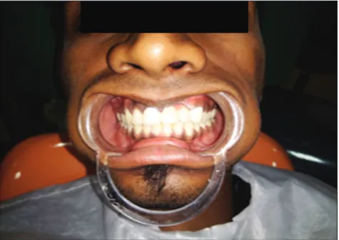
A 20-year-old male, healthy, non-smoker patient reported to the outpatient, department of prosthodontics with the chief complaint of his loose fixed prosthesis in the upper front tooth region. Intraoral examination revealed fixed metal ceramic restoration in relation to the left upper central incisor to the left upper canine 21–23, presence of high labial frenum with loss of labial bone up to the apex of 21 [Figure 1].
The patient revealed the history of trauma 5 years back in the upper anterior region following which tooth 22 was extracted and the fixed prosthesis was placed in relation to 21 and 23. Over a period of time, there was a loss of labial bone in relation to 21. Radiograph also showed complete loss of labial bone up to the apex of 21 [Figure 2]. The high frenal attachment could also be attributed in addition to trauma leads loss of labial plate of the bone in relation to 21. The presence of dehiscence was a matter of concern both esthetically, functionally, and oral hygiene.
The cause of dehiscence, however, was not explanatory. It could be attributed to the high frenal attachment along with the trauma. Hence, the treatment plan was the removal of loosened FPD along with the extraction of 21 followed by correction of high labial frenum.
Treatment was done in three stages 1. Preparatory phase
2. Surgical phase 3. Prosthetic phase.
Preparatory Phase
Preparation of the patient included oral prophylaxis and root planning of the entire dentition followed by frenectomy and oral hygiene instructions were given. The surgery was planned after 2 weeks.
SURGICAL TECHNIQUE
Under local anesthesia (Lignox 2%A), a full thickness mucoperiosteal flap was elevated with a no: 15 blades from the palatal surface with two Figure 1: Clinical picture showing dehiscence defect in relation
to left upper central incisor
vertical releasing incisions. On flap elevation, the recipient bed showed significant bone loss, which was planned to be treated by placement of PRF and bone graft (osseograft) to aid in bone regeneration [Figure 3].
Preparation of the PRF
The patient’s blood sample of 10 ml was taken in the operating room during the surgery. Immediately after the blood was drawn, the dried Monovettes (without anticoagulant) were centrifuged at 3000 rpm for 12 min in a table top centrifuge (REMI 3000rpm) [Figure 4a].
Due to the absence of an anticoagulant, blood begins to coagulate as soon as it comes in contact with the glass surface. Therefore, for successful preparation of PRF, speedy blood collection and immediate centrifugation, before the clotting cascade initiation is absolutely essential.
The PRF clot [Figure 4b] that was obtained was mixed with osseograft to form a homogenous graft material, and the fibrin is compressed on the defect site along with bone graft and sutured back [Figure 5]. The patient was given antibiotics (Amoxicillin 500 mg TID for 5 days) and analgesics (Ibuprofen 400 mg and Paracetamol 500 mg BD for
3 days), and post-operative instructions were given. Chlorhexidine (0.2%) mouth rinse was prescribed for 4 weeks.
The sutures were removed, 10 days after the procedure. The surgical site was examined for uneventful healing. There were no post-operative complications and healing was satisfactory. 6-months post-surgery check radiography was done and it showed bone fill at the defect site and the final prosthesis was planned [Figure 6].
Prosthetic Phase
The tooth number 11 was taken as an abutment. According to the principals of tooth preparation, the right central incisor was prepared and modifications were done in relation to 23. Equi gingival shoulder margins were given. Gingival retractions have been
Figure 5: Placing platelet-rich fibrin with bone graft
Figure 6: Intraoral periapical radiograph showing bone fill at the defect site after 6-months
Figure 4: (a) Collection of blood for platelet-rich fibrin (PRF), (b) PRF clot
b a
Figure 3: Clinical picture showing the surgical site with dehiscence
done with 3 gingival retraction cord. A putty and light body wash impression technique was used to make a complete both upper and lower arch impressions with vinyl polysiloxane impression material (Dentsply Aquasil Soft Putty Regular Set). The definitive casts were mounted in a Hanau Semi-Adjustable Articulator. Porcelain fused metal ceramic prosthesis (Vita, India) was fabricated. A modified ridge lap pontic design was given. A metal try-in was done to access the complete seating of the prosthesis. After all, modifications were made, a final prosthesis was cemented with glass ionomer cements–I (GC Gold label, GC Corporation, Tokyo, Japan) luting cement. Excess cement was removed. The occlusion was evaluated and necessary occlusion adjustments were made [Figures 7 and 8].
DISCUSSION
Cases of dehiscence and fenestration have been earlier reported in history. Most common occurrence reported have been in maxillary and mandibular incisors. The exact etiology is unknown, but a review of literature suggests that abnormally labially positioned tooth, very thin labial plates, high frenal pull, and sometimes long-standing trauma may cause the dehiscence and fenestration defects. Fuhrmann et al. have recently shown that the identification and evaluation of the vertical depth of infra-bony pockets were significantly better by axial high-resolution-CT than by intraoral radiography.[13,16] Previous studies showed that
gingival recessions damage to the root surface and the alveolar bone might occur with the proclination or retroclination of the mandibular incisors also. Literature review of the studies has proven that the use of platelet concentrates has technical benefits and may enhance bone regeneration when used in conjunction with bone grafts. The acceleration of platelet-derived growth factor and transforming growth factor beta is seen as an available and practical tool for increasing the rate of bone formation and the final quality of bone formed.[17]
PRF has many advantages over PFP. It eliminates the redundant process of adding anticoagulant as well as the need to neutralize it. It has been shown from literature that it increases the rate of clinical graft consideration and that the PRF enhanced grafts produce more mature and dense bone than grafts without PRF. Hence, PRF with bone graft was used as a biomaterial in closing the defect.
PRF was developed in France by Choukroun et al. in 2001. It is a fibrin matrix in which platelet, cytokines, and cells are entrapped. These are released after a certain time and can serve as a resorbable membrane. PRF is a healthy biomaterial that is used in oral implantology and its application has been advocated in various disciplines of dentistry.
Tanaka et al. did a study on additional effects of platelet-rich fibrin on bone regeneration in sinus augmentation with deproteinized bovine bone material. They found the additional effects of PRF in sinus augmentation, also a study shown that both the autogenous bone graft and PRF were effective in the treatment of the intrabony defects.
PRF is in the form of a platelet gel and can be used along with bone grafts which offer several Figure 8: Final prosthesis was cemented with glass ionomer
cement–I luting cement
Figure 7: Master impression was made using a putty and light body wash impression technique with vinyl polysiloxane
advantages including promoting wound healing, bone growth and maturation, graft stabilization, wound sealing, and hemostasis and improving the handling properties of graft materials. In some experimental studies, it was found that PRF had a superior influence over PRP.
PRF has immunological and antibacterial properties that may lead to leukocyte degranulation. It also has some cytokines that induce angiogenesis and anti-inflammatory reactions. PRF could also serve as a resorbable membrane for guided bone regeneration, preventing the migrations of non-desirable cells into bone defects and providing a space that allows the immigration of osteogenic and angiogenic cells. It also permits the underlying blood clot to mineralize.[18]
The concept of natural bone regeneration which includes regeneration through PRF membranes both the bone volume and gingival tissue, reported satisfactory clinical results related to reshaping the whole alveolar bone and the restoration of gingival bone and per-implant bone, achieving adequate mechanical, and esthetic properties. Tofler et al. recommended the use of PRF membrane to seal an undetected sinus membrane perforation during maxillary sinus lift procedures. PRF membrane helps in wound healing by shielding the surgical site, promoting soft tissue repair, when mixed with bone graft, it may also act as a biological connector, which attracts stem cells, favoring the migration of osteoprogenitor cells to the center of the graft and provides a neo-angiogenesis. Simonpieri et al. suggested using a mix of PRF with bone graft.
The combination of PRF with bone graft has shown better results in regenerative therapy.[19]
This may be due to better stability and improved proliferation of cells with PRF. Hence, it was chosen as a treatment modality to correct the defect and to improve the functional and esthetic outcome of the FPD by correcting the defect.
CONCLUSION
Management of anterior region with FPD is always challenging with respective of esthetics and function. The presence of dehiscence makes it more complex due to the defect which attracts food and affects esthetics and maintenance. Minor osseous defects such as fenestration and dehiscence were successfully treated with the use of PRF and bone graft. These defects should be corrected or eliminated during the pre-prosthetic phase
according to the esthetic and functional needs. The increasing benefits of PRF as a successful regenerative bio-material, management of osseous defects has proven to be a boon in both medicine and dentistry. In this case, the use of PRF is an acceptable and successful treatment in achieving esthetics and function with the multidisciplinary approach.
REFERENCES
1. Bains VK, Bains R, Gupta SJ, Mishra P, Loomba K.
Management of dehiscence and fenestration alveolar defects around incisors using platelet-rich fibrin: Report of two cases. J Interdiscip Dent 2015;5:92-6.
2. Singh S, Panwar M, Arora V. Management of mucosal fenestration by multidisciplinary approach: A rare case report. Med J Armed Forces India 2013;69:86-9.
3. Davies RM, Downer MC, Hull PS, Lennon MA. Alveolar
defects in human skulls. J Clin Periodontol 1974;2:107-11.
4. Keceli HG, Kamak G, Erdemir EO, Evginer MS, Dolgun A.
The adjunctive effect of Platelet rich fibrin to connective tissue graft in the treatment of buccal recession defects: Results of a randomized, parallel group controlled trial. J Periodontol 2015;86:1221-30.
5. Mathur A, Bains VK, Gupta V, Jhingran R, Singh GP. Evaluation of intrabony defects treated with platelet-rich fibrin or autogenous bone graft: A comparative analysis. Eur J Dent 2015;9:100-8.
6. Vijayalakshmi R, Rajmohan CS, Deepalakshmi D, Sivakami G. Use of platelet rich fibrin in a fenestration defect around an implant. J Ind Soc Periodontol 2012;16:108-12.
7. Borie E, Oliví DG, Orsi IA, Garlet K, Weber B, Beltrán V,
et al. Platelet-rich fibrin application in dentistry: A literature review. Int J Clin Exp Med 2015;8:7922-9.
8. Tanaka H, Toyoshima T, Atsuta I, Ayukawa Y, Sasaki M, Matsushita Y, et al. Additional effects of platelet-rich fibrin on bone regeneration in sinus augmentation with deproteinized bovine bone mineral: Preliminary results. Implant Dent 2015;24:669-74.
9. Mattoo KA, Khare S, Nagaraju K. Characterizing extreme dehiscence of a maxillary molar. Am J Med Case Rep 2015;3:13-5.
10. Larato DC. Alveolar plate fenestrations and dehiscences of the human skull. Oral Surg Oral Med Oral Path Oral
Rad 1970;29:816-9.
11. Tibbetts LS. Use of diagnostic probes for detection of
periodontal disease. J Am Dent Assoc 1969;78:549-55.
12. Eraydın F, Germec-Cakan D, Tozlu M, Ozdemir FI.
Three-dimensional evaluation of alveolar bone thickness of mandibular anterior teeth in different dentofacial types. Niger J Clin Prac 2018;21:103.
13. Fuhrmann RA, Wehrbein H, Langen HJ, Diedrich PR.
14. Preeja C, Arun S. Platelet-rich fibrin: Its role in periodontal regeneration. Saudi J Dent Res 2014;5:117-22.
15. Dohan DM, Choukroun J, Diss A, Dohan SL,
Dohan AJ, Mouhyi J, et al. Platelet-rich fibrin (PRF): A second-generation platelet concentrate. Part II:
Platelet-related biologic features. Oral Surg Oral Med Oral Pathol Oral Radiol Endod 2006;101:45-50.
16. Bhatsange A, Shende A, Deshmukh S, Japatti S. Management of fenestration using bone allograft in conjunction with platelet-rich fibrin. J Indian Soc Periodontol 2017;21:337-40.
17. Fuhrmann R. Three-dimensional interpretation of alveolar bone dehiscences. An anatomical-radiological study--Part I.
J Orofac Orthop 1996;57:62-74.
18. Carranza FA, Bernard GW, The tooth supporting structures.
In: Newman MG, Takei HH, Carranza FA, editors.
Carranza’s Clinical Periodontology. Philadelphia, PA: W.B.
Saunders Co.; 2002. p. 36-57.