www.fm.viamedica.pl
Address for correspondence: M.T. Demir, 19 Mayis University Medical Faculty, Anatomy Samsun, Turkey, e-mail: [email protected]
The double outlet right ventricle with
additional cardiac malformations:
an anatomic and echocardiographic study
M.T. Demir
1, Y. Amasyali
2, C. Kopuz
1, M.E. Aydin
1, U. Çorumlu
1119 Mayis University Medical Faculty, Anatomy Samsun, Turkey 2Anatolian Hospital, Cardiology Samsun, Turkey
[Received 26 September 2008; Accepted 26 February 2009]
Double outlet right ventricle (DORV) is a complex group of abnormal ventricu-loarterial connections where both the aorta and pulmonary artery rise from the right ventricle morphologically. It is a rare condition affecting 1–1.5% of pa-tients with congenital heart disease.
The patient’s cardiac measurements were taken by two-dimensional echocardio-graphy. The pathological findings of the defined case are illustrated in the figures. A 20-year-old male patient with palpitations, tachypnea, and tiredness with weak exercise capacity and back pain was diagnosed with heart murmur in different degrees; slight cyanosis, cachexia, and developmental retardation were found clinically. Echocardiography showed: DORV, perimembranous ventricu-lar septal defect, pulmonary stenosis, dilatated right ventricle, and dilatated left atrium. However, the left ventricle was normal. In addition, the septum interatriale filled the left atrium like an aneurysm.
DORV is an important and rare congenital cardiac malformation. This complex condition may result in different clinical findings and require different thera-peutic approaches. (Folia Morphol 2009; 68, 2: 104–108)
Key words: heart, malformation, double outlet right ventricle, echocardiography
INTRODUCTION
In normal cardiac anatomy, the aorta has its or-igin from the left ventricle and the pulmonary ar-tery has its origin from the right ventricle. Double outlet right ventricle (DORV) is a complex group of abnormal ventriculoarterial connections in which both the aorta and pulmonary artery emerge from the right ventricle morphologically [1, 12]. It is a rare condition, affecting 1–1.5% of patients with con-genital heart diseases with a frequency of 1 in each 10,000 live births [12]. A 20-year-old male patient with palpitations, tachypnea, and tiredness, with low exercise tolerance and back pain. Systolic heart mur-mur in different degrees (2o–3o/6o) in the
mesocar-diac region, slight cyanosis, cachexia, and develop-mental retardation were found clinically.
MATERIAL AND METHODS
A 20-year-old male patient was admitted to the cardiology clinic of the Büyük Anadolu Hospital with palpitations, tachypnea, and tiredness, with low exercise tolerance and back pain. In the physical examination, slight cyanosis at the limbs and nails, developmental retardation despite his age, and cachexia were determined. At auscultation, 2o–3o/6o
systolic murmur at the mesocardiac region and 1o–3o/6o systolic murmur at the pulmonary region
decided to evaluate the patient using echocardio-graphy. The patient’s cardiac measurements, taken by 2-dimensional echocardiography (HP Sonos 2000, 2-dimentional transthoracic echo-2.5 mHz cardiac prope) and the measured cardiac values, were com-pared with normal measurements. The causes of
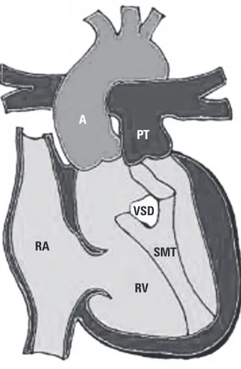
these conditions were discussed with knowledge of the patient’s anatomy and embryology. The patho-logical findings of the defined case and the normal form of the cardiac values as our specimens are illustrated in Figures 1 and 2. The patient did not have a familial history of cardiologic disease.
RESULTS
In our case, the echocardiographic findings are explained as below:
— Double outlet right ventricle (Fig. 3).
— Dilatation of the left atrium (Fig. 3). The mea-surement was made on the position of the parasternal long axis by M mode presentation. The diameter of the left atrium was measured as 48 mm (normal value: 0–40 mm) and on the parasternal longitudinal axis it was seen that the aorta did not emerge from the left ventricle. — Paradoxal motion of the interventricular septum
(Fig. 4). The dimensions of the left ventricle were normal. In addition, the interventricular septum had a paradoxal condition at the ventricular sys-tole. The ejection fraction (EF) of the left ventricle was normal at 67% (normal value of EF: 50–80%). The EF calculation was made according to Simp-son’s principle. Formula = end diastolic volume – – end systolic volume/end diastolic volume. — Dilatation of the right ventricle. The diameter of
the right ventricle was measured as 36 mm (nor-mal value: 0–26 mm).
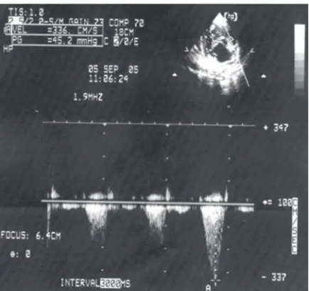
— Pulmonary stenosis (Fig. 5). The pressure gradi-ent on the pulmonary valve was measured as
Figure 1. Schematic diagram of a normal cardiac structure;
A — aorta, PT — pulmonary trunk, LV — left ventricle, RV — right ventricle, RA — right atrium, IVS — interventricular septum, SVC — superior vena cava.
Figure 2. Schematic diagram of our case; A — aorta, PT —
pul-monary trunk, VSD — ventricular septal defect, RA — right atrium RV — right ventricle, SMT — septomarginal trabecula, IVS — interventricular septum, SVC — superior vena cava.
Figure 3. Echocardiography showed dilatation of left atrium and
double outlet right ventricle. The measurement was made in the position of parasternal long axis by M mode presentation.
RA
RV SMT VSD
A
PT
RA SVC
A
PT
LV RV
A.
B.
45.2 mm Hg (slight stenosis: 0–20 mm Hg, me-dium stenosis: 20–50 mm Hg, severe stenosis: > 50). The visual images of diagnosis of pulmo-nary stenosis in transthoracic echocardiography on parasternal short axes were rendered. The
gradient between the right atrium and right ven-tricle was determined by using a simplified version of Bernoulli’s equation. The systolic pressure of the right ventricle, which is equal to the peak gradient of pulmonary stenosis by adding the pressure of the right atrium to this gradient, was calculated. — Perimembranous type ventricular septal defect (VSD). On echocardiography there was a shunt from left to right on the interventricular septum. The cause of this shunt was a defect in the inter-ventricular septum. The type of VSD was per-imembranous. In addition, the aorta emerged from the right ventricle.
— Aneurysm on the interatrial septum (Fig. 6). An-eurysm on the interatrial septum pouched to the left and filled up the left atrium.
The aortic arch had no obstruction; however, the pericardium had a normal appearance.
DISCUSSION
The origin of the aorta and pulmonary artery from the right ventricle is a complicated congenital cardi-ac malformation [4]. In normal cardicardi-ac anatomy, these great vessels have their origin of the aorta from the left ventricle as the pulmonary artery from the right ventricle [10, 14].
DORV can be classified according to the relation-ship between the VSD and blood vessels as: DORV with subaortic VSD, DORV with subpulmonary VSD, DORV with doubly-committed VSD, and DORV with noncommitted VSD [12]. The most frequent anatomic
Figure 4. Echocardiographic image showed ejection fraction of
left ventricle. The ejection fraction calculation was made accord-ing to Simpson’s principle. Formula = end diastolic volume – end systolic volume/end diastolic volume.
Figure 5. Echocardiographic view of pulmonary stenosis is in the
presented case. Doppler (continuous wave) measurement elevated gradient in main pulmonary artery — 45.2 mm Hg. The visual images of diagnosis of pulmonary stenosis in transthoracic echocardiography on parasternal short axes were rendered. The gradient between right atrium and right ventricle was determined using a simplified version of Bernoulli’s equation. Systolic pres-sure of the right ventricle, which is equal to the peak gradient of pulmonary stenosis by adding the pressure of right atrium to this gradient, was calculated.
Figure 6. Echocardiographic image showing aneurysm on
type is the subaortic VSD with posterior and right sided aorta [12]. The wide spectrum of anatomic variations found in DORV may result in different clin-ical findings and may require different therapeutic approaches [2, 3, 12]. DORV has traditionally been difficult to diagnose accurately [1]. In our case, DORV with perimembranous VSD has been described. Although many morphological variations of this con-dition have been described, DORV with normal left ventricle, perimembranous type VSD and aneurysm on the interatrial septum has not been reported
The additional anomalies were affected by the position of VSD, which was seen in DORV. When the VSD is subaortic; subpulmonary stenosis or sub-pulmonary-subaortic stenosis can be considered [12]. Appropriately to this knowledge, a perimembranous type VSD with pulmonary stenosis of middle degree was determined in our case
In any kind of VSD, atrial septal defect, or persis-tent ductus arteriosus can exist in this situation [12]. In support of this idea, a similar situation was con-sidered in our case. Conversely, there was not a com-plete defect in the interatrial septum but the occur-rence of an aneurysm which lay in the left atrium and filled the left atrium completely, which was determined by echocardiography.
The membranous part of the interventricular sep-tum is formed by the extension of the tissue from the endocardial cushion. This tissue merges with the aorticopulmonary septum and the muscular part of the interventricular septum. After the formation of the membranous part of the interventricular sep-tum, and at the same period after the closure of the interventricular foramen, as a result of these chang-es the pulmonary trunk communicatchang-es with the right ventricle and the aorta communicates with the left ventricle [11].
By the fifth week of development, mesenchymal cells proliferate in the walls of the bulbus cordis to form the bulbar ridges, and the truncal ridges are formed in the truncus arteriosus, which are the con-tinuous structures of the bulbar ridges. Both trun-cal and bulbar ridges have a spiral orientation as a result of the blood flow from the ventricles, and after the ridges fusion it again causes the spiral form of the aorticopulmonary septum [11]. This septum takes the main role in the division of the bulbus cor-dis and truncus arteriosus into two arterial ways, as the aorta and pulmonary trunk. The spiral form of the aorticopulmonary septum is the cause of the placement of the pulmonary trunk around the as-cending part of the aorta [11].
One single hypothesis cannot explain the origin of the defect during the embryological period, which may be the cause of this anomaly [1]. The late for-mation of the membranous part of the interventric-ular septum, and, as a result of this, the late com-munication of the great vessels with their original ventricles: aorta with the left ventricle and pulmo-nary artery with the right ventricle, may be the ori-gin of the positional anomaly of these great vessels. And again, the late formation or deformation of the spiral form of the aorticopulmonary septum may be the cause of the inadequate division of the truncus arteriosus and bulbus cordis as the aorta and pul-monary trunk [11]. Additional anomalies may be the result of these changes of embryologic period, such as VSD and pulmonary stenosis in our case. The hae-modynamic changes occurring as work against pres-sure may be the explanation of dilatation of the right ventricle, depending on these anatomic changes. In our case, the dilatation of the left atrium without dilatation of left ventricle may be the most interest-ing part of this variation.
The anatomic features of our case were clearly shown with two-dimensional echocardiography [8, 13, 15]. Two-dimensional echocardiographic find-ings have allowed the non invasive recognition of DORV, making haemodynamic studies unnecessary, and have the advantage of the demonstration of associated anomalies which were previously unrec-ognized preoperatively [4, 7, 9, 12]. The perimem-branous VSD, the left ventricle with normal stan-dard dimensions, and the interatrial septal aneurysm were the most interesting findings in our case. Sur-gical repair of this rare condition may be performed successfully, depending on the anatomic type of the condition, and the surgery may be planned by main-taining the normal anatomic origins of these great vessels, which may be obtained by echocardiography or by angiocardiography [5, 6, 12].
REFERENCES
1. Alehan D, Haliloglu O (2003) Two-dimensional echocar-diographic diagnosis of double outlet left ventricle with subaortic ventricular septal defect, pulmonary stenos-is, and a hypoplastic left ventricle. J Am Soc Echocar-diogr, 16: 91–93.
2. Anderson RH, Becker AE (1983) Abnormal ventriculo-arterial connexions. Cardiac pathology. An integrated text and colour atlas. 1st Ed. Churchill Livingstone,
London.
3. Aoki M, Forbess JM, Jonas RA, Mayer Jr JE, Castaneda A (1994) Result of biventricular repair for double outlet right ventricle. J Thorac Cardiovasc Surg, 107: 338– –350.
4. Berishvili II, Mitina IN, Ragimov FR (1992) Origin of the aorta and pulmonary artery from the right ventricle: anatomic and echocardiographic comparisons. Kardio-logiia, 32: 61–64.
5. Bitar FF, Kveselis DA, Smith FC, Byrum CJ (1998) Dou-ble-outlet right ventricle (tetralogy of Fallot type) as-sociated with anomalous origin of left coronary artery from the right pulmonary artery: report of successful total repair in a 2-month-old infant. Pediatr Cardiol, 19: 361–362.
6. De la Cruz MV, Cayre R, Aristo-Salado Martinez O, Sadowinski S, Serrano A (1992) The infundibular inter-relationships and the ventriculoarterial connection in double outlet right ventricle. Clinical surgical implica-tions. Int J Cardiol, 35: 153–164.
7. DiSessa TG, Hagan AD, Pope C, Samtoy L, Friedman WF (1979) Two dimensional echocardiographic
character-istics of double outlet right ventricle. Am J Cardiol, 44: 1146–1154.
8. Duro AC, Carbonero DR, Garcia ML, Menchaca EP, Miranda JMG (2003) Criss-cross condiscordancia auriculoventricular, acabalgamiento de la valvula auricu-loventricular derecha, doble salida del ventriculo derecho y preexcitacion. Anales de Pediatria Sabado, 59: 497–499. 9. Hagler DJ, Tajik AJ, Seward JB, Mair DD, Ritter DG (1981) Double-outlet riht ventricle: wide-angle two dimensio-nal echocardiographic observations. Circulation, 63: 419–428.
10. Moore KL, Dalley AF (1999) Clinically oriented anato-my. 4th Ed. Lippincott Williams and Wilkins,
Philadel-phia, pp. 120–130.
11. Moore KL, Persaud TVN (2004) The developing human: Clinically oriented embryology. 7th Ed. WB Saunders,
Philadelphia, pp. 368–369.
12. Peixoto LB, Leal SM, Silva CE, Moreira SM, Ortiz J (1999) Double outlet right ventricle with anterior and left-sided aorta and subpulmonary ventricular septal defect. Arq Bras Cardiol, 73: 441–450.
13. Piot JD, Rey C, Touchot A, Serraf A (1997) Secondary subaortic obstruction after correction of double outlet right ventricle. Echocardiographic aspects and surgical findings. Arch Mal Coeur Vaiss, 90: 639–643. 14. Standring S (2005) Gray’s anatomy. The anatomical
basis of clinical practise. 39th Ed. Elsevier Churchill
Livingstone, Edinburgh, London, New York, Oxford, Philadelphia, St. Louis, Sydney, Toronto.