Leukemia: A Method Comparison Study
Majid Teremmahi Ardestani PhD 1, Bahram Chahardouli PhD 2, Saeed Mohammadi PhD 2, Mohsen Nikbakht PhD 2, Bahareh Toosi PhD 1, Farhad Zaker PhD 1, Shahrbano Rostami PhD 1,*, Ahmad Kazemi PhD 1
1. Department of Hematology, Faculty of allied medical sciences, Iran University of medical sciences, Tehran, Iran. 2. Cell Therapy and Hematopoietic Stem Cell Transplantation Research Center, Tehran University of medical sciences, Tehran, Iran.
*Corresponding author: Shahrbano Rostami, PhD, Cell Therapy and Hematopoietic Stem Cell Transplantation Research Center, Jalale-Ale-Ahmad Street, Tehran University of medical sciences, Tehran, Iran. Email:[email protected].
Received: 14 January 2018 Accepted: 05 May 2018
Abstract
Background: Somatic mutations in the hotspot region of the DNA-methyltransferase 3A (DNMT3A) gene were recurrently identified in acute myeloid leukemia (AML). It is believed that DNMT3A mutations confer an adverse prognosis for AML patients. These lines of evidence support the need for a rapid and cost-efficient method for the detection of these mutations. The present study aimed to establish high resolution melting (HRM) curve analysis as a rapid and sensitive test to identify DNMT3A gene mutations in AML patients.
Materials and Methods: In this retrospective cohort study, a total of 220 AML patients who referred to hematology-oncology and stem cell transplantation centers (referral center) at Shariati hospital in Tehran, Iran, were included. AML-M3 and therapy-related AML patients were excluded. The HRM assay was used to identify R882 mutations in DNMT3A gene, and the results were compared with those of Sanger sequencing as the gold standard test for detection of such mutations.
Results: Among 220 samples from AML patients, Sanger sequencing detected 25 (11.4%) patients as having DNMT3A R882 mutations. HRM assay detected mutations in 23 (92%) samples and reported two false-negative results that were related to poor-quality DNA samples. There was an overall good agreement between direct sequencing and HRM assay (kappa value of 0.95) (p<0.001). Sensitivity assay showed that the analytical detection limits for HRM were 10% for the detection of R882H mutation compared with Sanger sequencing at 25%. Both Sanger sequencing and HRM assay reported no false-positive results.
Conclusion: HRM curve analysis can be considered as a sensitive, fast, and high-throughput method for the detection of DNMT3A R882 mutation in AML patients. These results validate HRM analysis as an alternative method to Sanger sequencing because of its simplicity along with the lower cost and less required time.
Keywords: Acute Myeloid Leukemia, DNA Methyltransferase 3A, Sequencing
Introduction
Acute myeloid leukemia (AML) is a genetically and epigenetically heterogeneous disease with clonal expansion of immature myeloid cells in bone marrow and peripheral blood. Gene fusion, cell signaling abnormalities, and epigenetic modification affect self-renewal and differentiation of hematopoietic stem cells which could lead to leukemogenesis (1, 2). Somatic mutations play an important role in the pathogenesis of AML. The prognostic risk stratification of
AML depends on cytogenetic and increased detection of molecular aberrations. Almost half of AML patients have normal cytogenetic and the role of molecular biomarkers in these patients is tangible. FMS-related tyrosine kinase 3 (FLT3-ITD), nucleophosmine1 (NPM1), C/enhancer binding protein α (CEBPA),
and DNA-methyltransferase 3A
(DNMT3A) mutations play an important role in the pathogenesis of AML and act as predictive tools for prognosis (3).
DNA methylation is a major mechanism of epigenetic regulation. Methyltransferase (MTase) is a key enzyme in methylation. DNMT3A is a member of the DNMTS family and plays an important role in the de novo methylation of cytosine residues in CpG islands. DNMT3A implements this process by adding a methyl group from S-adenosyl methionine to the fifth position of the cytosine residue in the CpG islands (4-6). DNMT3A gene comprises 23 exons for which various mutations have been described. More than 60% of mutations are localized on the R882 amino acid in the MTase domain. DNMT3A mutations are predominantly heterozygous and are strongly associated with FLT3-ITD, Isocitrate dehydrogenase 1(IDH1), and NPM-1 mutations.
The incidence of DNMT3A mutations detected in AML, ranges from 4.1% in a Japanese study to 25% in a Western study (7-9). Somatic mutations in the hotspot region of the DNMT3A gene (R882) were recurrently identified in AML. It is believed that DNMT3A mutations confer an adverse prognosis for AML patients. Clinically significant DNMT3A R882 mutations in AML patients support the need for a sensitive, rapid, and time-saving test for screening hotspot R882 mutations. In a comparative study to identify k-ras mutations, Jankik et al., showed that HRM analysis was superior to direct sequencing and pyrosequencing based on sensitivity basis and percentage of the detected mutations (10). In comparison with some advanced methods such as direct sequencing, high resolution melting (HRM) analysis has more sensitivity in detecting somatic mutations in tumoral tissues with identical sensitivity to that obtained by snapshot (11,12). HRM assay is a fast and dependable method for mutation scanning. It is a sensitive method for genotyping and mutation screening when technical considerations are observed carefully. The present study compared HRM assay with direct
sequencing for the detection of DNMT3A R882 mutations in AML patients.
Materials and Methods
Patient selection
In this retrospective cohort study, from August 2012 to September 2014, 220AML patients who referred to hematology-oncology and stem cell transplantation center (referral center) at Shariati hospital with an available Pre-treatment diagnostic samples were enrolled. The sample size was determined according to prevalence of DNMT3A mutations obtained by previously published data. AML-M3 and therapy-related AML constituted exclusion criteria in the present study. Written informed consent was obtained from all patients in agreement with the Declaration of Helsinki and the ethical guidelines of the Iran University of medical sciences in Tehran, Iran, which approved this study (IR.IUMS.REC.1394.26066).
Sample preparation
Genomic DNA was extracted from the
peripheral blood/bone marrow
mononuclear cell via the standard salting-out extraction method. DNA concentration was adjusted to 10ng/µL. To determine sensitivity and do comparison between HRM and direct sequencing, a dilution series of DNA from patients with DNMT3A R882H mutation was used and mixed with wild-type DNA. Mutant DNA was serially diluted into wild-type DNA at ratios of 1:1, 1:3, 1:9 and 1:19 (mutant/WT) to obtain 50%, 25%, 10%, and 5% of mutant DNA; respectively. Both direct sequencing and HRM analysis were performed on serially diluted DNA samples.
Mutation screening
High Resolution Melting analysis
Primer sequences to amplify DNA fragments spanning the codon R882 were as the following: Forward 5′-TTTGGTTTCCCAGTCCACTATAC-3′ and Reverse
3'.DNA was amplified in a final volume of 20µL containing 4µL of 1 ×HOT FIREPOL Eva Green QPCR mix plus (HOT FIREPOL DNA Polymerase, 5 × Eva Green QPCR buffer, 2.5 Mm Mgcl2, Evagreen dye, ROX dye) 0.2 pmol/L of each primer. PCR and HRM were performed on a Light Cycler LC96 Real Time analyzer (Roche Diagnostics, Basel, Switzerland). PCR conditions were as the
followings: initial activation 95 C for 15 min followed by 45 cycles of 15 s at 95 C, 20 s at 60 C and 20 s at 72 C. After PCR amplification, the PCR product was denatured at 95 C for 1 min and cooled to 40 C for 1 min. A high-resolution melt was immediately performed from 65 C to 97 C rising at 0.04 C/S.
Bidirectional sequencing
Exon 23 DNMT3A gene was amplified by PCR using the following primers: Forward 5′-GTGTGGTTAGACGGCTTCC-3′ and
Reverse 5′-
CTCTCCCACCTTTCCTCTG-3′
Polymerase chain reaction (PCR) conditions were as follows: One cycle of 95 °C for 3 min; 35 cycles of 94 °C for 30 s, 60 °C for 30 s and 72 °C for 30 s; and a final extension of 72 °C for 10 min. The PCR products were analyzed by electrophoresis on a 2% agarose gel, and then the PCR products were directly sequenced using the Big Dye Terminator v3.1 cycle sequencing kit (Applied Biosystems) and an automated DNA sequencer (Applied Biosystems® 3130 Genetic Analyzer). Positive mutations were identified by comparing bi-directional sequence data to a normal reference sequence.
Statistical analysis
Clinical characteristics of patients were compared using Mann-Whitney test for continuous variables and Pearson's chi-squared test for categorical variables. The level of agreement between HRM assay and direct sequencing was determined
using Chi-square and Cohen's kappa coefficient test using SPSS.
Results
Out of 220 patients, 25 cases (11.4%) were detected to harbor a DNMT3A R882 mutation, including c.2645G>A p.(R882H) (n = 14), c.2644C>T p.(R882C) (n =9), and c.2645G>C p.(R882P) (n = 2). The clinical characteristics of AML patients are summarized in Table I. No correlation was observed between DNMT3A status and median age, gender, white blood cell (WBC) count, hemoglobin, and platelet count. Analysis of DNMT3A mutations was done with the FAB subtypes of AML. Although, according to FAB classification, no significant differences were found between patients with and without DNMT3A R882 mutations, these mutations were found more frequently in the M4 and M5 subtypes.
Response to induction therapy
Among 220 AML patients, 157 (71.3%) achieved complete remission (CR),
39 (17.7%) were
resistant to chemotherapy, and 24 (10%) died before or during induction of chemotherapy. A total of 13 patients with DNMT3A mutations achieved CR, 9 were resistant, and 3 died during chemotherapy induction. There was a significant difference between the presence and absence of DNMT3A R882 mutation for achieving CR (p = 0.036). No significant correlation was found between NPM1 mutation status and response to induction chemotherapy (p=0.46). Regarding the FLT3-ITD mutation status and achieving CR, patients with FLT3-ITD mutation were more resistant to chemotherapy induction than those without mutations (p=0.008).
Comparison of HRM assay and Sanger sequencing
To evaluate the performance of both methods, DNA sample was prepared from
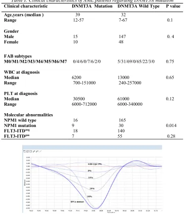
an AML patient with DNMT3A R882H mutation with more than 90% blast in bone marrow. DNA sample revealed a robust signal in sequencing analysis. After preparing the dilution serial in the context of wild-type DNA, 50% dilution produced a robust peak, wherase 25% represented a weak signal. By contrast, HRM detected the R882H mutation with a detection limit of 10% in the context of wild-type DNA (Figure 1). There was an overall good agreement between direct sequencing and
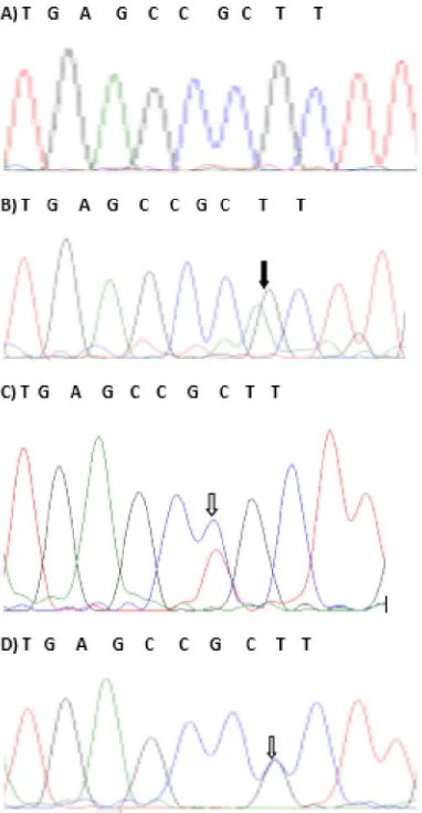
HRM assay (kappa value of 0.95) (p<0.001). In 218 cases (99%), complete agreement between assay findings was observed. The two cases (1%) with discordant findings resulted from the low quality of the DNA samples. The representative sequencing chromatograms and HRM curves for DNMT3A R882 mutations are shown in Figures 2 and 3.Samples containing of R882 mutations showed 2 peaks; whereas, wild-type sample showed only one peak at 82.8˚C.
Table I: Clinical characteristics of AML patients regarding DNMT3A mutation
Clinical characteristic DNMT3A Mutation DNMT3A Wild Type P value
Age,years (median ) Range
39 12-57
32
7-67 0.1
Gender Male Female
15 10
147 48
0. 4
FAB subtypes
M0/M1/M2/M3/M4/M5/M6/M7
0/4/6/0/7/6/2/0
5/31/69/0/65/22/3/0 0.75
WBC at diagnosis Median
Range
6200 700-151000
13000 240-257000
0.65
PLT at diagnosis Median
Range
30500 6000-712000
61000 6000-340000
0.12
Molecular abnormalities NPM1 wild type
NPM1 mutation FLT3-ITDneg
FLT3-ITDpos
16 9 18 7
165 30 140 55
0.014
0.28
Figure1. The sensitivity analysis of a dilution series of DNMT3A R882 mutant in a background of wild-type DNA detected by HRM
Figure 2. HRM curves analysis of three DNMT3A R882 mutations and wild-type
Figure3. Sanger sequencing analysis of DNMT3A R882 mutations (A: wild-type sequence; B: R882H mutation; C: R882C mutation; D: R882P mutation)
Iran J Ped Hematol Oncol. 2018, Vol 8. No 3, 172-179 177
Discussion
Molecular aberrations are important factors for risk-stratification of AML patients and molecularly targeted therapy. DNMT3A mutations have been attracting attention as markers for risk stratification in AML patients. Due to clinical significance and relatively high prevalence of DNMT3A R882 mutations in AML patients, progress in implementation of simple and rapid diagnostic methods is essential. HRM is a powerful technique for the detection of mutations and the scanning of epigenetic aberrations. It is cost-effective, time-saving, and more sensitive than Sanger sequencing (13). Detection of mismatched duplexes which is formed after amplification of heterozygous DNA makes HRM operation possible. Mutation scanning via the current method relies on the melting of heteroduplexes that twists the shape of the melting curve. Sanger sequencing is considered to be the gold standard test for mutational analysis. It provides a high degree of accuracy and long-read capabilities; however, it is laborious and expensive (14). Sequencing requires extensive automation and expensive instrumentation, and is time-consuming according to the cost for service setup. PCR amplification and mutation scanning can be performed in a single-step and closed-tube method by HRM technique and no other post-PCR processing is required. Therefore, limited sample handling reduces possible contamination. The present study analyzed DNA samples from 220 AML patients for DNMT3A R882 mutations using HRM and Sanger sequencing. HRM was more sensitive than Sanger sequencing (10% and 25%, respectively), but two samples were missed by HRM due to the low quality of DNA. To avoid false-negative results, it is very important to observe technical considerations. No false-positive results were detected. The results of the HRM
sensitivity limit for the detection of DNMT3A R882 mutations were in line with those of previous studies (15, 16). However, some studies as what mentioned in the following, reported a lower limit of sensitivity for HRM analysis compared to the present study. Berenstein et al., performed a comparative study on various PCR-based methods for the detection of DNMT3A and IDH1/2 mutations and described the HRM assay as being time-saving, cost-efficient, and sensitive. Their HRM assay had a sensitivity of 5.9% for the detection of DNMT3A R882 mutations (17). In determining recurrent R882 mutations in chinese patients with AML and MDS, Lin et al., revealed that HRM analysis could easily distinguish DNMT3A R882H mutation with maximal sensitivity of 2% in the background of wild-type DNA, wherease mutated R882H was detected at the maximal sensitivity of 10% by sanger sequencing (18). Singh et al., designed HRM analysis to identify high frequency DNMT3A mutations in AML patients. Sensitivity studies for exon 23 were performed by using DNA sample with R882H mutation and a blast count of 68% and the sensitivity was determined in 6.7% of mutant DNA in the context of wild-type DNA. They also showed that the false-negative rate for HRM was 0% (19). The discrepancy in the sensitivity limit for HRM between the studies could be due to the reagents (Real Time master mix) and type of equipment used. Therefore, each laboratory should justify HRM technique with its equipments before application. To setup the HRM assay to detect mutations, the availability of wild-type control samples and those with specific mutations are essential. The high sensitivity of HRM compared with Sanger sequencing makes it beneficial for the detection of molecular aberrations in AML patients with low cellularity in bone marrow or low allele burden of mutations (20).
178 Iran J Ped Hematol Oncol. 2018, Vol 8. No 3, 172-179
Conclusion
HRM curve analysis was confirmed as a sensitive, fast, and high-throughput method for the detection of DNMT3A R882 mutations in AML patients. HRM analysis was also validated as an alternative to Sanger sequencing because it is simple, relatively quick, and cost-effective.
Acknowledgment
This work was supported by a grant from Iran University of Medical Sciences [grant IR.IUMS.REC.1394.26066]. We would like to thank the professors and staff and the Hematology-Oncology and Stem Cell Transplantation Research Center at Tehran University of Medical Sciences.
Conflicts of interest
The authors declare that they have no conflict of interest.
References
1.Loghavi S, Zuo Z, Ravandi F, Kantarjian HM, Bueso-Ramos C, Zhang L, et al. Clinical features of De Novo acute myeloid leukemia with concurrent DNMT3A, FLT3 and NPM1 mutations. J Hematol Oncol 2014; 7(1):74-9.
2. Prada-Arismendy J, Arroyave JC, Röthlisberger S. Molecular biomarkers in acute myeloid leukemia. Blood Rev 2017;31(1):63-76.
3. Wang ML, Bailey NG. Acute myeloid leukemia genetics: risk stratification and implications for therapy. Arch Pathol Lab Med 2015; 139(10):1215-23.
4.Challen GA, Sun D, Jeong M, Luo M, Jelinek J, Berg JS, et al. Dnmt3a is essential for hematopoietic stem cell differentiation. Nat Genet 2012;44(1):23-31.
5. Shih AH, Abdel-Wahab O, Patel JP, Levine RL. The role of mutations in epigenetic regulators in myeloid
malignancies. Nat Rev Cance 2012; 12(9):599-612.
6.Suzuki MM, Bird A. DNA methylation landscapes: provocative insights from
epigenomics. Nat Rev
Genet2008;9(6):465-76.
7. Jia D, Jurkowska RZ, Zhang X, Jeltsch A, Cheng X. Structure of Dnmt3a bound to Dnmt3L suggests a model for de novo DNA methylation. Nature 2007; 449(7159):248-51.
8.Pezzi A, Moraes L, Valim V, Amorin B, Melchiades G, Oliveira F, et al. DNMT3A mutations in patients with acute myeloid leukemia in South Brazil. Adv Hematol 2012; 2012-15.
9. Russler-Germain DA, Spencer DH, Young MA, Lamprecht TL, Miller CA, Fulton R, et al. The R882H DNMT3A mutation associated with AML dominantly inhibits wild-type DNMT3A by blocking its ability to form active tetramers. Cancer cell 2014;25(4):442-54.
10.Jancik S, Drabek J, Berkovcova J, Xu YZ, Stankova M, Klein J, et al. A comparison of Direct sequencing, Pyrosequencing, High resolution melting analysis, TheraScreen DxS, and the K-ras StripAssay for detecting KRAS mutations in non small cell lung carcinomas. J Exp Clin Cancer Res 2012;31(1):79-84.
11.Magnin S, Viel E, Baraquin A, Valmary-Degano S, Kantelip B, Pretet J-L, et al. A multiplex SNaPshot assay as a rapid method for detecting KRAS and BRAF mutations in advanced colorectal cancers. The J Mol Diagn 2011;13(5):485-92.
12. Ramazanzadeh M, Salehi M, Salehi R.Assessment of high resolution melt analysis feasibility for evaluation of beta-globin gene mutations as a reproducible, cost-efficient and fast alternative to the present conventional method. Adv Biomed Res 2016; 5:71
13.Bilbao-Sieyro C, Santana G, Moreno M, Torres L, Santana-Lopez G, Rodriguez-Medina C, et al. High resolution melting analysis: a rapid and
Iran J Ped Hematol Oncol. 2018, Vol 8. No 3, 172-179 179
accurate method to detect CALR mutations. PloS one 2014; 9(7):e103511-14.
14. Buettner R, Froehner S, Merkelbach-Bruse S, Ney J, Roesler A. High resolution melting analysis as a prescreening tool. Google Patents 2016; 8: 33-40.
15.Gorniak P, Ejduk A, Borg K,
Makuch‐Lasica H, Nowak G,
Lech‐Maranda E, et al. Comparison of high‐resolution melting analysis with direct sequencing for the detection of
recurrent mutations in DNA
methyltransferase 3A and isocitrate dehydrogenase 1 and 2 genes in acute myeloid leukemia patients. Eur J Haematol 2016; 96(2):181-7.
16. Szarzyńska‐Zawadzka B, Kosmalska M, Sędek Ł, Sonsala A, Twardoch M, Kowalczyk JR, et al. Cost‐effective screening of DNMT3A coding sequence identifies somatic mutation in pediatric T‐cell acute lymphoblastic leukemia. Eur J Haemato 2017; 99(6):514-9.
17.Berenstein R, Blau IW, Kar A, Cay R, Sindram A, Seide C, et al. Comparative examination of various PCR-based methods for DNMT3A and IDH1/2 mutations identification in acute myeloid leukemia. J Exp Clin Cancer Res 2014; 33(1):44-7.
18.Lin J, Yao D-m,Qian J, Chen Q, et al. Recurrent DNMT3A R882 mutationsin chinese patients with acute myeloid leukemia and myelodysplastic syndrome. PLoS One 2011; 6(10):e26906
19. Singh RR, Bains A, Patel KP, Rahimi H, et al. Detection of high-frequency and novel DNMT3A mutations in acute myeloid leukemia by high-resolution melting curve analysis. J Mol Diagn 2012; 14(4):336-45
20. RH, Pham HT, Perkins SL, Prchal JT, Agarwal AM, Salama ME. Increased frequency of co-existing JAK2 exon-12 or MPL exon-10 mutations in patients with low JAK2V617F allelic burden. Leuk
Lymphoma2016; 57(6):1429-35.