International
J
ournal ofP
harmaceuticalB
iological andC
hemicalS
ciencese-ISSN: 2278-5191
International Journal of Pharmaceutical, Biological and Chemical Sciences (IJPBCS) | APR-JUN 2015 | VOLUME 4 | ISSUE 2 | 34-40
www.ijpbcs.net or www.ijpbcs.com
R
esearch
A
rticle
Pa
ge
34
DOCOSAHEXAENOIC ACID INDUCES APOPTOSIS
IN COLON CANCER CELL LINE, Caco-2
Entissar S. Al-Suhaibani 1
1
Faculty of Sciences, King Saud University, KSA.
*Corresponding Author Email: ealsuhaibani@hotmail.com
INTRODUCTION
Colorectal cancer is the third most common form of
cancer and the second leading cause of cancer deaths
in both men and women around the world.
Alarmingly, increasing numbers of reported cases of
colon cancer in recent years has made this form of
cancer a major health concern1. The current treatment
for colorectal cancer is generally surgical resection
combined with chemotherapy by cytotoxic drugs and
radiation. However, this therapy is just moderately
successful especially for late stage cancers; therefore
new approaches to the treatment of colorectal cancer
are required. In recent years, interest has increased in
using natural products for pharmacological purposes,
as a form of complementary or replacement therapy. It
is known that the risk of colorectal cancer increases
with dietary habits like high animal fat intake2.
Epidemiological and prospective studies have reported
several beneficial effects of bioactive compounds on
human health, particularly in protecting against
chronic degenerative diseases, such as cardiovascular
disease, diabetes mellitus and cancer.
Omega-3 fatty acids are long-chain polyunsaturated.
The principal dietary source of eicosapentaenoic acid
(EPA, 20:5n- 3) and docosahexaenoic acid (DHA,
22:6n-3) is from oily coldwater fish3. Epidemiological
studies have suggested that an increased fish oil intake
is associated with a reduced breast cancer incidence in
humans4. Consistent with this epidemiological
observation, laboratory studies have also shown that
omega-3 can suppress the formation and growth of
breast cancer in animal models5. A number of
mechanisms have been proposed for the anticancer
actions of omega-3, including suppression of
neoplastic transformation, inhibition of cell
ABSTRACT:
OBJECTIVES: The present study sought to further investigate the in vitro anticancer effects of a representative omega-3 fatty acid, docosahexaenoic acid (DHA), with a focus on assessing the induction of apoptosis as an important mechanism for
its anticancer actions. METHODS: Caco-2, cells were incubated with DHA at concentrations, 200µM, 400µM, 600µM and
800µM for 72h at 37°C and 5% CO2. MTT assay DNA fragmentation assay, cytological and immunocytochemical
investigations were investigated. RESULTS: DHA strongly reduces the viability and DNA synthesis of Caco-2 human
colorectal cancer cells in culture, and also promotes cell death via apoptosis. MTT assay was used to test DHA on the effect of the proliferation of the colorectal cancer cell line, Caco-2. IC50 DHA inhibited the proliferation of Caco-2 cells and induced Caco-2 cell death in a dose-dependent manner. Treated cells showed typical characteristics of apoptosis including inhibited the viability and proliferation of treated Caco-2 cells in vitro even by DNA fragmentation, cytological alterations
and downregulation of Bcl-2 activity. CONCLUSIONS: Results from this study show that DHA has antiproliferative effects
against cancer cells. fish oil as a dietary supplement can help to maintain good health and protect the body against disease. Future study will may deal with further investigations of fish oil as a dietary supplement possible usages as a new alternative or complementary chemotherapeutic agent for human cancer types especially colorectal cancer type.
KEYWORDS:
Entissar S. Al-Suhaibani * et al;DOCOSAHEXAENOIC ACID INDUCES APOPTOSIS IN COLON……
International Journal of Pharmaceutical, Biological and Chemical Sciences (IJPBCS) | APR-JUN 2015 |VOLUME 4 | ISSUE 2 | 34-40 | www.ijpbcs.net
Pa
ge
35
proliferation, enhancement of apoptosis, and
antiangiogenicity6.
Mitochondria are involved in a variety of key events,
including release of caspase-3 activators, changes in
electron transport, loss of mitochondrial membrane
potential, and participation of both pro- and
anti-apoptotic Bcl-2 protein7. Alterations in mitochondrial
structure and function have been shown to play a
crucial role in caspase-3-dependent apoptosis and
Bcl-2 expression8. Bcl-2 is the founding member of family
of genes that either prevents or promotes cellular
apoptosis. Bcl-2 itself is an antiapoptotic gene that
prevents initiation steps of apoptosis and programmed
cell death9. The aim of this study was to evaluate the
effect of DHA on the inhibition of proliferation of the
colorectal cancer cell line, Caco-2.
MATERIALS AND METHODS
Chemical reagents: DHA, MTT salt or
3-(4,5-dimethylthiazole-2-yl)-2,5-diphenyl tetrazolium
bromide, dimethylsulfoxide (DMSO), commercial
methanol, commercial ethanol, commercial acetone,
Tris-HCl, edetic acid, Triton-X100, RNase A,
proteinase K, NaCl, 2-propanol, phosphate-buffered
saline (PBS), ethidium bromide, agarose gel,
Peroxidase, trypsin, Hematoxylin and eosin (Hx & E)
stain, primary monoclonal antibody against Bcl-2 and
biotinylated immunoglobulin secondary antibody and
Tween 20 were purchased from Sigma-Aldrich, Egypt.
Cell line and cell culture: Caco-2 cell line, was
obtained from American Type Culture Collection
(ATCC, USA). They were sub-cultured as mono-layer
according to the instructions provided by ATCC in
Dulbecco modified Eagle medium (DMEM)
supplemented with 10% heat inactivated (56°C,
30min) fetal bovine serum, 2mmol/L L-glutamine,
100U/mL Penicillin-Streptomycin and 100U/mL
Amphotericin B at 37°C in a humidified atmosphere
of 5% CO2. Cells were used when monolayer reached
80% confluence in all experiments. Cell propagation
media was purchased from Invitrogen (Carlsbad, CA).
Methods: 1.Cell Viability Assay: In vitro
evaluation of antiproliferation effect: growth
inhibition was evaluated by MTT assay. MTT salt or
3-(4,5-dimethylthiazole-2-yl)-2,5-diphenyl tetrazolium
bromide was reduced by mitochondrial
dehydrogenases to water blue insoluble formazans10.
Viable cell number/well is directly proportional to
formazans production. 8.25×10³ cells were seeded into
each well of 96-well plate, incubated with culture
medium overnight (12h), replaced with fresh medium
containing DHA at concentrations: 200µM/L,
400µM/L, 600µM/L and 800µM/L for 72h at 37ºC in
an incubator with 5% CO2. After incubation, DHA
modified medium was replaced by 100μL of MTT
(0.5mg/mL) medium for incubation (3h at 37°C and
5% CO2). MTT medium was then replaced with
100μL of DMSO and left for 10min on a platform
shaker to solubilize converted formazan. The
absorbance values were determined at 570nm test
wavelength and 630nm reference wavelength (Spekol
1200 spectrophotometer). Untreated cells were as a
positive control cells and all values were correlated
with this set of data. The experiment was performed in
triplicates. Inhibition Percentage=[1–(net Absorbance
of treated well/net Absorbance of control
well)]x100%, then was plotted against DHA
concentrations.
2. Determination of DNA fragmentation by DNA
laddering assay: cells were seeded in 60-mm petri
dishes at density 4x10⁵ cells/plate (treated cells by IC50
concentration of DHA or positive control cells).
Adherent and floating cells were collected by
centrifugation at 1000×g/5min. Cell pellet was
suspended in cell lysis buffer (Tris-HCl 10mmol/L
pH7.4, edetic acid 10mmol/L pH8.0, Triton-X100
0.5%) and kept at 4ºC/10min then, lysate was
centrifuged at 25.000×g/20min. Supernatant was incubated with RNase A 40μg/L/1h (37ºC), incubated with proteinase K 40μg/L/1h (37ºC), mixed with NaCl
0.5mol/L and 50% 2-propanol overnight (-20ºC), then
Entissar S. Al-Suhaibani * et al;DOCOSAHEXAENOIC ACID INDUCES APOPTOSIS IN COLON……
International Journal of Pharmaceutical, Biological and Chemical Sciences (IJPBCS) | APR-JUN 2015 |VOLUME 4 | ISSUE 2 | 34-40 | www.ijpbcs.net
Pa
ge
36
was dissolved in buffer (Tris-HCl 10mmol/L pH7.4,
edetic acid 1mmol/L pH 8.0) and separated by
2%agarose gel electrophoresis at 100V for 50min.
DNA was visualized under ultraviolet light after
staining with ethidium bromide11.
3.Cytological changes investigation: detached and
trypsinized cells (IC50 concentration of DHA treated
cells and positive control cells) were collected and
centrifuged at 2000 rpm for 5min. Cell pellet was re-suspended with 100μL of PBS (pH7.3). 10μL of the
suspension were smeared on a glass slide, allowed to
air-dry, fixed with cool methanol for 5min before
proceeding by Hx&E stain and examined under light
microscope12.
4. Immunocytochemical investigations: bydetection
of Bcl-2 by immunocyto chemistry staining kits. The
procedure was done according to the manufacturer’s
instructions, simplified as follows: 1-2 drops of
Peroxidase was applied to cells (IC50 concentration of
DHA treated cells and positive control cells) on the
slide (10min), followed by blocking solution (10min).
Cells were fixed in ethanol:acetone (9:1) for 30min at
-20°C and then rinsed again with cold PBS at room
temperature. Cells were incubated overnight with
primary monoclonal antibody against Bcl-2 at dilution
of 1:75 at 4°C, then in Tris buffer and biotinylated
immunoglobulin secondary antibody was used13. The
slides were then mounted and examined under light
microscope.
5. Statistical analysis: results were presented as
mean±standard deviations (SD). Analysis of variance
(ANOVA) for two variables (Two Way-ANOVA) was
used together with student t-test. Significant analysis
of variance results were subjected to post hoc.
Statistical significance was set at P<0.05 and high
significance was set at P≤ 0.0114
.
RESULTS
1.Cell viability assay: In vitro evaluation of
antiproliferation effect.
Cytotoxic effect of different concentrations of DHA
(200µM, 400µM, 600µM and 800µM) for 72h on
Caco-2 cell line was determined by MTT assay
(Figure 1). Cells number started to reduce immediately
after treatment with DHA concentrations in a dose
dependent manner. All concentrations were found to
be high significantly different (P≤0.01) in respect to
their antiproliferative and apoptotic effects when
compared with positive control cells. Cell inhibition
percentage was gradually increased with DHA
concentration increasing and 100% of cell inhibition
was observed when cells were treated with
800µM/72h. Cell proliferation reduced about 30% and
45% when cells were treated with 200µM and 400µM
for 72h, respectively. Cells proliferation decreased to
60% when treated with concentration of 600µM/72h.
2. Determination of DNA fragmentation by DNA
laddering assay.
DNA degradation into multiple internucleosomal
fragments is a distinct biochemical hallmark for
apoptosis. Nuclear DNA isolated from Coca-2 cancer
cells was separated by agarose gel electrophoresis and
stained with ethidium bromide, and a typical ladder
formation was observed upon 72h when treated with
DHA concentration at 600µM whereas untreated cells
did not show typical ladder (Figure 2). Results
indicated that DHA induced DNA fragmentation
which was caused by apoptosis.
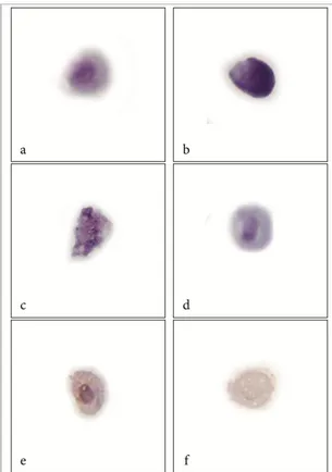
3. Cytological changes investigation.
Positive control cells group had round nuclei, distinct
small nucleoli and homogeneous chromatin with an
accentuated nuclear membrane (Figure 3a). After
Coca-2 cells treatment by DHA concentration at
600µM/72h, apoptotic cells were identified by a series
morphological changes as an important experimental
proof of underlying processes alterations appeared as:
bleb plasma membrane, cellular shrinkage, chromatin
condensation granules, vacuolated cytoplasm,
degrading nucleus and apoptotic bodies formation
were observed (Figure 3b, 3c and 3d).
Entissar S. Al-Suhaibani * et al;DOCOSAHEXAENOIC ACID INDUCES APOPTOSIS IN COLON……
International Journal of Pharmaceutical, Biological and Chemical Sciences (IJPBCS) | APR-JUN 2015 |VOLUME 4 | ISSUE 2 | 34-40 | www.ijpbcs.net
Pa
ge
37
After Coca-2 cells treatment by DHA concentration at
600µM/72h and regarding to the positive control
Coca-2 cells, Bcl-2 protein reaction was considered
positive (over expression of Bcl-2 protein) when over
55% of cells had nuclear membrane, mitochondrial
outer membrane and endoplasmic reticulum
membrane brown staining, with slight intensity
degrading in the same field(Figures 3e). After Coca-2
cells treatment by DHA concentration at 600µM/72h,
those fields that had necrotic or apoptotic nucleus as
sign for DHA apoptotic effect with Bcl-2 negative
reaction (faint to non-brown stain) (Figure 3f).
Figure 1: Effect of DHA with different concentrations on the cells viability of Caco-2 cells. The experiment was
performed in triplicates and values means were calculated [mean ± SD, n (for each concentration) =4].
Figure 2: DNA fragmentation by DNA laddering assay of extracted DNA from DHA treated cells and positive
control cells. DNA laddering, typical for apoptotic cells, which were visible in treated Coca-2 cells (T), and there
Entissar S. Al-Suhaibani * et al;DOCOSAHEXAENOIC ACID INDUCES APOPTOSIS IN COLON……
International Journal of Pharmaceutical, Biological and Chemical Sciences (IJPBCS) | APR-JUN 2015 |VOLUME 4 | ISSUE 2 | 34-40 | www.ijpbcs.net
Pa
ge
38
Figure 3: Cells in different apoptosis stages in treated cells are easily distinguishable. Cell with normal
morphology (a). Complete apoptotic cell (b). Degradation of nucleus, vacuolated cytoplasm with apoptotic
bodies (c). Nuclear condensation is evident in cells (dark, condensed and irregular rounded nucleus), bleb
membrane and cell shrinkage (d). Immunocytochemistry of Bcl-2 protein as control positive cell showing Bcl-2
protein nuclear membrane, mitochondrial outer membrane and endoplasmic reticulum membrane showing
brownish positive reaction (e). Treated cell showing negative reaction of apoptotic cell (f).
DISSCUTION
The results of our present study showed that DHA, an
omega-3, has a strong anticancer activity in cultured
Caco-2, human colorectal cancer cells through a
combination of multiple actions, including inhibition
of DNA synthesis, suppression of cell viability, and
induction of apoptotic cell death. It was reported
earlier that while omega-3 could selectively inhibit
tumor cell proliferation, they were significantly less
cytotoxic in normal cells15. The results of our present
study showed that the anticancer effect of DHA
Apoptosis, as programmed cell death, is a highly
organized cell death process characterized by an early
obvious condensation of nuclear chromatin, loss of
plasma membrane phospholipid asymmetry, activation
Entissar S. Al-Suhaibani * et al;DOCOSAHEXAENOIC ACID INDUCES APOPTOSIS IN COLON……
International Journal of Pharmaceutical, Biological and Chemical Sciences (IJPBCS) | APR-JUN 2015 |VOLUME 4 | ISSUE 2 | 34-40 | www.ijpbcs.net
Pa
ge
39
oligonucleosomal fragments and segmentation of the
cells into membrane-bound apoptotic bodies16. DNA
fragmentation, a hallmark of apoptosis, is regulated by
a specific nuclease called caspase-activated DNase and
its inhibitor17. Apoptosis has specific signals
instructing the cells with specific morphological
change as plasma and nuclear membrane blebbings,
chromatin condensation, proteases activation and
DNA fragmentation that are considered as landmarks
of the apoptotic process18. That was agreed with the
results of recent study after treatment by DHA. DHA
decreased the viable percentage of cell number (dose
dependent effect) and induced apoptosis of Caco-2
cells.
Therefore, we may presume that as primary
mechanism involved in DHA growth-inhibitory effects
as it considered main apoptotic signals. Caco-2 cells
which were treated with DHA exhibited down
regulated levels of Bcl-2 expression at concentration
of 600µM/72h, which suggested that Bcl-2 involved in
DHA-induced Caco-2 cell death as mitochondrial
pathway was involved in DHA-induced Caco-2 cell
death19.
CONCLUSION
In this study, we have demonstrated that fish oil as a
dietary supplement inhibited proliferation and induced
apoptosis in colon cancer (Caco-2) cells which
depended on down-regulation of Bcl-2 protein. Future
in vitro and in vivo study will may deal with further
investigations of the possible usages of fish oil as a
dietary supplement as a new alternative
chemotherapeutic agent but in limit doses for human
colorectal cancer suggested treatment and other types
of cancer.
ACKNOWLEDGMENTS
We are grateful for all of Nile center for experimental
researches team specially Miss. Noha T. Badawy
(Department of cell culture) for her kindly support in
the cell culture and drug induction stage.
REFERENCES
1. Mayer, R. (2009). Targeted therapy for advanced
colorectal cancer-more is not better. J. Med.,
360:623-625.
2. Murillo, G.; Hoard, M.; Naithani, R. and Mehta,
R. (2008). Efficacy of herbal products in
colorectal cancer prevention. Current Colorectal
Cancer Reports., 4:34-42.
3. Bartram, H.; Gostner, A.; Scheppach, W.; Reddy,
B. and Rao, C. (1993). Effects of fish oil on
rectal cell proliferation, mucosal fatty acids, and
prostaglandin E2, release in healthy subjects.
Gastroenterology., 105:1317-1322.
4. Hardman, W. (2002). Omega-3 fatty acids to
augment cancer therapy. J Nutr.,
132:3508S-3512S.
5. Kaizer, L.; Boyd, N.; Kriukov, V. and Tritchler,
D. (1989). Fish consumption and breast cancer
risk: an ecological study. Nutr Cancer., 12:61-68.
6. Bagga, D.; Anders, K..; Wang, H. and Glaspy, J.
(2002). Long-chain n-3-to-n-6 polyunsaturated
fatty acid ratios in breast adipose tissue from
women with and without breast cancer. Nutr
Cancer., 42:180-185.
7. Adams, J. and Cory, S. (1998). Bcl-2 protein
family: arbiters of cell survival. Science.,
281:1322-1325.
8. Droin, N. and Green, R. (2004). Role of Bcl-2
family members in immunity and disease. BBA.,
1644:179-188.
9. Wolvetang, E.; Perez, J.; Roig, T.; Manzano, A.;
Larm, J. and Moutsoulas, P. (1996). Apoptosis
induced by inhibitors of plasma membrane
NADH-oxidase involves Bcl-2 and calcineurin.
Cell Growth Differ. 7:1315-1318.
10. Mosmann, T. (1983). Rapid colorimetric assay
for cellular growth and survival: application to
proliferation and cytotoxicity assays. J. Immunol.
Entissar S. Al-Suhaibani * et al;DOCOSAHEXAENOIC ACID INDUCES APOPTOSIS IN COLON……
International Journal of Pharmaceutical, Biological and Chemical Sciences (IJPBCS) | APR-JUN 2015 |VOLUME 4 | ISSUE 2 | 34-40 | www.ijpbcs.net
Pa
ge
40
11. Zhang, Y.; Chen, X.; Liu, J. and Wong, T.
(2003). DNA excision repair system of highly
radioresistant bacterium Deinococcus
radiodurans is facilitated by pentose phosphate
pathway. Mol. Microbiol., 48:1317-1323.
12. John, A. and Abraham, S. (1991). Cytological
changes produced by red pepper in mitotic cells
of Vicia faba L. Caryologia., 44:325-331.
13. Yoon, K.; Nakamura, Y.; Doubek, D. and
Arakawa, H. (2004). Identification of ALDH4 as
p53, caspase-3 and Bcl-2-inhibitor gene and its
protective role in cellular stresses. J. Hum.
Genet., 49:134-140.
14. Snedecor, G. and Cochran, W. (1980). Statistical
Methods, 7th ed Iows State unive. Press, Iowa,
U.S.A.
15. Rose, D. and Connolly, J. (1999). Omega-3 fatty
acids as cancer chemopreventive agents.
Pharmacol Ther., 83:217-44.
16. Wolvetang, E.; Perez, J.; Roig, T.; Manzano, A.;
Larm, J. and Moutsoulas, P. (1996). Apoptosis
induced by inhibitors of plasma membrane
NADH-oxidase involves Bcl-2 and calcineurin.
Cell Growth Differ., 7:1315-1318.
17. Enari, M.; Jen, K.; Cheung, V.; Sakahira, H. and
Yokoyama, H. (1998). A caspase-activated
DNase that degrades DNA during apoptosis, and
its inhibitor ICAD. Nature., 391:43-46.
18. Kidd, V. (1998). Proteolytic activities that
mediate apoptosis. Annu. Rev. Physiol.,
60:533-538.
19. Moll, U. and Zaika, A. (2001). Nuclear and
mitochondrial apoptotic pathways of Bcl-2.
FEBS. Lett., 493:65-9.
*Corresponding author Email address: