60 | P a g e
International Journal of Phytopharmacology
www.onlineijp.com
e- ISSN 0975 – 9328 Print ISSN 2229 – 7472
HEPATOPROTECTIVE ACTIVITY OF
GUETTARDA SPECIOSA L.
AGAINST
CARBON
TETRA
CHLORIDE
(CCl
4)-INDUCED
HEPATOTOXICITY IN WISTAR ALBINO RATS
P. Vennela Priya and A. Saravanakumar*
Department of Pharmacology, Sri Venkateswara College of Pharmacy, R.V.S. Nagar, Tirupathi Road, Chittoor-517127, Andhra Pradesh, India.
ABSTRACT
The present study investigate the phytoconstituents, acute oral toxicity and hepatoprotective activity of methanol extract of inner bark of Guettarda speciosa L. using CCl4induced hepatotoxicity in male Wistar albino rats. The MEGS at doses of 200 and 400mg/kg, p.o and the standard drug Silymarin (100mg/kg, p.o.) were administered for three times at 12h intervals. After 36hrs of carbon tetrachloride treatment, blood was collected from all groups of rats by puncturing the heart. Serum was separated by centrifugation at 2500rpm at 370C for 15min and analyzed for various biochemical parameters. The hepatoprotective activity was assessed by using various biochemical parameters like SGOT, SGPT, ALP, γ-GT, total bilirubin and Total protein along with histopathological studies of liver tissue. The animals treated only with CCl4 exhibited a significant increase (P<0.001) the levels of SGOT, SGPT, ALP, γ-GT and total bilirubin as well as decrease in
the levels of TP when compared to the normal control group after 36hrs of CCl4 treatment, indicating hepatocellular damage. The MEGS afforded significant protection against CCl4induced hepatocellular injury.
Key words: Hepatotoxicity, CCl
4, Hepatic Enzymes, Guettarda speciosaL., Hepatoprotective.
Corresponding Author: Dr.A. Saravanakumar Email: saravanacology@gmail.com
INTRODUCTION
The liver is the largest internal organ of the body weighing 1200-1500gm. It is a key organ in regulating homeostasis within the body. It regulates several important functions including protein synthesis, storage and metabolism of fats and carbohydrates, detoxification of the drugs and other toxins, metabolism of hormones and excretion of bilirubin. Liver diseases are associated with distortion of these metabolic functions (Ward FM and Daly MJ, 2005).
Access this article online Home page:
http://onlineijp.com/
DOI:
http://dx.doi.org/10.21276/ijp.2017.8.2.3
Quick Response code
Received:04.02.17 Revised:10.03.17 Accepted:19.03.17
Liver produces and secretes bile; it also produces blood clotting factors prothrombin, fibrinogen and heparin. The liver converts sugar into glycogen (Sharma B and Sharma UK, 2009).
The liver has considerable reserve capacity, can often maintain function in spite of significant disease and is one of the few human organs capable of regeneration. However, in cases of severe injury or disease of the liver, a diverse range of physiological roles are impaired, with potentially serious consequence for the individual concerned (Ward FM and Daly MY, 2005).
The liver is exposed to drugs in higher concentration than other organs of the body, because most drugs are administered orally and are absorbed from the gastrointestinal tract (GIT). Thus the whole dose must pass through the liver to reach systemic circulation. Because of this, the liver is a vulnerable target for injury from drugs and chemicals and disordered hepatic function is an important cause of
61 | P a g e
abnormal drug handling and response (Bennett PN and Brown MJ, 2006).
Liver diseases have become one of the major causes of morbidity and mortality in man and animals all over globe and hepatotoxicity due to drugs appears to be the most common contributing factor (Sharma B and Sharma UK, 2009). About 20000 deaths found every year due to liver disorders. Hepatocellular carcinoma is one of the ten most common tumors in the world with over 250000 new cases each year (Gupta AK and Misra N, 2006).
TYPES OF LIVER DISEASES
There are two main type of liver disease, namely acute and chronic;
Acute Liver Disease
Acute liver disease is usually a self-limiting episode of liver cell inflammation or damage which in most cases resolves without clinical sequence. In some cases, however, the hepatocyte damage is so severe that it affects the whole liver and leading to hepatic failure with an associated high mortality. Alternatively, the patient can develop chronic liver disease (Ward FM and Daly MJ, 2005).
Chronic Liver Disease
Chronic liver disease occurs when permanent structural changes within the liver, leads secondary to long-standing cell damage with the consequent loss of normal liver architecture. In most cases this progresses to cirrhosis, where fibrous scars divide the liver cells into areas of regenerative tissue called nodules. This process is irreversible and eventually leads to liver failure. Cirrhosis may be secondary to autoimmune hepatitis or may result from chronic alcohol ingestion (Ward FM and Daly MJ, 2005).
Various Types Of Liver Disorders
The sheer complexity and varied nature of its interactions continually exposes it to a variety of toxins, therapeutic agents, etc., making it susceptible to literally hundreds of diseases. Some of these diseases are rare; others are more common, such as;
1. Hepatitis 2. Cirrhosis
3. Jaundice
4. Fatty liver
5. Portal hypertension 6. Drug induced liver disease.
Hepatitis
Hepatitis is an inflammation of liver and usually characterized as viral hepatitis or non-viral hepatitis. Viral hepatitis (A, B, C, D and E) is a contagious infection of liver usually caused by one of the
three different organisms.
Hepatitis-A formerly known as infectious hepatitis, can be contracted by consuming contaminated water or food, most notably shellfish. The virus is eliminated in stool and though seldom serious it can cause severe liver failure and death. It does not cause chronic hepatitis and does not lead to cirrhosis or other long-term liver problems.
Hepatitis-B formerly known as serum hepatitis and it is found in blood and other body fluids such as urine, tears, semen, breast milk and vaginal secretions. It is usually transmitted in blood via transfusion or through illicit injectable-drug use.
Type-C hepatitis virus is also contracted through contact with infected person (Mohammad O et al., 2006).
Cirrhosis
In cirrhosis, liver cells are damaged and replaced by scar tissue which as it accumulates, hardens the liver, diminishes blood flow and causes even more cells to die. The loss of liver function that accompanies this degenerative condition results in gastrointestinal disturbances, jaundice, enlargement of liver and spleen, maceration and accumulation of fluid in the abdomen and other tissues. Alcohol abuse, hepatitis, chemical poisoning, excess of iron or copper, other viruses and blockages of the bile duct can cause the disease. Cirrhosis is the third leading cause of death in adults aged between 25 and 59 and seventh leading cause of death overall (Mohammad O et al., 2006).
Jaundice
Jaundice is a yellow coloration of the sclerae (white of the eyes), skin and mucous membranes due to the build up of a bilirubin. After bilirubin is formed from the break down of the haem pigment in aged red blood cells and it is transported to the liver, where it is processed and eventually excreted into bile (Gerard JT and Bryan D, 2006).
Fatty Liver
Fatty liver is a common and generally beginning condition. The majority obese patients (60-90%) and up to 50% of type-2 diabetics have fatty liver. In general the lipid accumulation is characterized by macro vesicular steatosis. In a minority cases hepatic seatosis is associated with an inflammation in filtrate. The histological appearance described as steato hepatitis may be caused by alcohol misuse. However in some patients with other causes there is no history of excessive alcohol consumption is known as non-alcoholic steato hepatitis and may progress to cirrhosis in small proportion of patients (Hayes PC et al., 2002).
62 | P a g e
Portal hypertension occurs when there is increased pressure within the portal venous system secondary to increased resistance to flow through the damaged liver. Portal hypertension is an import contributory factor to the formation of ascites and the development of encephalopathy due to by passing of blood from the systemic circulation. The major, potentially life threatening complication of portal hypertension is torrential venous haemorrhage from the thin walled veins in the oesophagus and stomach (Ward FM and Daly MJ, 2005).
Drugs induced Liver Disorders
After oral administration, the entire absorbed dose of drug is exposed to the liver during the first pass through the body, the drug itself or its metabolites may affect liver function. Metabolic pathways may become saturated at high concentration and drug or metabolites may accumulate and leading to toxicity. The drugs like paracetamol, salicylates, tetracyclines, rifampcin, etc., causes hepatotoxicity at excessive doses (James MR, 2006).
Causes Of Liver Disease
Liver diseases are mainly caused by viral infections (Hepatitis-A, B, C, D and E), excess alcohol consumption, drug and toxins (For example, certain antibiotics, chemotherapeutics, peroxidised oil, aflatoxin, carbon-tetrachloride, chlorinated hydrocarbons, etc.), immune disorders, inherited metabolic disorders, biliary tract diseases, infectious diseases, vascular abnormalities and gilbert’s syndrome (Ward FM and Daly MJ, 2005).
Clinical Manifestation Of Liver Disease Symptoms of Liver Disease
Patients frequently complain of being easily fatigued and generally run down with loss of appetite and weight loss.
In chronic liver disease there may be loss of muscle from the arms and legs and swelling of the abdomen and lower body due to fluid retention.
Individuals with enlarged liver may complain of abdomen pain and tenderness.
Patients with chronic liver disease and associated abnormal clotting of the blood or low platelet counts often complain of increased bleeding from the gums and nose and easy bruising.
Signs of Liver Disease
Many of the signs associated with chronic liver disease are related to the failure of the liver to carryout normal synthetic, metabolic and excretory functions. The most common signs are;
Cutaneous Signs
There are a number of cutaneous signs that indicate the presence of chronic liver disease, although none are entirely specific. They include finger clubbing (swelling of the finger ends), spider naevi (small vascular malformation in the skin), palmor erythema (reddening of the palm of the hand) and white
nails (associated with low serum albumin).
Abdominal signs
Various changes may be detectable on examination of the abdomen. An enlarged liver (hepatomegaly) is a common finding in acute liver disease, indicating inflammation and regeneration of the liver tissue.
Jaundice
Jaundice indicates impaired liver cell function and is a common finding in acute liver disease, but may be absent in chronic liver disease until the terminal stages of cirrhosis are reached.
Ascites
A swollen abdomen (ascites) is caused by the accumulation of fluid within the abdominal cavity.
Portal Hypertension
Portal hypertension occurs when there is increased pressure within the portal venous system secondary to increased resistance to flow through the damaged liver.
Gynaecomastia
Hormonal changes may occur in patients with liver disease, partly due to the inability of the cirrhotic liver to metabolize oestrogen, which may even lead to the development of gynaecomastia in males.
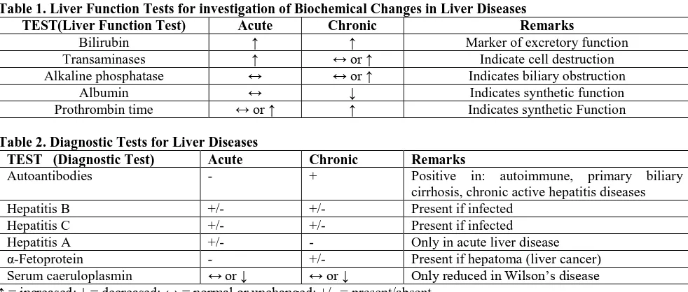
INVESTIGATIONS
Most of the investigations to determine the cause and extent of the liver disease are similar for the both acute and chronic liver disease, and are summarized in the table 1 and 2.
Imaging Techniques
Visualization of the liver texture and bile ducts are important to the impaired liver function. Ultrasound is the investigation of first choice, allowing investigation of possible liver tumours and the extent of splenomegaly and portal hypertension. Computed tomography is used to enable more detailed examinations.
Liver Biopsy
63 | P a g e
Hepatitis SerologyTests for hepatitis A, B and C are mandatory in unexplained liver disease.
Biochemical Liver Function Test
Biochemical liver function tests (LFTs) are simple, inexpensive and easy to perform, but provide a relatively crude measure of liver function which cannot be used in isolation to make a diagnosis. They are most useful in monitoring disease progression and response to treatment. Serum elevations greater than twice the upper limits of the reference range are usually considered significant changes. Usually more than one LFT tends to be raised in patients with liver disease.
TREATMENT
Conventional Medicines for Liver Diseases
Immunoglobulin (Ig) is quite effective against hepatitis-A when administered to anyone exposed to the virus as soon as possible or within two weeks after jaundice appears.
Vaccines for hepatitis are now a common feature of immunization programs the world over.
Treatment for acute hepatitis consists of rest and small, nourishing MEGSs, fluids and sometimes anti-nausea drugs such as trimethobenzamide. Chronic cases of hepatitis-B and C are treated with
interferon.
The problem of gallstones is usually solved by surgical operation. Chenoidiol, a recently available drug that dissolves gallstones are an alternative to surgery, but trouble some side-effects have been reported.
In the treatment of cirrhosis elimination of the underlying cause is emphasized, if possible, to avoid further damage, and abstinence from alcohol is supportive measures. For extreme cases a liver transplant is an option, though risky.
Herbal Medicines for Liver Diseases
Despite advancements in modern medicine, no hepatoprotective medicine is available. Treatment options for cirrhosis, fatty liver and chronic hepatitis are limited as well as problematic. The conventional drugs used in such treatments are inadequate and inconsistent at best as well as these drugs may themselves cause damage of hepatocytes. Alternative treatments for liver diseases to replace the currently used drugs need to be given impetus in light of current findings from research studies and publications in the field of herbal treatment of liver diseases, especially during the last quarter of the twentieth century. Indigenous medicines, especially of plant origin, are used extensively for the treatment of various diseases. With lack of safe and effective treatment for liver diseases, researchers have been
looking for alternative therapies that curb symptoms with minimum adverse effects on patients.
Plant drugs are known to play a vital role in the management of liver diseases. There are numerous plants and polyherbal formulations claimed to have hepatoprotective activity. Several hundreds of plants are reported to have hepatoprotective properties, and a number of studies have been conducted taking in to consideration valid scientific, clinical and research parameters. These plants include Cochlospurmum planchonii, Zingiber officinale, Nardostachs jatamansi,
Swertia chirata, Cichorium intybus, Teprosiapurpurea,
Plumbago zelanicam, Solanum nigrum, Tinospora cardifolia, Eclipta alba, Allium sativam, Camellia sinensis, Curcuma longa, Cassia occidentalis and
Capparis spinosa (Mohammad Owaiset al., 2006). Nearly 160 phytoconstituents from 101 plants belonging to 52 families have been claimed to possess liver protecting activity (Baskar RG and Chezhiyan N, 2002). In India, more than 87 medicinal plants are used in different combinations in the preparation of 33 patented herbal formulations. Most commonly use 12 plants in herbal formulations are; Andrographis paniculata, Boerhaavia diffusa, Eclipta alba, Picrorhiza kurroa, Oldenlandia corymbass, Asteracantha longifolia, Apium graveolens, Cassia occidentalis,
Cichorium intybus, Embelia ribes, Tinospora cordifolia, and Trachyspermum ammi (Subramoniam A and Pusphangadan P, 1999). Some of the constituents isolated from these hepatoprotective plants and reported to have anti-hepatotoxic activity includes kaempferol, caffeic acid, ferulic acid, p-cumaric acid, azelaic acid, α-amyfrin, taraxerone, baurenyl acetate, β-sitosterol, daucosterol, oleanolic acid, silybin, kutkoside, picroside I & II, ursolic acid, curcumin and fumaric acid (Mohammad O et al., 2006).
The most important phytoconstituents used for liver diseases, like Silymarin from Silybum marianum
(milk-thistle) is now currently used worldwide. Silymarin may benefit the liver by promoting the growth of certain types of cells, demonstrating a protective effect, inhibiting inflammation and fighting oxidation. Similar to silymarin some other plant constituents are also promising hepatoprotective agents. These include andrographolides (Andrographis paniculata), kaempferol (Capparis spinosa), glycerrhizin (Glycyrrhizaglabra), catechin (Anacardium occidentalis) and picroliv (Picrorhiza kurroa) (Mohammad O et al., 2006; Baskar RG and Chezhiyan N, 2002).
64 | P a g e
MATERIALS AND METHODS Materials Used
Extract Sample
Methanol extract of the inner bark of Guettarda speciosa L. (Rubiaceae) (MEGS).
Inducing Agent
Carbon tetrachloride (CCl4).
Standard Drug Silymarin.
Animals
Male Wistar albino rats (150-200g) were obtained from the animal house of Sri Venkateswara College of Pharmacy, Chittoor, Andhra Pradesh. The animals were maintained in a well-ventilated room with 12:12 hour light/dark cycle in polypropylene cages. The animals were fed with standard pellet feed (Hindustan Lever Limited., Bangalore) and water was given ad libitum. Ethical committee clearance was obtained from
IAEC (Institutional Animal Ethics Committee) of CPCSEA.
Methodology
Carbon tetrachloride induced hepatotoxicity in rats The liver protective effect was evaluated using the carbon tetrachloride (CCl4) model described by Rao and Mishra (Rao and Mishra, 1998). Rats were divided into five groups, each consisting of six rats and were subjected to the following treatments;
Group-I: Served as normal control; received vehicle. Group-II: Served as untreated group; received only CCl4 (1ml/kg, p.o), to assist assessing the severity of toxicity produced by carbon tetrachloride administration.
Group-III: Served as treated group; received methanol extract of Guettarda speciosa L. (Rubiaceae) (MEGS) dissolved in distilled water, at the dose of 200mg/kg, p.o. Group-IV: Served as treated group; received methanol extract of Guettarda speciosa L. (Rubiaceae) (MEGS) dissolved in distilled water, at the dose of 400mg/kg, p.o. Group-V: Served as treated groups; received standard drug Silymarin at a dose of 100mg/kg, p.o.
The extract (200 and 400mg/kg) and the standard drug were administered orally to rats of the respective groups (III, IV and V) three times at 12h intervals. Then carbon tetrachloride diluted with liquid paraffin (1:1) was administered in dose of 1ml/kg, p.o, daily once for 2 days, to all the animal groups except for normal control. After 36hrs of carbon tetrachloride treatment, blood was collected from all groups of rats by puncturing the heart. Serum was separated by centrifugation at 2500rpm at 370C for 15min and analyzed for various biochemical parameters.
Biochemical estimation
The separated serum was subjected to estimate SGOT and SGPT by Reitman and Frankel method (Reitman S and Frankel F, 1957), alkaline phosphatase (ALP) by Kind and King method (Kind PRN and King EJ, 1954) and bilirubin by Malloy and Evelyn method (Malloy HT and Evelyn, 1937).
Histopathological studies
The rats were sacrificed and the liver was carefully isolated. The tissues of liver were fixed in 10%
formalin and embedded in paraffin wax. Sections of 4-5 microns thickness were made using rotary microtome and stained with haematoxylin-eosin and histological observations were made under light microscope (Luna LG, 1966; Galigher and Kozloff, 1971).
Statistical analysis
The data were expressed as mean ± standard error mean (S.E.M). The Significance of differences among the group was assessed using one way and multiple way analysis of variance (ANOVA). The test followed by Tukey-Kramer multiple comparison tests, the p values less than 0.05 were considered as significance.
RESULTS
Effect of MEGS on CCl4 – induced hepatotoxicity
The results of MEGS on Carbon tetrachloride-induced hepatotoxicity were represented in Table 1.The animals treated only with CCl4 exhibited a significant increase (P<0.001) the levels of SGOT, SGPT, ALP,
γ-GT and total bilirubin as well as decrease in the levels of TP when compared to the normal control group after 36hrs of CCl4 treatment, indicating hepatocellular damage. The MEGS at tested doses (group-III & IV) produced a significant reduction (P<0.001) in the CCl4 induced elevated levels of SGOT, SGPT, ALP, γ-GT and total bilirubin as well as increases the TP when compared to the animals treated only with CCl4 (group-II) after 36hrs of CCl4 treatment. Overall, MEGS at tested doses significantly reduced the levels of hepatic enzymes and total bilirubin.
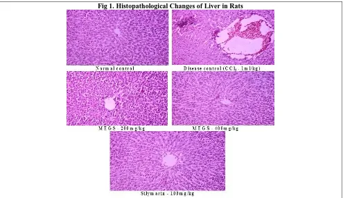
Effect of MEGS on Histopathological Changes of Liver
65 | P a g e
absence of necrosis and vacuoles in a dose dependent manner. Almost similar sign of protection was shown in
the liver sections of Silymarin at a dose of 100mg/kg, p.o, treated rats.
Table 1. Liver Function Tests for investigation of Biochemical Changes in Liver Diseases
TEST(Liver Function Test) Acute Chronic Remarks
Bilirubin ↑ ↑ Marker of excretory function
Transaminases ↑ ↔ or ↑ Indicate cell destruction
Alkaline phosphatase ↔ ↔ or ↑ Indicates biliary obstruction
Albumin ↔ ↓ Indicates synthetic function
Prothrombin time ↔ or ↑ ↑ Indicates synthetic Function
Table 2. Diagnostic Tests for Liver Diseases
TEST (Diagnostic Test) Acute Chronic Remarks
Autoantibodies - + Positive in: autoimmune, primary biliary
cirrhosis, chronic active hepatitis diseases
Hepatitis B +/- +/- Present if infected
Hepatitis C +/- +/- Present if infected
Hepatitis A +/- - Only in acute liver disease
α-Fetoprotein - +/- Present if hepatoma (liver cancer)
Serum caeruloplasmin ↔ or ↓ ↔ or ↓ Only reduced in Wilson’s disease
↑ = increased; ↓ = decreased; ↔ = normal or unchanged; +/- = present/absent.
Table 3. Effect of MEGS on Hepatic enzyme and serum bilirubin in rats after 36hrs of CCl4 treatment
Groups (n=6)
Biochemical Parameters
SGOT (U/L) SGPT (U/L) ALP (U/L) -GT (IU/L) TP (gm/dl) Total Bilirubin
(mg/dl)
Group-I
(Normal Control) 32.52± 0.05 17.58±0.33 175.84±2.32 49.26±1.17 7.85±0.05 0.82±0.00 Group-II
(CCl4: 1ml/kg)
65.22± 0.24***a 38.64±0.42***a 435.29±3.65***
a 99.52±1.28***a 2.19±0.04***a 3.94±0.05***a
Group-III (MEGS-200mg/kg)
45.37±0.14***b 30.19±0.28***b 265.46±3.52
***
b 64.14±1.52***b 3.54±0.17***b 1.33±0.02***b
Group-IV (MEGS-400mg/kg)
35.69±0.42***b 23.54±0.19***b 219.17±2.41
***
b 58.33±1.24***b 5.67±0.05***b 0.97±0.04***b
Group-V (Silymarin-100mg/kg)
30.02±0.01***b 20.22±0.02***b 197.48±0.06
***
b 52.37±0.13***b 7.12±0.07***b 0.79±0.02***b
Values are expressed as mean ± SEM of 6 rats in each group. SGOT = Serum glutamate oxaloacetate transaminase, SGPT = Serum glutamate pyruvate tranaminase, ALP = Alkaline phosphatase, γ-GT = Gamma glutamyltranspeptidase, TP = Total proteins. *** p<0.001, as compared to CCl4-treated group.a – Group I Vs Group II; b – Group II Vs Group III – V.
DISCUSSION
Liver is the vital organ of metabolism and excretion. It produces and secretes bile as well as produces the fibrinogen, prothrombin, heparin and sulfuric acid ester. The liver converts sugar into glycogen (Nadeem MPC et al., 1997). Any changes in anatomy or functions of liver are characterized by cirrhosis, jaundice, tumors, liver cell necrosis and hepatitis, metabolic and degenerative lesion etc. The management of hepatic diseases is still a challenge to the modern medicines (Harsh M, 2002; Meyer and Kulkarni, 2001). Herbal medicines play a major role in the treatment of liver disorders. A number of medicinal
plants and their formulations are widely used for the treatment of these disorders (Subramoniam A and Pushpangadan P, 1999; Thyagarajan SP et al., 2002). However, there were not enough scientific investigations on the hepatoprotective activities conferred to these plants. One of the plants from Indian flora is Guettarda speciosa L. (Rubiaceae). The present studies were performed to investigate the hepatoprotective activity of methanol extract of inner bark Guettarda speciosa L.
66 | P a g e
Fig 1. Histopathological Changes of Liver in Rats
Carbon tetrachloride (CCl4) is one of the most commonly used hepatotoxins in the experimental study of liver diseases (Johnson and Kroening, 1998). CCl4 is potent hepatotoxin producing centrilobular hepatic necrosis. It is accumulated in hepatic parenchyma cells and metabolized to trichloromethyl free radicals (CCl3.)
by liver cytochrome P-450 dependent monooxygenases. This CCl3 free radical combined with cellular lipids and proteins in the presence of oxygen to produce lipid peroxides (Recknagel RO et al., 1989). Thus, antioxidant or free radical generation inhibition is important in protection against CCl4 induced liver lesion (Sujaet al., 2004). The flavonoids constituents possess free radical scavenging properties (Hesham R et al., 2007).
In general, the extent of liver damage is assessed by histopathological evaluation and levels of hepatic enzymes such as ALP, SGOT, SGPT and also Bilurubin release in circulation (Plaa and Charbonnean, 1994; Portmann et al., 1975). The estimation of gamma glutamyltranspeptidase (γ-GT) is an important screening test with a high negative predictive value for hepatic disease (Nemesanszky E, 1996).
Administration of hepatotoxins (CCl4) elevated the serum levels of SGOT, SGPT, ALP, γ-GT and bilurubin as well as decreases total serum proteins (TP) significantly (Singh B et al., 1998; Kim NK et al., 1977). The rise in serum enzymes level and bilurubin has been attributed to the damaged structural integrity of the liver, because they are cytoplasmic in location and released
into circulation after cellular damages (Sallie R et al., 1991).
In our investigation, the biochemical changes were observed after 36hrs of CCl4 treatment. Thereby, it was found that the animal groups which are pretreated with MEGS at the dose of 200 and 400mg/kg (groups-III and IV) as well as silymarin at the dose of 100mg/kg (group-V), for three times at 12hrs intervals, resulted in significantly decreases the hepatic enzymes such as SGOT, SGPT, ALP and γ-GT and also total bilurubin; as well as increases the total serum proteins (TP) as compared to animals treated only with CCl4 (group-II). These results give us the suggestion that, the animals which are pretreated with MEGS as well as silymarin, showed a protection against the injurious effects of CCl4 that may results from the interference with cytochrome P-450. These biochemical restoration may be due to the inhibitory effects on cytochrome P-450 or/and promotion of its glucuronidation (Wesley GC et al., 1992; Gilman
et al., 1992). Silymarin is a known hepatoprotective drug. It is reported to have a protective effect on the plasma membrane of hepatocytes (Ramellini G and Meldolesi J, 1976).
In histopathological assessment, it was found that the normal liver architecture was disturbed by CCl4 intoxication. In the liver section of rats treated with MEGS showed the ability of MEGS to prevent hepatocellular necrosis, thereby further confirming the significant hepatoprotective effect of inner bark of
67 | P a g e
It is well documented that the phytoconstituents comes under the category of flavonoids, alkaloids, glycosides, carotenoids, phenols, coumarins, lignans, essential oil, lipids, monoterpenes, xanthenes and organic acids are reported to have hepatoprotective activity (Sharma GRK et al., 2002). In our investigation, the preliminary phytochemical screening of the methanol extract of the inner bark of Guettarda speciosa L.
(Rubiaceae) revealed that the presence of alkaloids, carbohydrates, triterpenoids, flavonoids, and tannins. The hepatoprotective activity of Guettarda speciosa L.
(Rubiaceae) may be attributed due to presence of these constituents. This study supports the traditional claims and the MEGS could be added in traditional preparations for the various liver diseases.
CONCLUSION
The results indicate that the methanol extract of inner bark of Guettarda speciosa L. (Rubiaceae)
possesses significant hepatoprotective property and may prove to be effective for the treatment of liver disorders. However, longer duration studies on chronic models are necessary to elucidate the exact mechanism of action so as to develop it as a potent hepatoprotective drug.
ACKNOWLEDGMENTS
The authors highly acknowledge the facility provided by Sri Venkateswara College of Pharmacy, RVS Nagar, Chittoor. Also thank Dr. Ravuri Venkataswamy, Chairman and Mr.R.V.Srinivas, Vice Chairman for their support to develop in this paper.
CONFLICT OF INTEREST No Conflict of interest.
REFERENCES
Agrawal SS and Paridhavi M. Herbal Drug Technology, 1st edition, Universities Press (India) Private Limited, Hyderabad, 2007, 1-512.
Alok S, Shanker C, Lalit K, Mahendra S, RaoCh V. Herbal Medicine for Market Potential in India: An Overview.
Academic Journal of Plant science, 1 (2), 2008, 26-36.
Aneesh TP, Mohamed H, Sonal SM, et al.. International market scenario of traditional Indian herbal drugs- India declining.
Int J Green Pharm, 3, 2010, 184-190.
Ansari SH. Essentials of Pharmacognosy, 2nd edition, Birla Publications Pvt. Ltd., New Delhi, India, 2008, 582-583. Augustin S, Clavdine M, Christine M, Christian R. Dietarypolyphenols and the prevention of diseases. Rev. Food sciences,
2005, 287-306.
BabuBH, Shylesh BS, Padikkala J. Antioxidant and hepatoprotective effect of Alanthusicicifocus. Fitoterapia, 72, 2001, 272-277.
Baskar RG and Chezhiyan N. Strength and Wealth of Therapeutic medicinal plants in India, In: Irfan A Khan, AtiyaKhanum, Role of Biotechnology in Medicinal and Aromatic plants, Ukaaz Publications, Hyderabad, 2002, 151-152.
Bennett PN and Brown MJ. Clinical Pharmacology, 9th edition, Elsevier - a division of Reed Elsevier India private Limited, New Delhi, 2006, 651.
Blois MS. Antioxidant determinations by the use of a stable free radical. Nature, 181, 1958, 1199-1200.
Brent RL. Utilization of animal studies to determine the effects and human risks of environmental toxicants (drugs, chemicals and physical agents), Pediatrics, 113 (4), 2004, 984-995.
Chaudhri RD. Herbal Drug Industry, 1st edition, Eastern publishers, New Delhi, India, 2004, 1-491.
Dekkers J, Van DLJP and Hen KCG. The role of antioxidant vitamins and enzymes in the prevention of Exercise induced muscle damage. Sports Med, 21, 1996, 213-238.
Dubey NK, Rajesh K and Pramila T. Global promotion of herbal medicine: India’s opportunity. Current Science, 86 (1), 2004, 37-41.
Evans WC, Trease and Evans Pharmacognosy, 5thedition, Elsevier, a division of Reed Elsevier India Private Limited, Noida, 2008.
Frindivich I. The biology of oxygen radicals. Science, 201, 1978, 875-880.
Galigher AE, Kozloff EN. Essential Practical Microtechnique, 2nd edition, Lea and Febiger, Philadelphia, 1971, 77–210. Gandhimathi R, Saravana KA, Senthil KKK, Kusuma P, Uma MJ. Pharmacological studies of anti-diarrhoeal activity of
Guettarda speciosa (L.) in experimental animals. J. Pharm. Sci. & Res., 1(2), 2009, 61-66.
Gerard JT and Bryan D. Principles of Anatomy and Physiology, 11th Edition, JhonWeily and Sons, Inc., Hoboken, NJ, 2006, 918-921.
German J. Food processing and lipid oxidation, AdvExp Med Biol, 459, 1999, 23-50.
68 | P a g e
Gupta AK and Misra N. Hepatoprotective Activity of Aqueous Ethanolic Extract of Chamomile capitula in Paracetamol intoxicated albino rats,. American Journal Pharmacology and Toxicology, 1 (1), 2006, 17-20.
Harborne JB. Phytochemical Methods, 3rd edition, Springer (India) Private Limited, New Delhi, 1998. Harsh M. Text book of Pathology, 4th edition, Jaypee Publisher, 2002, 569-630.
Hayes PC, Simpson KJ, Garden OJ. Liver and Bilary track disease, In: Davidson’s, Principles and practice of medicine, 19th edition, Churchill Livingstone Elsevier, 2002, 831-869.
Hesham R, et al. Chemistry of Bioflavonoids. Indian J Pharm Edu, 39, 2007, 172.
Iqbal A, Farrukh A, Farah A, Mohammad O. Herbal Medicines: Prospects and Constraints, In: Iqbal Ahmad, FarrukhAqil, Mohammad Owais, Modern Phytomedicine, 1st edition, WILEY-VCH Verlag GmbH & Co. KGaA, Weinheium, 2006, 59-77.
Jacob R. Three eras of vitamin C discovery. SubcellBiochem, 25, 1996, 23-50.
James MR. Test book of Clinical Pharmacology, 9th edition, Churchill Livingstone, a division of Reed Elsevier India, Private Limited, New Delhi, 2006, 384.
Jayaprakash GK, Singh RP and Sakariah KK. Antioxidant activity of grape seed extracts on peroxidation models invitro. J. Agric Food Chem, 55, 2001, 1018-1022.
Johnson DE and Kroening C. Mechanism of early carbon tetrachloride toxicity in cultured rat hepatocyte.
Pharmacoltoxicol, 83, 1998, 231-239.
Kannare J, et al. Natural antioxidants in grapes and wines. J. Agric. Food. Chem., 42, 1994, 64-69. Karisson J. Exercise, muscle metabolism and antioxidant defence. World Rev Nutri Diet, 81, 1997, 81-100. Khandelwal KR. Practical Pharmacognosy, 10th edition, Nirali Prakashan, Pune, India, 2003, 149-156.
Kim NK, Vasmineh WG, Frejar EF, Goldman AI, Theologides A. Value of alkaline phosphatase, 5’-nucleotidase, -glutamyltransferase and glutamate dehydrogenase activity measurements is serum in diagnosis of metastases to the liver. Clinical Chemistry, 23, 1977, 2034-2038.
Kind PRN and King EJ. Determination of Serum Alkaline Phosphatase. Journal of Clinical Pathology, 7, 1954, 132-136. Kinsella JE, Franeel E, German B, Kanner J. Possible mechanisms for the protective role of antioxidants in wine and plant
foods. Food Technology, 47(4), 1993, 85-90.
Kokate CK, Purohit AP, Gokhale SB. Text book of Pharmacognosy, 27th edition, Nirali Prakashan, Pune, India, 2004. Koleva II, Van Beek TA, Linssen JPH, De Groot A, Evstatieva LN. Screening of plant extracts for antioxidant activity, a
comparitive study on three testing method. Phytochemical analysis, 13, 2002, 8-17.
Luna LG. Manual of Histological Staining Methods of Armed Forces, Institute of Pathology, London, 1966, 1–31.
Makari HK, Haraprasad N, Patil HB, Ravi K. In vitro antioxidant activity of the hexane and methanolic extracts of Cordial wallichiand Celastruspaniculata. The internet J. Aesthetic and antiaging medicine, 1, 2008, 1-10.
Malloy HT and Evelynl KA. Determination of Bilurubin with the Photoelectric Colorimeter. Journal of Biological Chemistry,119 (2), 1937,481-485.
Mathiesen L, Malterud KE, Sund RB. Antioxidant activity of fruit exudate and methylated dihydrochalcones from myrice gale. Plenta med, 61, 1995, 515-518.
Matill HA. Antioxidants. Annu Rev Biochem, 16, 1947, 177-192.
Meyer SA, Kulkarni AP, Hepatotoxicity. In: Hodgson E, Smart RC, Introduction to biochemical toxicology, 3rd edition, John Wiley and Sons, New York, 2001,487-490.
Mohammad O, et al. An Alternative Holistic Medicinal Approach to the total Management of Hepatic Disorders : A Novel Polyherbal Formulation, Modern Phytomedicine, 1st edition, , Weinheim, 2006, 233-242.
Moreau and Dufraisse. comptesrendus des seanceset memories de la societe de. Biologie, 86, 1922, 322.
Nadeem MPC, Dandiya PC, Pasha M, Imran D, Balani K, Vohora SB. Hepatoprotective activity of Solanumnigrumfruit.
Fitoterapia, 68, 1997, 245-251.
Naznin A and Hasan N. In vitro antioxidant activity of methanolic leaves and flowers extracts of Lippia alba. Research journal of medicine and medical agencies, 4 (1), 2009, 107-110.
Nemesanszky E. Enzyme testing hepatobiliary disease, In: Donald W Moss, Sidney B rosarki, Enzyme test in diagnosis, Oxford University Press, New York, 1996, 25-59.
OECD. Acute oral toxicity, Acute oral toxic class method guidelines 423 adopted 23.03.1996, In: Eleventh Addendum to the OECD guidelines for the testing of chemicals organization for economical co-operation and development, Paris, 2000.
Okuda T, Kimura Y, Yoshida T, Hatano T, Okuda H, Arichi HS. Studies on the Activities of Tannins and Related Compounds from Medicinal Plants and Drugs. I. Inhibitory Effects on Lipid Peroxidation in Mitochondria and Microsomes of Liver. Chem. Pharma. Bull, 31, 1991, 1625-1631.
69 | P a g e
Polterait O. Antioxidants and free radical scavengers of natural origin. Current org. Chem, 1, 1997, 415-440.
Portmann B, Talbot IC, Day DW, Davidson AR. Histopathological changes in the liver following a paracetamol over dose; Correlation with clinical and Biochemical parameter. Journal of Pathology, 117, 1975, 169-180.
Pulok KM. Quality Control of Herbal Drugs, 1st edition, Business Horizons, New Delhi, India, 2008, 2-125.
Ramellini G and Meldolesi J. Liver protection by silymarin; In vitro effect on dissociated rat hepatocytes. Drug Research, 26, 1976, 89-73.
Recknagel RO, Glende EA, Dolak JA, Walter RL. Mechanism of Carbon tetrachioride toxicity, Pharmacology and Therapeutics, 43, 1989, 139-154.
Reitman S, Frankel S. A colorimetric method for the determination of SGPT and SGOT. American Journal of ClinicalPathology, 28, 1957,56-62.
Rice-Evans C and Miller NJ. Total antioxidant status in plasma and body fluids. Methods Enzymol, 243, 1994, 279-293.
Robak J and Gryglewski RJ. Flavonoids are scavengers of superoxide anions.
Biochem. Pharmcol, 37, 1998, 837-841.
Sallie R, Tredger JM, William R. Drug and Liver. Biopharmaceutical Drug Disposition, 12, 1991, 251-259.
Sanjay Jachak and Aravind S. Challenges and opportunities in drug discovery from plants. Current Science, 92 (9), 2007, 1251-1257.
Saravanakumar A, Amudha P, Gandhimathi R, Dhanapal R. Study on the Antiseizure Activities of Inner Bark of Guettarda speciosa (L.). Iranian Journal of Pharmacology & Therapeutics, 8(2), 2009, 73-76.
Seis H. Oxidative stress: Oxidants and antioxidants. ExpPhysiol, 82(2), 1997, 291-5.
Sharma B and Sharma UK. Hepatoprotective activity of some indigenous plants. International Journal Pharm Tech Research, 1 (4), 2009, 1330-1334.
Sharma GRK. Commercial usage of Medicinal Plants and, Traditional Knowledge in India, Medicinal Plants Traditional Knowledge, I.K. International Publishing House Pvt.Ltd, 118-137, 2006.
Sharma SK, Ali M, Gupta J. Plants having Hepatoprotective activity. Phytochemistry and Pharmacology, 2, 2002, 253-270.
Shui GH, Leong LP. Analysis of polyphenolic antioxidants in star fruit using liquid chromatograjhy and mass spectrometry. J Chromatogr A, 1022, 2004, 67-75.
Singh B, Saxena AK, Chandan BK, Suri OP, Suri KA, Sathi NK. Hepatoprotective activity of Verbenalinon experimental liver damage in rodents. Fitoterapia, 60, 1998, 135.
So FV, Guthrie N, Chambers AF, Moussa M. Inhibition of human breast cancer proliferation and delay of memory tumorigenesis by flavonoids and citrus juices. Nutr Cancer, 26(2), 1996, 167-81.
SoumyaPrakash Rout, Choudary KA, Kar DM, et al. Plants in Traditional Medicinal System- Future Source of New Drugs.
International Journal of Pharmacy and Pharmaceutical sciences, 1(1), 2009, 1-23.
Subramoniam A and Pushpangadan P. Development of Phytomedicines for Liver Diseases. Indian Journal of Pharmacology, 31, 1999, 166-175.
Suja SR, Latha PG, Pushpangadan P, Rajasekharan S. Evaluation of hepatoprotective effects of
HelminthostochysZeylanica(L) Hook against carbon tetrachloride-induced liver damage in Wistar rats. Journal of Ethnopharmacology, 92, 2004, 61-66.
Sunil Kumar Reddy T, A. Saravana Kumar, R. Gandhimathi. Evaluate the antiulcerogenic properties of Guettarda speciosa
(L.) in experimental animals. International Journal of Biopharmaceutics, 1(1), 2010, 1-6.
Tanaka M, Kuie CW, Nagashima Y, Taguchi T. Application of antioxidativemaillard reaction products from histidine and glucose to sardine products. Nippon Suisan Gakkaishi, 54, 1998, 1409-1414.
Thamizhvanan K, Pavan Kumar P. Antibacterial and antifungal activities of various extracts of Guettarda speciosa L.
International Journal of Phytopharmacology, 1(1), 2010, 20-22.
Thyagarajan SP, Jayaram S, Gopalakrishnan V, Hari R, Jayakumar P, Sripathi MS. Herbal medicines for liver diseases in India. Journal of Gastroenterol Hepatol, 17, 2002, 370-376.
Trivedi PC. Medicinal Plants; Traditional Knowledge, 1st edition, I.K. International Publishing House Pvt. Ltd, New Delhi, India, 2006, 1-135.
Tsao R and Akhtar MH. Nutraceuticals and functional foods: I. Current trend in phytochemical antioxidant research. J Food Agri Environ, 3(1), 2005, 10-17.
Vani T, Rajani M, Sarkar S and Shishoo CJ. Antioxidant properties of the ayurvedic formulation triphala and its constituents. Inter. J. Pharmacognosy, 35, 1997, 313-317.
Wangsteen H, Samuelsen AB, Malterud KE. Antioxidant activity in extracts from coriander. Food Chem, 88, 2004, 293-297.
70 | P a g e
Watkins JB. Exposure of rats to inhalational anaesthetics alters the hepatobiliary clearance of cholephilicxenobiotics.
Journal of Pharmacol. Exp. Ther., 250 (20), 1989, 421-427. Weiner MA. Secrets of Fijian Medicine.Govt. Printer, Suva, Fiji, 1984, 93. Weiner MA. Ethnomedicine in Tonga. Econ.Bot, 25, 1971, 423-450.
Wesley GC, Brater CC, Alice RJ. Cloth’s medical pharmacology, Mosby year Book, US, 1992.
Wickramasinghe MB. Quality Control, Screening, Toxicity and Regulation of Herbal Drugs, Modern Phytomedicne, 1st edition, Weinheim, 2006, 25-57.
Cite this article:
Vennela Priya P and A. Saravanakumar. Hepatoprotective Activity of Guettarda speciosa L. Against Carbon tetra Chloride (CCl4)-Induced Hepatotoxicity in Wistar Albino Rats. International Journal of Phytopharmacology, 8(2), 2017, 60-70. DOI: http://dx.doi.org/10.21276/ijp.2017.8.2.3