Advanced Ceramics Progress
J o u r n a l H o m e p a g e : w w w . a c e r p . i r
Effect of Surface Roughness on Coating SiO
2-P
2O
5-CaO-ZrO
2Upon Zirconium
by Sol-Gel Method
S.M. Rabiee*, L. Karamzadeh, H. Jamshidi-Aval
Department of Mechanical Engineering, Babol Noshirvani University of Technology, Babol, Iran
P A P E R I N F O
Paper history:
Received 30 January 2017
Accepted in revised form 22 May 2017
Keywords:
Glass-ceramic Zirconium Sol-gel Bioactive Coating
A B S T R A C T
Zirconium and its alloys have many applications in orthopedic medicine and have higher strength and corrosion resistance compared to stainless steel, titanium and other metals which are being used for manufacturing implants. This research shows that the method of preparation and surface modification before coating process has a significant impact on improving the metal implants among which is the sandblasting operation. The SiO2-P2O5-CaO-ZrO2 is coated on zirconium by sol-gel method and the
impact of surface modification procedure on the grip is assessed. First, the chemical composition was determined by the quantometric test and then the surface roughness and morphology were analyzed before and after coating in two sandblasted and non-sandblasted modes by AFM. Coating morphology, structure and creating a structure without separation at the interface was investigated by Scanning Electron Microscope (SEM). Phase analysis and evaluation of chemical groups of the prepared samples before and after immersion in SBF solution were performed by XRD and FTIR, respectively. The results showed better coating of the surface after the sandblasting which isdue to increasing roughness from 2.940 to 19.74 nm for the sandblasted specimen and apatite formation of the coating after placement in the SBF solution for 21 days.
1. INTRODUCTION1
Implants in addition to have desirable mechanical properties, should be also compatible with living tissues. Because of the good mechanical properties as well as easy production and shaping methods to the main body of the implants, metal alloys are used in the production of implants [1]. Due to good mechanical properties such as strength and corrosion resistance and lower cytotoxicity and magnetic sensitivity, Zirconium and its alloys are better candidates for knee joints and medical purposes compared to metals such as titanium and steel. On the other hand, inability of the metal to be combined with the bone and in accelerating the formation of hydroxyapatite, is its disadvantage [2-4]. Finding a proper alternative that is bioactive and has appropriate mechanical properties is done through coating metal implants by using bioactive materials. Some of the methods of coating metal implants include plasma spraying and sol-gel electrophoresis [5]. Sol-gel method with atomic-scale coating structure control,
1*Corresponding Author’s Email: [email protected] (S.M. Rabiee)
provides a homogeneous structure with a good adhesion between coating and substrate. The coating enjoys a good uniformity and can reduce the possibility of cell death by creating a nanoscale structure, increasing reactivity and reducing the bone-building cells’ adhesion [6].
High reactivity of the surfaces coated with SiO2-P2O5
-CaO-ZrO2 glass ceramic will be followed by increased
apatite mineralization at planting surfaces, biological stability and fast implantation in the body [7]. Due to differences in thermal expansion coefficient of the metal and ceramic during the sintering and cooling processes, the heat stresses lead to formation of cracks thus it is necessary to seek a way to reduce the density of the cracks; accordingly, it is possible to use additives such as zirconium with high fracture strength and enhanced mechanical properties as a strengthening factor [2]. When a substance is placed in the body, the surface is in contact with the cells and body fluids. The first step in the development of tissue along with the biomaterials, is its sticking to the surface and this is the beginning of the next stage of cell growth and development. As the better context is selected, the cell growth will be more and in fact the biomaterials will be more easily accepted in the
body. Importance of surface and surface properties of materials has led to significant efforts to improve the surface properties to achieve the targets. Biomaterials’ surface modification methods to increase the biocompatibility are divided into four general classes of biological, mechanical, chemical and physical methods. Many studies have been done considering the effects of surface topography on adhesion and have facilitated understanding of the mechanism [8].
Adhesion is one of the major properties which determines quality of the coating. The coating’s ability to remain on the substrate under the desired working conditions is called coating adhesion or adhesion strength [9].
Detachment or crushing of the surface layer during the usage a basic defect and problem of the coatings. This can lead to economic losses and poisoning of the body implants. In general, coating adhesion strentgh to the substrate depends on metallurgical, mechanical, physical and chemical mechanisms. Forces between atoms have various bond strengths. Forces between atoms with van der Waals bonds have the lowest bond strengths. The hydrogen, metal and ionic bonds are located after it while the highest bond strength is associated with covalent bonds. The adhesion strength of the coatings depends on several parameters including coating method, coating material, substrate material, coating conditions and so on. Since the adhesion is one of the most important parameters which determines the quality of the coatings and in case that the coated piece requires high adhesion strength while being used, the applied method for coating should provide the necessary adhesion strength. Improvement of the biocompatibility of metals and alloys is done through surface science and technology and creating coating on the surface or surface corrective action [10].
At the same time beceause roughness is increased by sandblasting the metal before adding the silica layer, a mechanical bond is created and the created grip in the system is chemical-mechanical. The aim of this study was to evaluate the effect of surface roughness by the sandblasting process on the adhesion and coating created by SiO2-P2O5-CaO-ZrO2 glass ceramic upon
Zirconium substrate by sol-gel method.
2. MATERIALS AND METHODS
In order to determine the chemical composition of the studied zirconium substrate, the emission spectroscopic analysis of the substrate was conducted. The Foundry Master UV device was used for chemical composition analysis of zirconioum plate. Then the samples were cut in the size of 5×5 cm2 and the corners were cut by the grinding operations. The initial wash was done with distilled water and ethanol. Then the specimen surface was exposed to sandblasting mechanical operation (equivalent to sandpaper 150). Then the specimens were
washed in distilled water and ethanol and dried slowly. For degreasing number of operations, the specimens were exposed to ultrasonic waves in ultrasonic device for 20 minutes. After the above mentioned steps, the specimens were kept in ethanol until coating moment. The study was laboratory experimental and the specimens were grouped into sandblasted and pure zirconium specimens.
Sandblasted specimen:
The particles utilized for sandblasting the alloy were aluminum oxide with the average diameter of 3 microns. Sandblasting was done as automatic blowing with a specific pressure and on the other hand the specimens were rotated in the sandblasting area at a rate of 1 rpm.
Non- sandblasted zirconium specimen:
According to the standard ASTM B350 / B 350M - 02, the desired compound is within the alloy range UNS R60904 of Zirconium.
Table 1 shows the results of the quantometric test results for the zirconium substrates used in this study. Sol–gel solution was prepared with 35 mol% CaO, 5 mol% P2O5, 57 mol% SiO2 and 3 mol% ZrO2.
Ca(NO3)2·4H2O and tetraethyl orthosilicate (Merck,
99%) were added to the distilled water and nitric acid (1mol). The mixture was stirred for an hour until a clear solution was obtained. The mixing ratio of raw materials was H2O:HNO3:TEOS≈ 2:1:5. Then triethyl
phosphate was added drop wise to the solution which was being stirred; for obtaining the full solubility, the solution was left for 45 min in the working stirrer. All the raw materials had high purity and were purchased from Merck, Germany. The ethylene glycol (PEG, Merck) with a molecular mass of 4000 and the ratio of PEG/TEOS = 0.01 were added to the solution to obtain a structure in the nanometer range and the solution was stirred for one hour. The zirconium chloride by Merck and Yttrium(III) nitrate hexahydrate by Dajung were used as the zirconium and yttrium sources. Each of these two substances were dissolved separately in 50°C water and then mixed with each other and then different amounts of stabilized zirconia with 5mol% yttrium were added to the initial sol.
Zirconium-Niobium alloy plates (UNS R60904) with dimensions of 5 × 5 cm2 were used as substrates. Dip coating was conducted in the sol. The speed of entering and removing the specimens from the solution was 5 cm per minute. After 6 hours remaining at room temperature and 5 hours at 80°C, they were dried. Then dried specimens were subjected to heat treatment at 700°C based on the differential thermal analysis [11].
TABLE 1. the chemical composition of the used zirconium based on the quantometric test / element w%.
3. RESULT AND DISCUSSION
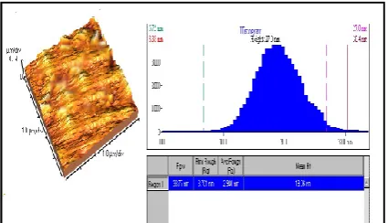
In order to improve adhesion of the coating to the zirconium substrate, the substrate was prepared by sandblasting and then the specimens’ surface roughness and morphology were analyzed by using atomic force microscopy technique. Figs. 1 and 2 present the zirconium substrate before and after sandblasting. According to these images, it is understood that after sandblasting the roughness and unevenness are increased. It seems that this would facilitate the mechanical connection of the coating to substrate mechanism.
According to the results, the mean roughness is increased from 2.940 nm for the unprepared specimen to 19.74 nm for the sandblasted specimen. This increase in roughness increases the likelihood of mechanical connection of the coating to the zirconium substrate. After coating and heat treatment, a glass-ceramic layer will cover all over the surface and fills almost all pores on the surface which results in the reducing surface roughness to 10.43 nm. At the end and after immersion in SBF as there is the possibility of corrosion and chipping off in some areas, the roughness number was increased to 11.85 nm. Since the porous structure is among the features of glass ceramic coatings obtained by sol–gel, adhesion of the coating to the substrate was increased at the contact area between the coating and the substrate due to this porous structure and surface roughness. Increased roughness, increases the active area and makes chemical and polar adhesion to the surface. The increase in the substrate surface after implantation will cause bone growth over time. Coating adhesion happens as the result of mechanical locking and chemical bonding between coating and substrate [12]. Ups and downs is an area for the grip between coating and substrate and increase coating density. The created coating density is somewhat evident in Figs. 3 and 4.
Fig. 1-4 is the atomic force microscope image of the specimens’ coating surface before and after the surface preparation and sandblasting
Fig. 5 is image related to the specimen before and after the sandblasting. Based on the images it can be observed that the coating consists of very fine particles and by roughening operation of the surface, a homogenous coating with less porosity is obtained. Since the porous structure is among the features of glass ceramic coatings by sol-gel method, adhesion of the coating to the substrate increases at the contact area between the coating and the substrate due to this porous structure and surface roughness. An increase in roughness was followed by increasing the active surface which makes chemical and polar adhesion to surface possible. The increase in the substrate surface after implantation will cause bone growth over time [8]. Coating adhesion happens as the result of mechanical
locking and chemical bonding between coating and substrate.
Figure 1. AFM image of zirconium specimen’s atomic force.
Figure 2. AFM image of zirconium specimen’s atomic force after sandblasting.
Figure 3. AFM image of zirconium specimen’s atomic force after coating.
Figure 5. SEM image of the coating surface after coating and heat treatment process A) before sandblast, and b) after sandblast.
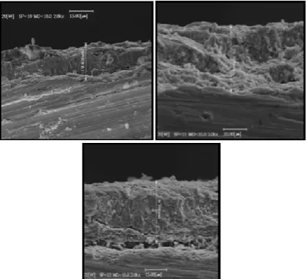
In Fig. 6, thickness and form of the section deposited on zirconium by sol-gel method was analyzed by SEM. Based on these images, it can be observed that the coating thickness in the sandblasted specimen is higher and connection is obvious. According to the interface between the coating and substrate, it seems that the connection mechanism is a combination of chemical and physical connections between the substrate and the coating. Lack of full adhesion of the coating to the substrate in the specimens obtained by the cross section, is quite evident in SEM images. While increasing the thickness the coating connection, mechanism to the substrate will be changed from chemical to mechanical connections.
Figure 6. SEM image of the cross section of the coated specimens A) before sandblast, B) after sandblast, and C) the sandblasted specimen after being left in SBF solution for 21 days.
Fig. 7 presents the XRD pattern of sandblasted and coated specimen after being left in SBF solution for 21 days. Comparison of this model with X-ray diffraction pattern of sandblasted specimen prior to placement in SBF indicates that in addition to the amorphous
structure due to glass phase, hydroxyapatite structure can be identified. Zirconium ions can form zirconium phosphate phase by absorbing phosphate groups and forming solid solution ZrO2.2P2O5 at the diffraction
angles 22.73, 29.45 and 39.38° that due to the small amount and overlap with Hydroxyapatite in XRD it is not traceable. The peaks available at the diffraction angle of 25.72 and 29.35° are related to the hydroxyapatite phase.
Figure 7. XRD pattern of sandblasted specimen after being left in SBF solution for 21 days.
According to infrared spectroscopy of the sandblasted and coated specimen after exposure to simulated body solution (SBF) for 21 days, it can be observed that there are peaks at the ranges of 779, 802, 1437 and 1445 cm-1 that can be associated to carbonate foundation. Peaks in the range 1300-1650 cm-1 are due to V3 vibration mode
that is available in all specimens. Occupancy of these positions depends on the competition between OH- and carbonate groups on the surface of the growing crystal (this type of carbonate hydroxyapatite is called type A carbonate hydroxyapatite).
Figure 8. infrared spectroscopy related to the sandblasted and coated specimen after exposure to simulated body solution (SBF) for 21 days.
The peak at 873 cm-1 is due to the vibration mode V2 of
carbonate which represents carbonate in phosphate ions network. Occupancy of these positions depends on the competition between phosphate and carbonate ions. Presence of these carbonate vibrational modes may lead to the reduction in the hydroxyl bands in the spectrum (This type of carbonate hydroxyapatite is called type B carbonate hydroxyapatite). Bands in the range of 1078, 1083, 570 and 571 cm-1 are due to the phosphate group that are available in a specimens which were exposed to SBF for 21 days which confirms the presence of apatite structure. The area under the graph of the bands 540-680 cm-1 and 900-1350 cm-1 of phosphate group is good for the formation of hydroxyapatite [11, 12].
After exposing the glass ceramic coating to the human body simulated environment, hydroxyapatite was produced; carbonate apatite and zircon phosphate phases. Presence of these phases after exposure to simulated body solution represents the biocompatibility of the created coating.
4. CONCLUSION
Based on the results of this study it can be concluded that the strength of materials associated with sandblast technique creates the best combination. The sandblasted surface provided the best surface in terms of adhesion compared to other specimens. The prepared specimens before and after immersion in SBF were analyzed by XRD and infrared spectroscopy techniques. The results indicate that the better coating of the surface is obtained by sandblasting and apatite is formed after placing the specimen in the SBF solution.
REFERENCES
1. Galletti, P.M., and Boretos, J.W., "Report on the Concensus Development Conference on Clinical applications of Biomaterials", Journal of Biomedical Material Research, Vol. 17, (1983), 539-55.
2. Basso, A., Roy, S. and Mermoud, A., "Biocompatibility of an x-shaped zirconium implant in deep sclerectomy in rabbits",
Graefe's Archive for Clinical and Experimental Ophthalmology, Vol. 246, (2008), 849–855.
3. Bornstein, M.M., Berner, S. and Buser, D., "bone Opposition to a Titanium –Zirconium Alloy Implant, as compared two other Titanium –Containing Implants", European cells & Materials, Vol. 23, (2012), 273-286.
4. Tsutsumi, Y., Nishimura, D., Doi, H., Nomura, N. and Hanawa, T., "Cathodic alkaline treatment of zirconium to give the ability to form calcium phosphate", Acta Biomaterials, Vol. 6, (2010), 4161-4166.
5. Zhitornirsky, I., "Electrophoretic hydroxyapatite coatings and fibers", Materials Letters, Vol. 42, (2000), 262.
6. Tang, H., Xin, T. and Wang, F., "Calcium Phosphate/Titania Sol-Gel Coatings on AZ31 Magnesium Alloy for Biomedical Applications", International Journal of Electrochemical Science, Vol. 8, (2013), 8115 – 8125.
7. Nabian, N., Jahanshahi, M., and Rabiee, S.M., "Synthesis of nano-bioactive glass–ceramic powders And its in vitro bioactivity study in bovine serum albumin protein", Journal of Molecular Structure, Vol. 998, (2011), 37-41.
8. Paital, S.R. and Dahotre, B.N. "Calcium Phosphate Coatings for Bio-Implant Applications: Materials, Performance Factors, and Methodologies," Materials Science and Engineering: R, Vol. 66, (2009), 1–70.
9. Sridhar, T.M., karnachi Mudali, U. and Subbaryan, M., "Sintering atmosphere and tempreture effects on hydroxyapatite coated type 316L stainless steel", Corrosion Science , Vol. 45, (2003), 2337-2359.
10. Gallardo, J., and Galliano, P., "Bioactive and Protective Sol-Gel Coatings on Metals for Orthopaedic Prostheses", Journal of Sol-Gel Science and Technology, Vol. 21, (2001), 65–74. 11. Rabiee, S.M., and Azizian, M. "Effect of Zirconia
Concentration on the Growth of Nanowires in Bioactive Glass– Ceramic Coatings", International Journal of Applied Ceramic Technology, Vol. 10, (2013), 33–39.