doi: 10.5455/2319-2003.ijbcp20150434
IJBCP International Journal of Basic & Clinical Pharmacology
Research Article
Anti-proliferative effect of
Ficus pumila
Linn. on human leukemic
cell lines
Christopher Larbie
1*, Regina Appiah-Opong
2, Felix Acheampong
1, Isaac Tuffour
2,
Takuhiro Uto
3, Gideon Agyare Yeboah
1, Offeibea Abboah-Offei
1, Doris N. K. Tagoe
1,
Samuel E. Inkabi
1INTRODUCTION
Cancer is one of the most severe health problems in both developing and developed countries worldwide. Among the most common types of cancers are lung, stomach, colorectal, liver, and breast. Lung cancer is the most commonly
diagnosed cancer in men whereas breast cancer is the most common type in women.1 Curative surgery is the first option
for patients with early-stage cancer while radiotherapy and chemotherapy have proven to be effective treatments for patients in the advanced stages. However, the curative effect of traditional chemotherapeutic drugs is limited and their ABSTRACT
Background: Cancer is one of the many diseases of global concern due to its high mortality rate with drug resistance becoming a major challenge to chemotherapy and this have propelled many cancer patients to seek alternative and complementary methods of treatment. The objective for this study was, therefore, to determine the antiproliferative activity as well as phytochemical, total phenolic content (TPC), and antioxidant activity of the stem and leaf extracts (FPS and FPL) of Ficus pumila (L.) using standard methods.
Methods: The 3-(4,5-dimethylthiazol-2-yl)-2,5-diphenyltetrazolium bromide assay was used to evaluate anti-proliferative effect and spectrophotometric-based assays for antioxidant and TPC. Phytochemical constituents were accessed by standard methods.
Results: The hydroethanolic extracts of the leaves and stems were rich in tannins,
general glycosides, saponins, terpenoids, alkaloids, flavonoids (leaves only), and
sterols (stem only). Strong total antioxidant activities were observed with FPL and FPS with EC50 values of 0.07 mg/ml and 0.089 mg/ml, respectively. All the crude
extracts showed anti-proliferative effect towards the three human leukemic cell lines used (Jurkat, CEM, and HL-60). However, FPL gave the strongest inhibition concentration at 50% values of 130.97 µg/ml (Jurkat) and 56.31 µg/ml (HL-60).
Conclusion: These findings suggest that crude extracts of FPS and FPL have
anti-proliferative effect on the leukemia cells. The antioxidant properties of the plant including phenolics may be partly responsible for the anti-proliferative activity. Further studies are required to isolate chemical components of the plant and establish their anti-proliferative activities and mechanism of action.
Keywords: Anti-proliferative, Antioxidant, Ficus pumila, Phenolics, Phytochemical
1Department of Biochemistry
and Biotechnology, Kwame Nkrumah University of Science and Technology, Kumasi, Ghana, 2Department
of Clinical Pathology, Noguchi Memorial Institute for Medical Research, College of Health Sciences, University of Ghana, Legon, Accra, Ghana, 3Department
of Pharmacognosy, Nagasaki International University, 2825-7 Huis Ten Bosch-Cho, Sasebo-shi Nagasaki, 859-3298, Tokyo, Japan
Received: 10 February 2015
Revised : 12 February 2015
Accepted: 07 March 2015
*Correspondence to:
Dr. Christopher Larbie, Email: ekowlarbie@gmail. com
side effects, such as neurological and/or renal and cardiac toxicity, are serious.2
Ficus pumila, synonymous to Ficus repens, is an ornamental plant from the family Moraceae. It is native to East Asia,
specifically South China through to Malaysia.3 Ornamental
plants, such as F. pumila are grown for only their aesthetic value. According to Yong et al.,4 the leaves of this plant
have antimicrobial, antileishmanial, and anti-inflammatory effects among many other medicinal benefits. These benefits
could be attributed to the wide array of chemicals contained in it. This plant, like all other ornamental plants, is very likely to contain certain important phytochemicals with pharmacological properties that could be useful.3
The objective of the current study was to assess the anti-proliferative effect of 50% hydroethanolic extracts of F. pumila leaves and stem, phytochemical and total phenolic content (TPC) and antioxidant activity.
METHODS
Cell lines and reagents
The cell lines used (CEM, Jurkat, HL-60 and PNT2) were obtained from RIKEN BioResource Centre Cell Bank
(Japan). Culture media (RPMI and α-MEM), 96 well plates,
3-(4,5-dimethylthiazol-2-yl)-2,5-diphenyltetrazolium bromide (MTT) dye, isopropanol, HCl, trypan blue solution, absolute ethanol, foetal bovine serum (FBS), antibiotics (penicillin and streptomycin), 2, 2-diphenyl-1-picryl hydrazyl (DPPH), and phosphate buffer saline were obtained from Sigma-Aldrich Company (St. Louis, MO, USA).
Plant and preparation
The leaves and stems of F. pumila were collected in November, 2013 (before 9 am) from the lawns of the Republic Hall of KNUST Kumasi campus. The plant was
certified at the Department of Herbal Medicine (KNUST,
Kumasi), and a specimen was deposited at the department’s herbarium (voucher number KNUST/HM1/2014/L093). The different parts of the plant were then washed, air-dried, pulverized and packaged in zip-locks for further use. Preparation of 50% hydroethanolic extraction of the plant leaves and stems were carried out separately, by suspending 50 g of the powder of each part in 500 ml of 50% ethanol (50:50 v/v). The extraction was done by cold maceration for 24 hrs at room temperature on a shaker. The extracts
were filtered through cotton wool, concentrated using a
rotary evaporator5 and freeze-dried to obtain the F. pumila
hydroethanolic leaf (FPL) and stem (FPS) extract.
Phytochemical screening
Qualitative analysis for phytochemicals was carried out to determine the presence of phytochemicals in both the
powdered samples and extracts of F. pumila using standard methods.6-8 Phytochemicals tested were tannins, saponins,
general glycosides, alkaloids, flavonoids, triterpenes, and
sterols.
Determination of total phenols
Total phenolic content (TPC) of FPL and FPS was determined using the Folin–Ciocalteau assay9 with slight modification.
To a volume of 10 µl of sample, 790 µl of distilled water was added. The concentration of the FPL and FPS extracts tested was 5 mg/ml. 50 µl of Folin–Ciocalteau reagent was added to the diluted samples and thoroughly mixed. The mixtures were incubated in the dark for 8 mins. Subsequently, 150 µl of 7% Na2CO3 was added before incubation of the mixture for 2 hrs
in the dark at room temperature. Triplicate experiments were performed. The absorbance was read at a wavelength of 750
nm using a microplate reader (Tecan Infinite M200, Austria).
Gallic acid (GA) was used as the standard phenolic compound. A GA calibration curve was plotted and used to determine the phenolic content. The results were expressed in milligrams of GA equivalents per 100 g dry mass (mg GAE/100 g DM).
Antioxidant assay
The antioxidant activity of FPL and FPS extracts was determined using the free radical scavenging activity
by DPPH method with some modification.10 Methanolic
solution of DPPH (0.5 mM) was added to equal volumes of various concentrations of each extract (concentration range 0-5 mg/ml). After 20 mins incubation at room temperature, the absorbance was read at a wavelength of 517 nm (Tecan
Infinite M200 Pro plate reader, Austria). The inhibition
concentration at 50% (IC50) value of each extract was calculated from the following formula:
% Antioxidant activity = [(A0−A1)/Ao × 100]
Where A0 is the absorbance of negative control (methanol),
and A1 is the absorbance of test sample with DPPH.
Butylated hydroxytoluene (BHT) was used as standard control. Triplicate experiments were performed. The EC50
value, which is the concentration of the extracts that can cause 50% free radical scavenging activity was determined.
MTT assay
L-RPMI and α-MEM culture media, respectively,
supplemented with 10% FBS, containing penicillin, streptomycin, and L-glutamine were maintained in culture
at 37°C in the presence of humidified 5% CO2 atmosphere. The tetrazolium-based colorimetric assay (MTT) was used to determine the cytotoxicity of F. pumila on the cancer and normal cell lines.11 Cells were seeded into the 96-well
plates at the concentration of 1×10⁴ cells/well, treated with
plate was also setup for each extract including the positive control, curcumin. MTT solution (0.5 mg/ml) was added to each well on the plate, and incubation continued for further
4 hrs. The reaction was stopped with acidified isopropanol
solution, and the plate incubated in the darkness overnight at room temperature before reading the absorbance at 570 nm
using a microplate reader (Tecan Infinite M200 Pro, Austria).
The percentage cell viability was determined as follows: % Cell viability A A
A A
0 1
0
=[ − ]× %
− 100
Where A0 = Mean absorbance of control wells
A1 = Mean absorbance in test wells
A = Mean absorbance of blank wells
Percent the IC50 values were determined from the plot
of percent cell viability on the y-axis against extract concentrations on the x-axis.
Statistical analysis
Data were analyzed using GraphPad Prism 5 for Windows and Microsoft Excel 2010. The experimental results were expressed as the mean±standard error of the mean. Data were assessed by one-way ANOVA followed by Newman–Keuls multiple comparison test. Values for which p<0.05 was
considered as statistically significant.
RESULTS
[image:3.595.315.537.551.728.2]Phytochemical constituents
Table 1 shows the phytochemical constituent of the powdered raw sample and hydroethanolic extract. The solvent system was effective in extracting majority of the phytochemical contents of raw samples. The extracts contained tannins, saponins, general glycosides, alkaloids,
flavonoids, triterpenes, and sterols.
TPC
The TPC of extracts, as assessed against GA standard is as shown is Figure 1. A linear plot of GA standard produced a straight line (y=0.029x + 0.0657, R2=0.9978). The TPC of
5 g each of FPS was significantly higher (p<0.001) compared
with an equal amount of FPL.
Antioxidant activity
The antioxidant activity of FPS, FPL and standard BHT is shown in Figure 2. FPS and FPL showed a dose-dependent increase in activity similar to that of standard, BHT. Both FPS and FPL showed strong antioxidant activities which were similar.
Anti-proliferative activity of curcumin and extracts
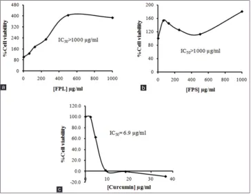
The activity of extracts and the standard, curcumin on the Jurkat cells are as shown in Figure 3. FPL had stronger inhibitory activity against the cells with IC50
value of 130.97 µg/ml compared to FPS. A concentration-dependent effect was observed for both extracts as was observed for curcumin. The effect of the extracts on human promyelocytic leukemic cells (HL-60) is shown in Figure 4. Similarly, FPL showed a stronger inhibitory activity with IC50 value of 56.31 µg/ml compared to FPS. The
anti-proliferative effect of the extracts and standard curcumin on human T-lymphoblastoid leukemia (CEM) cells is shown in Figure 5. Both FPL and FPS showed weak inhibitory activity towards the proliferation of CEM cells (IC50>1000 µg/ml).
The anti-proliferative effect of curcumin and extracts on normal human prostate cells (PNT2) are shown in Figure 6. Curcumin inhibited the growth of normal cells (IC50=15.01 µM) whereas hydroethanolic extracts of
[image:3.595.53.287.606.750.2]F. pumila had no significant inhibitory effect.
Table 1: Phytochemical content of powdered samples and hydro ethanolic extracts.
Phytochemical Leaves Stem
Powdered
sample FPL Powdered sample FPS
Tannins + + + +
Saponins + + + +
General glycosides + + + +
Alkaloids + + + +
Flavonoids + + − −
Triterpenes + + + +
Sterols − − + +
−: Absent, +: Present, FPS: Ficus pumila stem extract, FPL: Ficus pumila leaf extract
Figure 1: Total phenolic content of Ficus pumila stem and F. pumila leaves extracts. Each column represents a
DISCUSSION
Novel therapies are needed to overcome the challenges to cancer therapy. In the present study, we investigated the anti-leukemic cancer activity of the hydroethanolic extracts
of the leaves and stems of F. pumila. A wide range of phytochemicals which include tannins, saponins, glycosides,
alkaloids, flavonoids, triterpenes, and sterols were detected in both leaf and stem extracts. However, sterols and flavonoids
[image:4.595.121.476.72.361.2]were absent from the leaves and stems, respectively. The Figure 3: Anti-proliferative activity of (a) Ficus pumila leaves, (b) F. pumila stem, and (c) curcumin, on Jurkat cell.
Each point represents a mean of three determinations.
c
[image:4.595.121.478.400.672.2]b a
Figure 2: Antioxidant activity of (a) Ficus pumila leaves, (b) F. pumila stem, and (c) standard, butylated hydroxytoluene. Each point represents a mean of three determinations.
c
phytochemicals present in the plants powdered sample (without extraction) were the similar to those hydroethanolic extracts. Thus, it appears 50% ethanolic solution was an appropriate solvent for the extraction process.
The phenolic content of the plant was very significant
(p<0.001) especially in the stem. This is comparable to previous work done,12 which revealed that the TPC in acetone
[image:5.595.121.477.72.367.2]extract of Ficus capreifolia and Ficus coralata were 4.73 Figure 4: Anti-proliferative activity of (a) Ficus pumila leaves, (b) F. pumila stem, and (c) standard, curcumin on
HL-60 cell line. Each point represents a mean of three determinations.
c
b a
Figure 5: Anti-proliferative activity of (a) Ficus pumila leaves, (b) F. pumila stem, and (c) curcumin on CEM cell line. Each point represents a mean of three determinations.
c
[image:5.595.122.476.419.693.2]and 8.23 mg GAE/g dry weight, respectively. The differences in the TPC could be due to the type of solvent used in the extraction and the specie variety.
From Figure 2, it was observed that the mean % antioxidant activity increased as concentration increase. The EC50 values
of 0.07 and 0.089 mg/ml for FPL and FPS respectively, suggest that the crude extracts possess strong free radical scavenging activity. Work done has shown that Ficus species (Ficus virosa and Ficus ingens) had EC50 values
of 0.03 mg/ml and >2.5 mg/ml, respectively.13 This
confirms that the F. pumila may have comparatively higher antioxidant activity (EC50 value of 0.07 mg/ml for the
leaves and 0.089 mg/ml for the stem) compared to the other species. Ficus species are a rich source of polyphenolic compounds, which are responsible for strong antioxidant properties that help in prevention and therapy of various oxidative stress related diseases including cancers.14 Thus,
the phenolic content of the crude extracts measured in this study may be partly attributed to the antioxidant properties of the extracts. Accordingly,15 phenolic compounds increase
plasma antioxidant capacity, which is required in cancer chemotherapy. Antioxidants play a very relevant role in the control of many health disorders relating to oxidative stress and free radical activities of which cancer is major outcome.16
From the MTT data, it was observed that all the crude extracts had anti-proliferative activity against the three human leukemic cancer cell lines (Jurkat, CEM and HL-60), which was concentration-dependent. The HL-60 cell line was more sensitive to the anti-proliferative activity of the
F. pumila extracts. The FPL was more cytotoxic exhibiting three-fold stronger anti-proliferative activity towards the cell line than the stems (FPS). Similarly, the leaves showed stronger anti-proliferative activity towards Jurkat cells than the stem cells. Both crude extracts showed rather weak anti-proliferative activity against CEM cell line compared to the other cell lines. This can be attributed to the fact that CEM cell line is a multidrug-resistant leukemia cell line,17
therefore, the cells were resisting the inhibitory action of the extracts. Interestingly the extracts showed strong antioxidant properties. The free radical scavenging activity is crucial in cancer therapy since reactive oxygen species are closely involved with various pathological events such as cancers,
aging and, inflammation among others.
Similar cytotoxic effects have been observed in previous work done.18 Their study revealed that crude extracts of
F. pumila and Flemingia strobilifera were cytotoxic (IC50
values of 131 and 81 µg/ml respectively) against MT-4 human leukemia cancer cell lines which is comparable to that observed for FPL on JURKAT and HL-60 cell lines. However, in the same study, better anti-proliferative effect was achieved for F. pumila with chloroform extraction (IC50
value of 23 µg/ml). This is suggestive of the fact that different solvents may affect the concentration of anticancer agents in the extracts differently.
It was however, observed that the extracts especially the
leaf extracts had no significant anti-proliferative effect on
[image:6.595.116.481.462.743.2]the normal prostate cell line in that in the presence of the extract and even at higher concentrations, the normal prostate
Figure 6: Anti-proliferative activity of (a) Ficus pumila leaves, (b) F. pumila stem, and (c) standard curcumin on normal prostate cell line (PNT2). Each point represents a mean of three determinations.
c
cell lines were still viable. In the case of the stem extracts, the cell viability increased at higher extract concentrations which were the exact opposite in the case of the standard drug curcumin. The selectivity indices of both leaf and stem extracts in Jurkat and HL-60 cell lines was in the range 4.9-17, indicating rather a good selectivity of the extracts for the human leukemia cell lines compared to normal human cells.
CONCLUSION
This study has shown that hydroethanolic extracts of F. pumila have selective anti-leukemic activity towards two human leukemia cell line and thus contains constituents with potential as anti-leukemic agent in humans. F. pumila is also a rich source of phenols and antioxidants, and these may partly account for the anti-leukemic activity of the extracts. Besides its anti-proliferative effect on leukemic cells, the plant has minimal cytotoxic effect on normal cells which most cancer drugs lack. Bioassay-guided fractionation of extracts and isolation of pure compounds are warranted to identify the anticancer principle of F. pumila.
Funding: No funding sources Conflict of interest: None declared
Ethical approval: The study was approved by the Institutional Ethics Committee.
REFERENCES
1. Fernando W, Rupasinghe HV. Anticancer properties of phytochemicals present in medicinal plants of North America. Using Old Solutions to New Problems - Natural Drug Discovery in the 21st Century. Croatia: InTech; 2013: 161-73.
2. Zhu X, Wang J, Ou Y, Han W, Li H. Polyphenol extract of Phyllanthus emblica (PEEP) induces inhibition of cell proliferation and triggers apoptosis in cervical cancer cells. Eur J Med Res. 2013;18:46-50.
3. Starr F, Starr K, Loope L. Ficus pumila. United States Geological Survey, Biological Resources Division Haleakala Field Station, Maui, Hawaii; 2003.
4. Yong YK, Zakaria ZA, Kadir AA, Somchit MN, Ee Cheng Lian G, Ahmad Z. Chemical constituents and antihistamine activity of Bixa orellana leaf extract. BMC Complement Altern Med. 2013;13:32-8.
5. Ahmed SA, Hanif S, Iftkhar T. Phytochemical profiling
with antioxidant and antimicrobial screening of Amaranthus viridis L. leaf and seed extracts. Open J Med Microbiol. 2013;3:164-71.
6. Harborne JB. Phytochemical Methods: a Guide to Modern
Techniques of Plant Analysis. 3rd Edition. London: Chapman and Hall; 1998: 235.
7. Trease GE, Evans WC. Pharmacognosy. 12th Edition. London: Balliere-Tindall; 1989: 241-60.
8. Sofowora A. Phytochemical Screening of Medicinal Plants and Traditional Medicine in Africa. 2nd Edition. Nigeria: Spectrum Books Limited; 1993: 150-6.
9. Marinova D, Ribarova F, Atanassova M. Total phenolics and
total flavonoids in Bulgarian fruits and vegetables. J Univ
Chem Technol Metal. 2005;40(3):255-60.
10. Blois MS. Antioxidant determinations by the use of a stable free radical. Nature. 1958;181:1199-200.
11. Ayisi NK, Appiah-Opong R, Gyan B, Bugyei K, Ekuban F. Plasmodium falciparum: assessment of selectivity of action of chloroquine, Alchornea cordifolia, Ficus polita, and other drugs by a tetrazolium-based colorimetric assay. Malar Res Treat. 2011;816250.
12. Olaokun OO, McGaw LJ, Eloff JN, Naidoo V. Evaluation of the inhibition of carbohydrate hydrolysing enzymes, antioxidant activity and polyphenolic content of extracts of ten African Ficus species (Moraceae) used traditionally to treat diabetes. BMC Complement Altern Med. 2013;13:94.
13. Chauke AM, Shai LJ, Mphahlele PM, Mogale MA. Radical scavenging activity of selected medicinal plants from Limpopo province of South Africa. Afr J Tradit Complement Altern Med. 2012;9(3):426-30.
14. Sirisha N, Sreenivasulu M, Sangeeta K, Madhusudhana C. Antioxidant properties of Ficus species – A review. Int J Pharm Tech Res. 2010;2(4):2174-82.
15. Pandey KB, Rizvi SI. Plant polyphenols as dietary antioxidants in human health and disease. Oxid Med Cell Longev. 2009;2(5):270-8.
16. Valko M, Leibfritz D, Moncol J, Cronin MT, Mazur M, Telser J. Free radicals and antioxidants in normal physiological functions and human disease. Int J Biochem Cell Biol. 2007;39(1):44-84.
17. Cochrane CB, Nair PK, Melnick SJ, Resek AP, Ramachandran C. Anticancer effects of Annona glabra plant extracts in human leukemia cell lines. Anticancer Res. 2008;28(2A):965-71. 18. Ramcharan G, Clement YN, Maxwell AR. Cytotoxic
activity of selected West Indian medicinal plants against a human leukaemia cell line. West Indian Med J. 2010;59(6):597-601.
doi: 10.5455/2319-2003.ijbcp20150434
Cite this article as: Larbie C, Appiah-Opong R,