2014 NRITLD, National Research Institute of Tuberculosis and Lung Disease, Iran ISSN: 1735-0344 Tanaffos 2014; 13(1): 15-19
Upper Third to Lower Third Width Ratio on Chest X-Ray
May Predict Severity of Obstruction in Obstructive Lung
Disease
Ali Alavi Foumani, Sahand Hamidi, Maryam
Shakiba, Sara Massahnia
Respiratory Diseases Research Center of Guilan University of Medical Sciences (GUMS), Rasht, Iran
Received: 12 December 2013
Accepted: 10 February 2014
Correspondence to: Alavi Foumani A
Address: Respiratory Diseases Research
Center, Razi Hospital, Rasht- Iran.
Email address: [email protected]
Background: The symptoms and functional limitations due to obstructive lung disease (OLD) are the direct results of airway and lung parenchymal destruction. In these conditions, airflow obstruction leads to increased work of breathing, and gas exchange abnormalities. Hyperinflation, which is inferred from a standard chest radiograph (CXR), may imply increased total lung capacity that can be seen in patients with OLD. Based on experimental observations in OLD patients, we proposed that upper third width in posterioranterior (PA) CXR could be used as a rapid screening method for suggestion of OLD.
Materials and Methods: In this cross-sectional study, 99 patients admitted to the Respiratory Ward of Razi Medical Center, a teaching referral hospital affiliated to Guilan University of Medical Sciences (GUMS), were entered in the study. The inclusion criteria were any FEV1 with FEV1/FVC <70% or FEV1/FVC>70% with MMEF 75/25 <65%. All cases with diagnostic possibilities other than OLD were excluded. The PA and lateral CXR were performed and 13 measurements – including previous well-known measurements and our proposed new ones- were made by an ordinary ruler on the films.
Results: There was no significant correlation between the upper third width and superior/inferior (sup/inf) ratio with spirometric indices in patients. When considering only patients with FEV1/FVC <70%, middle third proportion width had a significant correlation with FEV1/FVC. In subgroup analysis when considering sup/inf ratio > 0.8, superior and inferior third widths were correlated with FEV1/FVC and when considering sup/inf ratio > 0.9, sup/inf ratio was significantly correlated with FEV1/FVC and FEV1.
Conclusion: The sup/inf ratio >0.9 in PA CXR, may be a predictor of obstructive pattern in OLD patients. For better correlation determination, larger and more extensive studies are needed.
Key
words: Obstructive Lung Disease, Severity, Chest X-Ray
INTRODUCTION
Obstructive pulmonary diseases are a large group of disorders determined by airflow limitation. Airflow limitation frequently occurs because of abnormal inflammatory responses of the airways to many different materials leading to airways narrowing. These diseases
include asthma, chronic obstructive pulmonary disease (COPD) and bronchiectasis (the most prevalent ones). Both asthma and COPD are widespread chronic lifetime diseases affecting patient’s quality of life and imposing high burdens on health organizations. Millions of people
are suffering from asthma and COPD in the world. However, It seems that asthma and COPD are under-diagnosed because the symptoms and signs may not be sufficient for bringing the patients to medical centers or reaching a diagnosis by the physicians. Thus, we need diagnostic methods like spirometry to detect obstructive ventilatory defects. Permanent chronic airway obstruction may lead to hyperinflation. This condition, inferred from a standard chest radiograph (CXR), may be seen in COPD, asthma, bronchiectasis and bronchiolitis patients. However, CXR is not often considered a quantitative tool in routine practice but it seems that in some cases it can be (1). Hyperinflation on chest radiography is usually marked with lowered diaphragm (the diaphragm in the midclavicular line is at or blow the anterior end of the seventh rib), flattened diaphragm (determined by drawing a line between the costophrenic and cardiopherenic angles and the diaphragm height less than 1.5 cm), or increased retrosternal space (distance between the ascending aorta and the posterior sternum= 2.5-4.5 cm). Among these, the best indicator is the flattened diaphragm along with the concavity of the upper surface of the diaphragm. Another sign is increased width of the sternum; however, it has less sensitivity (2-4). Pulmonary function tests (PFT) and particularly spirometry are used for diagnosis, determining the severity of obstruction and tracking the disease progression (5,6). There is an agreement with the correlation between radiographic findings obstruction indicators and spirometry (7-10). In posteroanterior film assessments in obstructive lung disease especially in the severe and long-term cases, we experimentally found that the ratio of lung width in upper third of lung length to the lung width in lower third tends towards one .The lung shape tends to change to a cylindrical or barrel shape. Therefore, this study was designed to assess the correlation between radiographic findings on PA films and spirometric obstruction indicators in patients with hyperinflation.
MATERIALS AND METHODS
This cross-sectional study was conducted on 99 patients
admitted to the Respiratory Ward of Razi Medical Center (a teaching referral hospital affiliated to Guilan University of Medical Sciences) with spirometric data in favor of obstructive pattern: FEV1 /FVC(VC) <70 with any FEV1,or
FEV1/FVC(VC) >70% with MMEF75/25 <65%; they did not have any other diseases. Posteroanterior and lateral chest radiographs were obtained by an analog radiography system and the measurements were made by an ordinary
ruler (Figures 1,2).
Fourteen different radiographic measurements-including previous well-known measurements and our proposed new ones- were made on the films. Previous
indices included: (A)– lung length, (B)- lung width at the level of right diaphragm, (C)- retrosternal space width, (D)– right diaphragm level on a PA film, (E)-right diaphragm level on a lateral film, (F)-lung width in upper
third proportion of lung length, (G)-lung width in middle third proportion of lung length, (H)- lung width in lower third proportion of lung length, (I)- lung width at retrosternal width, (J)- Distance between parameter (B) and
parameter (I). Our proposed indices included: (K)-The ratio of lung width in upper third proportion of lung length to lung width in middle third proportion of lung length, (L)- The ratio of lung width in middle third proportion of lung
length to lung width in lower third proportion of lung length (M)-The ratio of lung width in upper third proportion of lung length to lung width in lower third proportion of lung length (N).
measures and spirometric indices were analyzed by SPSS 19, using Pearson’s correlation method.
Figure 1. Radiographic indices in Posteroanterior chest x ray
Figure 2. Radiographic indices in Lateral film.
RESULTS
In correlation analysis with cut-off-point correlation ratio more than (r>04), a correlation was observed between lung length on a PA film and obstructive index of FEV1/FVC with r=-0.403. No correlation was observed between other conventional or newly proposed radiographic indices with spirometric indices according to
Table 1. A correlation was detected only between some radiographic findings or proposed measurements and the spirometric results.
The aim of this study was to assess the correlation
between the proposed radiographic indices and the ratio of upper third to lower third lung width in PA films (indicating barreling or cylindrical shape of the chest) with obstructive pulmonary disease in spirometry. In order to
determine the cut–off-point for the correlation of this index to obstructive indices in spirometry, only patients whose index N (sup/inf ratio) was more than 0.8 were entered in the second analysis. This way, 54 patients were eligible.
After studying the results with r>04, significant correlations were found between the diaphragm height index on PA films with obstructive index of FEV1/FVC with r=-0.440, anteroposterior diameter of chest index on
lateral films with FEV1/FVC with r =0.576 and proposed index of distance between index E and index C with FEV1
(r=-451). In the third analysis, only cases with index N(sup/inf ratio) greater than 0.9(sup/inf > 0.9) were
studied. Eleven patients were eligible who underwent correlation analysis between spirometric and radiographic indices. Statistically, there was a significant correlation between conventional and proposed measurement with
obstructive pulmonary indices in spirometry including MMEF75/25, FEV1 and FEV1/FVC (Table 2). There was a significant correlation between the conventional and newly proposed radiographic measurements (table 3). There was
no significant correlation between the index N (sup/inf ratio) and spirometric indices in patients. When considering only patients with FEV1/FVC <70%, middle third width had a significant correlation with FEV1/FVC.
In subgroup analysis when considering index N (sup/inf ratio) > 0.8, superior and inferior third widths were correlated with FEV1/FVC and when considering index N(sup/inf ratio) > 0.9, sup/inf ratio was found to be
Table 1. Pearson’s coefficient in patients who underwent spirometry.
Radiographic indices Lung length in PA films Lung width in PA films Diaphragm height in PA films
Lung width in upper third 0.580 0.712 -
Lung width in middle third 0.547 0.711 -
Lung width in lower third 0.614 - 0.416
Distance between parameter B and I 0.470 - -
Lung width in parameter E - - 0.531
Table 2. Pearson’s coefficient in patients with Sup/Inf ratio more than 0.9
Radiographic Indices FEV1/FCV FEV1 MMEF 75/25
Lung length in PA films - - 0.730
Lung width in PA films - - 0.660
Retrosternal width - 0.521 0.578
Diaphragm height in PA films -0.649 - 0.673
AP diameter - 0.739 0.663
Lung upper third width - 0.620
-Lung middle third width - 0.782
-Lung lower third width - - 0.768
Sup/inf - 0.594 -
Sup/mid - - 0.798
Mid/inf - 0.594 -
Distance between parameter B and I - -0.475 0.569
Lung width in parameter E - 0.712 -
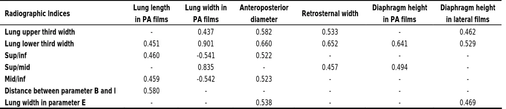
Table 3. The Pearson’s correlation coefficient between old radiographic indices and proposed new radiographic indices only in patients with Sup/Inf ratio more than 0.9.
Radiographic Indices Lung length
in PA films
Lung width in PA films
Anteroposterior
diameter Retrosternal width
Diaphragm height in PA films
Diaphragm height in lateral films
Lung upper third width - 0.437 0.582 0.533 - 0.462
Lung lower third width 0.451 0.901 0.660 0.652 0.641 0.529
Sup/inf 0.460 -0.541 0.522 - - -
Sup/mid - 0.835 - 0.457 0.494 -
Mid/inf 0.459 -0.542 0.523 - -
-Distance between parameter B and I 0.580 - - -
Lung width in parameter E - - 0.538 - - 0.469
DISCUSSION
In a study done by Dernaika and colleagues in 2005 in Oklahoma city (7), the predictability of the physiological impairment of pulmonary function test was assessed by CXR in COPD patients. Radiological evidence of emphysema was seen in 62% of patients. Flattening of the diaphragm (68%) was the most prevalent finding followed by bullous disease (59%) and increased retrosternal air space (46%). This study showed that CXR was an accurate
the fourth analysis, the height of diaphragm on the lateral film was only correlated with FEV1 (r=0.408). In addition, increased retrosternal space had a correlation with FEV1 after the fourth analysis (r=0.521).
In a retrospective study done by Rothpearl and colleagues between 1980 and 1987 in New York city (8), the accuracy of radiological assessment of hyperinflation in 44 patients with emphysema and 39 control subjects and its correlation with ABG and PFT were assessed. The best indices that had a correlation, were pulmonary height, decreased diaphragmatic angle, total lung capacity (TLC) and heart size. In our study, no significant correlation was found between pulmonary height and spirometric indices in any analysis. We used height of diaphragm dome instead of angle, which was correlated with spirometric indices in the third and fourth analyses.
Reich and his colleagues in their study in California in 1984 (10), showed that the quantitative changes of the right lung length in PA radiographs and the arc of the right diaphragm in lateral films (with or without adjustment for body surface area), and the level of the diaphragm relative to posterior ribs (when adjusted for body surface area) had the best correlation with abnormal PFT, showing that CXR can help the radiologist detect COPD. It may also be helpful in cases where clinical evaluation is not possible.
In our study, we assessed the relative correlation between conventional and proposed CXR indices and spirometric data, and quantitative measurement correlations were not determined. In sub-analysis, the ratio of upper third to lower third width on PA radiographs (index N: sup/inf ratio>0.8 and >0.9) was correlatively assessed with conventional radiographic and spirometric indices and a correlation was observed between index N and obstructive spirometric indices with ratio >0.9.
We proposed that N index (sup/inf ratio>0.9) in CXR could be a valuable parameter for detecting patients with obstructive lung disease. This index could be helpful as a rapid diagnostic tool for patients with obstructive lung disease when other evaluation methods are not available. Although this index correlated with an obstructive pattern,
this study was performed on a small group of patients. Hence, a more comprehensive study with a larger sample size is needed in order to achieve high validity and assess the correlations in various cut-off-points.
REFERENCES
1. Steven D .Shapiro, John J Reily Jr. Stephen I. Rennard. Chronic bronchitis and emphysema.In Murray and Nadels Text book
of respiratory medicine.5th edition .United states of America:
2010: 919-921
2. Peter Armstrong, Martin L. Wastie. Diagnostic Imaging.
Fourth edition. Blackwell publishing ltd. 1998; Page 754.
3. Robert J. Mason, V. Murray and Nadel's Textbook of Respiratory Medicine: 2-Volume Set (Broaddus, V. Courtney.
Murray, John F. Nadel, . Jay A.). Saunders; 4th edition. 2005;
Volume 1. Page 1118
4. Fraser, Richard S, Colman, Neil. Muller, Nestor L, Pare PD.
Synopsis of diseases of the chest. Third edition. Saunders.
2005; Page 131-132.
5. Rennard SI. Chronic obstructive pulmonary disease:
Definition, clinical manifestations, diagnosis, and staging;
UpToDate version 17.1: January 2009
6. Dewar M., M.D., J.D., Curry RW Jr. Chronic obstructive
pulmonary disease: diagnostic considerations. American
Family Physician 2006; 73(4), 669-76.
7. Dernaika TA, Keddissi JI, Younis WG, Kinasewitz GT. M.D.
Radiographic abnormalities predict functional impairment in
patients with chronic obstructive pulmonary (abstract). Chest
supplement 2005; 128(4):, 249s
8. Rothpearl A, Varma AO, Goodman K. Radiographic measures
of hyperinflation in clinical emphysema. Discrimination of
patients from controls and relationship to physiologic and
mechanical lung function. Chest 1988; 94 (5): 907- 13.
9. Kilburn KH, Warshaw RH, Thornton JC. Do radiographic
criteria for emphysema predict physiologic impairment? Chest
1995; 107 (5): 1225- 31.
10. Reich SB, Weinshelbaum A, Yee J. Correlation of radiographic
measurements and pulmonary function tests in chronic
obstructive pulmonary disease. AJR Am J Roentgenol 1985;