O R I G I N A L R E S E A R C H
In vitro
–
in vivo evaluation of chitosan-PLGA
nanoparticles for potentiated gastric retention and
anti-ulcer activity of diosmin
This article was published in the following Dove Press journal: International Journal of Nanomedicine
Walaa Ebrahim Abd El Hady Elham Abdelmonem Mohamed Osama Abd El-Aazeem Soliman Hassan Mohamed EL-Sabbagh
Department of Pharmaceutics, Faculty of Pharmacy, Mansoura University, Mansoura 35516, Egypt
Background: Diosmin showed poor water solubility and low bioavailability. Poly(d,l-lactide-co-glycolide) (PLGA) nanoparticles were successfully used to improve the drugs solubility and bioavailability. Coating of PLGA nanoparticles with chitosan can ameliorate their gastric retention and cellular uptake.
Methodology:PLGA nanoparticles of diosmin were prepared using different drug and poly-mer amounts. Nanoparticles were selected based on entrapment efficiency% (EE%) and particle size measurements to be coated with chitosan. The selected nanoparticles either uncoated or coated were evaluated regarding morphology,ζ-potential, solid-state characterization, in vitro release, storage stability, and mucoadhesion. The anti-ulcer activity (AA) against ethanol-induced ulcer in rats was assessed through macroscopical evaluation, histopathological exam-ination, immunohistochemical localization of nuclear factor kappa-light-chain-enhancer of activated B cells (NF-κB) and transmission electron microscopic examination of gastric tissues compared to free diosmin (100 mg/kg) and positive control.
Results: Based on EE% and particle size measurements, the selected nanoparticles, either uncoated or coated with 0.1% w/v chitosan, were based on 1:15 drug-PLGA weight ratio and 20 mg diosmin employing methylene chloride as an organic phase. Examination by scanning electron microscopy (SEM) and transmission electron microscopy (TEM) revealed nanoscopic spherical particles. Drug encapsulation within the selected nanoparticles was suggested by Fourier transform-infrared, differential scanning calorimetry (DSC) and X-ray diffractometry results. Chitosan-coated nanoparticles were more stable against size enlargement probably due to the higher
ζ-potential. Only coated nanoparticles showed gastric retention as revealed by SEM examination of stomach and duodenum. The superior AA of coated nanoparticles was confirmed by significant reduction in average mucosal damage, the majority of histopathological changes and NF-κB expression in gastric tissue when compared to positive control, diosmin and uncoated nanoparticles as well as insignificant difference relative to normal control. Coated nanoparticles preserved the normal ultrastructure of the gastric mucosa as revealed by TEM examination.
Conclusion: The optimized chitosan-coated PLGA nanoparticles can be represented as a potential oral drug delivery system of diosmin.
Keywords:diosmin, poly(d,l-lactide-co-glycolide), chitosan-coating, polymeric nanoparticles, gastric retention, anti-ulcer activity
Introduction
There are some endogenous aggressive factors that can cause gastric ulcer such as
overproduction of hydrochloric acid and pepsin, leukotrienes, refluxed bile, and
stress oxygen species.1The defensive endogenous mechanisms against the damage
Correspondence: Elham Abdelmonem Mohamed
Department of Pharmaceutics, Faculty of Pharmacy, Mansoura University, Gomhoreyah St., Mansoura 35516, Egypt Tel +20 106 569 0987
Fax +20 50 224 7496
Email elham.mabdelmonem@gmail.com
International Journal of Nanomedicine
Dove
press
open access to scientific and medical research
Open Access Full Text Article
International Journal of Nanomedicine downloaded from https://www.dovepress.com/ by 118.70.13.36 on 22-Aug-2020
of the gastric mucosa include the surface mucus, the
regulation of gastric mucosal blood flow, bicarbonate,
antioxidants, surface active phospholipids, the acceleration of epithelial regeneration, and the preservation of epithe-lial hemostasis. Excessive gastric acid secretion was con-sidered to be the major reason of the gastric ulcer for decades; thus, anti-cholinergic drugs, antacids, histamine
H2-receptor antagonists, and proton pump inhibitors were
the main therapy regimens. Nevertheless, the limited effi
-cacy and the adverse effects of most of the current thera-pies limited their application.2Therefore, there is a great necessity for safe and effective anti-ulcer agents.
Diosmin (3,5,7-trihydroxy-4-methoxyflavone
7-rutino-side) is a natural flavonoid glycoside that can be obtained
from different plant sources or derived from theflavonoid
hesperidin.3 Diosmin has been widely used as a vascular
protector for the treatment of hemorrhoids and venous leg ulcers.4 It also exhibited anti-inflammatory, free-radical
scavenging,5 and anti-ulcer activities.6 This drug showed
gastro-protection against ethanol-induced gastric ulcer in rats by inhibiting the mitochondrial damage and MMP-9
upregulation.7 However, diosmin is poorly soluble, thus
low dissolution rate and impaired gastrointestinal absorption
were observed.8 Following oral administration, diosmin is
quickly hydrolyzed by enzymes produced by intestinal
microflora into its aglycone diosmetin that is absorbed
through the intestinal wall to be then enzymatically esterified
to its metabolite of 3,7-O-diglucuronide.8 Consequently, a
large oral dose (500 mg twice daily) is usually required.9
However, the amount of diosmetin detected in plasma after a single oral administration of diosmin is low and highly inconsistent. The variability of absorption could be reduced by adherence to the gastrointestinal wall to allow a rapid replenishment of the absorbed drug. Small particles tend to adhere well to the mucus layer and then penetrate this layer to bind to the underlying epithelium.10It has been reported that oral administration of diosmin in micronized form can ame-liorate its plasma concentrations due to the larger surface area
and subsequent improved intestinal absorption.11Different
strategies have been attempted to improve diosmin solubility, such as complexation withβ-cyclodextrin,6as well as parti-cle size reduction by formulation into nanosuspension with
hydroxypropyl methylcellulose9 and electrospinning to
nanofibers.5
Poly(d,l-lactide-co-glycolide) (PLGA) is a synthetic copolymer that has been approved by FDA for various medical and pharmaceutical applications including drug
delivery.12 PLGA is biocompatible and biodegradable
since it is hydrolyzed into non-toxic oligomer and mono-mer of lactic and glycolic acids that are hydrophilic and
finally eliminated as carbon dioxide and water.13,14 In
addition, the degradation rate of this copolymer can be
modified by controlling the molar ratios of lactic and
glycolic acids in the polymer chain and the degree of crystallinity, as well as the molecular weight and
stereo-chemistry of the polyester.15 PLGA nanoparticles can
increase the drug penetration across the different biologi-cal barriers, such as the blood–brain barrier,
gastrointest-inal mucosa, nasal mucosa, and ocular tissue.16Therefore,
this copolymer has been extensively used as nanoparticu-late drug delivery system to enhance the biological activ-ity, water solubilactiv-ity, and bioavailability of drugs.13PLGA produces negatively charged, smooth surfaced, and sphe-rical particles that are relatively resistant to salt- and pH-induced instability, and can slowly release the entrapped drugs by polymer hydrolysis. Yet, unsuccessful results have been observed in some cases possibly due to the
lack of mucoadhesiveness and immune-stimulating
factors.17 Also, the negative surface charge of PLGA
nanoparticles can hinder the interaction with the nega-tively charged plasmids and hence limit their intracellular uptake and the bioavailability of the loaded drugs.18
Chitosan is a naturally occurring linear amino poly-saccharide (poly 1, 4-day-glucoamine) which can be obtained from crustacea shells, insects cuticles, and cell
walls of some fungi.19 Chitosan is an interesting
bioma-terial for entrapment of bioactive mabioma-terials in nanoparti-culate delivery systems due to its water solubility under
acidic conditions, biocompatibility, non-toxicity as
well as its mucoadhesive and permeability-enhancing properties.18–20Modification of PLGA nanoparticles sur-face with a mucoadhesive polymer, such as chitosan, can offer several advantages. Among these, increased stabi-lity of macromolecules such as proteins, providing a positive surface charge that can promote cellular adhe-sion and delivery system retention at the target site, as well as conjugating targeting ligands to chitosan free
amine groups.21Mucoadhesive delivery systems increase
the residence time of dosage forms at the delivery site which may lead to improved bioavailability, lower drug
dose, less dosing frequency, and minimal side effects.22
The successful use of chitosan-based gastrointestinal mucoadhesive delivery systems has been reported in
some studies.18,23 Longer residence time in the stomach
is advantageous in the treatment of gastric ulcer.24
International Journal of Nanomedicine downloaded from https://www.dovepress.com/ by 118.70.13.36 on 22-Aug-2020
Therefore in this study, it was worth to prepare, char-acterize, and optimize polymeric nanoparticulate delivery systems of diosmin with the biodegradable and biocompa-tible PLGA. Coating of the selected PLGA nanoparticles with chitosan was attempted and its effects on the gastric retention of diosmin were assessed. Additionally, the effects of the selected PLGA nanoparticles either uncoated or coated on cytoprotective activity of diosmin against ethanol-induced ulcer in rats were investigated through macroscopical, histopathological, and transmission elec-tron microscope examination of gastric tissues as well as immunohistochemical localization of gastric nuclear factor
kappa-light-chain-enhancer of activated B cells (NF-κB).
Materials and methods
Materials
Diosmin was purchased from Sigma-Aldrich, St. Louis,
MO, USA. PLGA (lactide–glycolide 50:50; Viscosity 0.8–
1.2 dL/g) was kindly supplied by Purac Biomaterials,
Holland. Chitosan (molecular weight 100,000–300,000)
and polyvinyl alcohol (molecular weight 31,000–50,000
Da) were purchased from Acros organics, Belgium. Methylene chloride, dimethyl sulfoxide, and glacial acetic acid were purchased from Adwic, EL Nasr Pharmaceutical
Chemicals, Co., Egypt. All other chemicals were of fine
analytical grades.
Preparation of diosmin nanoparticles
The effects of several formulation parameters including drug:polymer weight ratio, loaded drug amount, and the organic phase nature on the particle size and entrapment
efficiency% (EE%) of diosmin nanoparticles were studied
to select PLGA nanoparticles to be coated with chitosan aiming to enhance their gastric retention and the anti-ulcer activity (AA) of diosmin. Different diosmin:PLGA weight ratios (1:10, 1:15, 1:20) were used. The loaded drug was either 10 or 20 mg. The organic solvents employed were methylene chloride and ethyl acetate individually since they are water immiscible with higher vapor pressure, and hence their removal is facilitated.25 In addition, they are good solvents for PLGA and much less toxic than chloroform and acetonitrile.26
Nanoparticles were prepared according to the
emul-sion–solvent evaporation method.27 Briefly, accurately
weighed PLGA was dissolved in 4 mL of either methylene
chloride (F1–F7) or ethyl acetate (F8–F13) (Table 1).
Diosmin was dissolved in 1 mL dimethylsulfoxide. The
resulting organic solutions were vortexed (Model VM-300, Gemmy Industrial Corp, Taipei, Taiwan). The organic phase was then poured into 30 mL aqueous phase contain-ing 2% w/v polyvinyl alcohol with ultrasonic homogeni-zation (Ultrasonic homogenizer, Cole-Parmer Instrument Co., Chicago. IL, USA) at 90% amplitude for 2 mins. The organic solvent was evaporated overnight under a moder-ate magnetic stirring (Heidolph, IL, USA). Nanoparticles were recovered by centrifugation at 13,000 rpm (Heraeus, GmbH, Osterode, Germany) for 1 h and washed with double deionized water. The washing was performed in triplicate and then the nanoparticles were resuspended in double deionized water and lyophilized (SIM, FD8-8T, Newark, NJ, USA).
Chitosan-PLGA nanoparticles of diosmin (F14–F16)
were prepared following the same procedure except that chitosan was dissolved in an aqueous solution of 0.2% v/v acetic acid at three different concentrations (0.10%, 0.15%, and 0.30% w/v) and then mixed with the aqueous solution of 2% w/v polyvinyl alcohol to obtain 30 mL aqueous phase (Table 2).28
Characterization of diosmin nanoparticles
Determination of entrapment efficiency%The lyophilized nanoparticles (SIM, FD8-8T, Newark, NJ, USA) were mixed with 10 mL dimethylsulfoxide and properly diluted to be assayed spectrophotometrically at 269 nm (ultraviolet/visible [UV/VIS] spectrophotometer; JASCO, Tokyo, Japan) against a blank of the plain nano-particles that were treated the same. The experiments were
carried out in triplicate. Entrapment efficiency% (EE%)
was calculated as follows:29
EE %¼ Drug weight in nanoparticles
Weight of drug and polymer added100%
Particle size measurements
Dynamic light scattering technique (Malvern Instruments Ltd., Malvern, Worcestershire, UK) was used to evaluate the size distribution of the prepared nanoparticles. The lyophilized nanoparticles were reconstituted in double deionized water, properly diluted, and sonicated to obtain uniformly distributed particles.
Scanning electron microscopy (SEM)
The shape and surface characteristics of diosmin as well as the lyophilized uncoated and chitosan-coated PLGA nano-particles were recorded using SEM (JSM-6510 LV; JEOL, Tokyo, Japan). Samples were mounted on metal stub using
International Journal of Nanomedicine downloaded from https://www.dovepress.com/ by 118.70.13.36 on 22-Aug-2020
double sided adhesive carbon tapes. Samples were coated with a gold layer and visualized under SEM at 30 kV.
Transmission electron microscopy (TEM)
The morphology of the selected uncoated and coated PLGA nanoparticles was studied using transmission elec-tron microscope (TEM-2100, JEO, Tokyo, Japan) operated at an accelerating voltage of 160 kV. The lyophilized nanoparticles were reconstituted with double deionized water, properly diluted and sonicated for 2 mins. One drop was added on Formvar-coated copper grid (200 meshes, Science Services, Munich, Germany) and the
excess material was removed with a filter paper. After
the complete drying at room temperature, the image was captured and the analysis was done using imaging viewer software.
ζ-potential determination
ζ-potential of the selected uncoated and coated PLGA
nanoparticles was measured using photon correlation spec-troscopy-based instrument at 25°C (Malvern Instruments).
ζ-potential was estimated on the basis of electrophoretic
mobility under an electricfield.30To determine nanoparti-cles surface charge, the lyophilized samples were recon-stituted and properly diluted with double deionized water.
Solid-state characterization
Fourier transform-infrared (FT-IR) spectra, differential scanning calorimetry (DSC) curves, and X-ray diffracto-metry (XRD) patterns of the selected PLGA nanoparticles either uncoated or chitosan-coated were recorded in com-parison with diosmin, the polymer(s), the corresponding physical mixture (PM), and the plain nanoparticles.
FT-IR spectroscopy
FT-IR spectra of samples homogeneously mixed with KBr and compressed into discs were traced in the region of
400–4000 cm−1 employing FT-IR spectrophotometer
(Madison Instruments, Middleton, WI, USA).
Differential scanning calorimetry (DSC)
DSC thermograms were recorded using differential
scan-ning calorimeter (model DSC-4, PerkinElmer Inc.,
Waltham, MA, USA). Five milligrams of each sample were separately sealed in aluminum pans. Then, they were heated at the range from 35°C to 400°C at a heating rate of 10°C/min under a constant purging of dry nitrogen at 20 mL/min. Indium with purity of 99.99% and melting point of 156.6°C was used to calibrate the temperature.
X-ray diffractometry (XRD)
XRD patterns of the examined samples were obtained using X-ray diffractometer (Diano, Woburn, MA, USA)
operated at 45 kV, 9 mA, and at an angle of 2θ.
In vitro drug release study
In vitro release of diosmin from the selected uncoated and chitosan-coated PLGA nanoparticles was studied in com-parison with free diosmin using USP apparatus II (paddle
method) (Dissolution Apparatus USP Standards, Scientific,
DA-6D, Bombay, India). A dissolution medium consisting of 500 mL borate buffer pH 10.5 was kept at 37±0.5°C and stirred at 100 rpm. Accurately weighed sample of the lyo-philized nanoparticles equivalent to 20 mg diosmin were introduced into the dissolution tester cells. At predetermined time intervals (0.5, 1, 2, 3, 4, 6, 8, 24, 48, and 72 h), 3 mL of the dissolution medium was withdrawn and replaced with an equal volume of a fresh dissolution medium. The
collected aliquots werefiltered and spectrophotometrically
analyzed for drug concentration at 269 nm (ultraviolet/visi-ble [UV/VIS] spectrophotometer; JASCO). The blank was the corresponding plain nanoparticles that were treated the same as the medicated. Each experiment was done in tripli-cate and the average percentage drug released was calcu-lated to construct in vitro release curves.
Release kinetics
First-order and zero-order kinetics31 as well as diffusion-controlled release model32were applied to analyze in vitro
release data. To verify the release mechanism, Korsmeyer–
Peppas kinetic model (mt/m∞= ktn) was also used as a
logarithmic relation of the fraction of drug released (mt/
m∞)and the release time (t), where k is the kinetic constant
and n is the diffusional exponent of the drug release
calculated as the slope of the plot.33 The model showing
the greatest correlation coefficient (r2) was proposed to explain the drug release mechanism from the studied nanoparticles.
Stability study
The stability of the lyophilized selected uncoated and chitosan-coated PLGA nanoparticles was evaluated during the storage period of 3 months at refrigerator (4±1°C) and room (25±1°C) temperatures. Nanoparticles were
exam-ined regarding particle size, EE%, and ζ-potential, as
described above, at 0, 1, 2 and 3 months. Average drug retention (%) of the stored nanoparticles was also calcu-lated over the storage period.
International Journal of Nanomedicine downloaded from https://www.dovepress.com/ by 118.70.13.36 on 22-Aug-2020
In vivo evaluation
AnimalsAll animal handling and procedures were performed in accordance with US National Institute of Health Guide for the Care and Use of Laboratory Animals (NIH
pub-lication No. 85–23, revised 1996). Approval of the
proto-col by the Ethical Committee of Faculty of Pharmacy, Mansoura University, Egypt, was accomplished. Animals were kept under regular 12 h light/12 h dark cycles at a temperature of 25±1°C and a relative humidity of 55±5%. The animals had free access to a standard laboratory food and water.
Mucoadhesion study
Thirty-six male Sprague-Dawley rats weighting 200±20 g were used to perform the mucoadhesion study. They were randomly divided into six groups of six rats each. Before the day of the experiment, all rats were fasted overnight but had free access to water. Animals of groups I and II orally received diosmin suspension in 1% w/v carboxymethylcel-lulose (CMC) at a dose of 100 mg/kg. The selected uncoated PLGA nanoparticles dispersion at a dose equiva-lent to 100 mg/kg of diosmin was orally given to rats of groups III and IV. Rats of groups V and VI orally adminis-tered chitosan-coated PLGA nanoparticles dispersion at a dose equivalent to 100 mg/kg of the drug. Oral administra-tion was accomplished by use of feeding tube. After 2 h, animals of groups I, III, and V were euthanized under deep ether anesthesia. Euthanization under deep ether anesthesia of rats of groups II, IV, and VI was performed after 8 h. Euthanization was followed by immediate laparotomy to collect stomachs and small intestines in 2.5% (w/v) aqueous glutaraldehyde to be quickly soaked in water before
freeze-drying and SEM (JSM-6510 LV; JEOL) examination.34
Induction of gastric ulcer and treatment protocol
Thirty male Sprague-Dawley rats (200±20 g) were
ran-domly assigned into five groups (six rats per each).
Animals of groups I (normal control) and II (positive con-trol) did not receive any treatment for 5 days. Diosmin at a dose of 100 mg/kg displayed a protection against ethanol-induced gastric injury in rats.7 Thus, oral pretreatment of the other three groups was continued for successive 5 days with free diosmin at a dose of 100 mg/kg (group III), uncoated PLGA nanoparticles (equivalent to 100 mg/kg of diosmin, group IV), or chitosan-coated PLGA nanoparticles
(equivalent to 100 mg/kg of diosmin, group V). On thefifth
day, all rats had free access to water but were deprived of
food for 24 h. On the sixth day, gastric mucosal injury was induced in rats of groups II (positive control), III, IV, and V by a single intragastric instillation of 70% ethanol (10 mL/ kg).35
Tissue collection and preparation
The rats were euthanized under deep ether anesthesia 2-h post-intragastric instillation of 70% ethanol. Immediate laparotomy was followed by stomach separation that was opened along the greater curvature and rinsed with normal saline to get rid of gastric contents and blood clots. Tissue specimens were collected from glandular and non-glandu-lar portions of each stomach and immersed in 10%
buf-fered formalin. All fixed specimens were routinely
processed to prepare two sets of 5 μm thick paraffi
n-embedded sections for histopathological examination and
immunohistochemical assessment of NF-κB.
Macroscopic evaluation
The gastric segments were examined by an observer who was blinded to the identity of samples. Stomachs were blotted dry and photographed to be inspected for gross gastric injury. Paul’s index and AA were calculated to assess the anti-ulcer potential of diosmin either as free drug or encapsulated
within PLGA or chitosan-PLGA nanoparticles.29 Paul’s
index was calculated by multiplying the average ulcers num-ber by the percentage incidence of animals with ulcers to be then divided by 100. In addition, AA was estimated by dividing Paul’s index of the positive control group (II) by that of each of the treatment groups. AA was indicated if it was two units or more.29
Histopathological examination
Gastric tissue samplesfixed in 10% (v/v) buffered
forma-lin solution were washed then dehydrated by alcohol,
cleared in xylene, and embedded in paraffin in hot air
oven (56°C) for 24 h. Paraffin blocks were cut into 5μm
sections to be then stained with H&E according to a
previously reported method.36 The stained sections were
examined under a light microscope (Leica Microsystems). The histopathological examination was performed by a
qualified pathologist unaware of the specimens’ identity
in order to prevent any bias.
Gastric microscopic damage was scored based on the criteria described in the literature.37 One centimeter seg-ment of glandular portion of each stomach was examined
for epithelial cell loss (score: 0–3), edema in submucosa
(score: 0–4), congestion in submucosa (score: 0–4), and
the presence of inflammatory cells (score: 0–3). Also,
1-International Journal of Nanomedicine downloaded from https://www.dovepress.com/ by 118.70.13.36 on 22-Aug-2020
cm segment of non-glandular portion of each stomach was examined for submucosal edema and congestion (score: 0–4).
Immunohistochemical loclization of NF-κB
The second set of paraffin-embedded sections was used for
immunohistochemical detection of NF-κB. After removal
of paraffin with graded xylene followed by rehydration in
ethanol, blocking of the endogenous peroxidase activity
was obtained by 3% hydrogen peroxide (H2O2) for 5 min
at room temperature. For antigen retrieval, tissue sections were put in glass jars containing 0.01 M sodium citrate buffer (pH 6.0) and boiled in a microwave oven twice for 5 mins each to enhance immunoreactivity and reserve the antigenicity that was masked by some epitopes in
forma-lin-fixed paraffin-embedded tissues. The slides were
allowed to cool and then rinsed with phosphate buffer saline pH 7.2. Immunohistochemical staining was done
according to the manufacturer’s instructions using ready
to use primary polyclonal rabbit anti-NF-κB (Santa Cruz
Biotechnology Inc., CA, USA) at a concentration of 1μg/
mL in 5% bovine serum albumin in tris-buffered saline overnight at 4°C. The slides were then washed with tris-buffered saline and incubated with secondary anti-rabbit IgG using Vector Elite ABC kit (Vector Laboratories, Burlingame, CA, USA). Finally, the sections were washed with tris-buffered saline and the immunoreaction was visualized using 3,3-diaminobenzidine tetrahydrochloride (Substrate Kit, Vector Laboratories Inc., CA, USA). Sections were washed under running tap water for 10
mins, and then counterstained with Mayer’s hematoxylin.
Intensity of positively stained cells was evaluated using a digital camera (Olympus Corporation, Tokyo, Japan) placed on a light microscope (Leica Microsystems, Wetzlar, Germany). The intensity of immunohistochemical staining was scored as follows: 0, negative; 1, weak; 2, moderate; and 3, strong staining. All readings were blindly performed by a pathologist.
TEM examination of ultrastructure of the gastric mucosa
Gastric specimens (2 mm×2 mm) close to the gastric
antrum were fixed in 2.5% glutaraldehyde prepared in
phosphate buffer at room temperature. After 2 h, the
samples were postfixed in 1% osmium tetroxide prepared
in phosphate buffer for 1 h. Samples were washed with
buffer, dehydrated in gradual ethanol (30–100°), and
finally embedded in epoxypropyl ether of glycerol
(EPON 812, Carl Roth GmbH, Karlsruhe, Germany). Ultrathin sections were contrasted with saturated uranyl acetate and lead citrate to be examined under TEM.
Statistical analysis
Statistical analysis using one-way ANOVA, followed by
Tukey–Kramer multiple comparisons test was carried out
employing GraphPad Prism version 5.00 (GraphPad soft-ware, San Diego, CA, USA). The results were statistically analyzed at the significance levels of P<0.05, <0.01, and <0.001.
Results and discussion
Characterization of diosmin nanoparticles
Entrapment efficiency% (EE%)The results indicated that drug:polymer weight ratio, loaded drug amount and the organic phase nature affected EE% of
uncoated diosmin-PLGA nanoparticles (F1-F13,Table 1). At
the same drug-loaded amount (10 or 20 mg), the increase in the polymer content from 1:10 to 1:15 drug:polymer weight ratio resulted in a rise in EE%. This behavior was more pronounced on the use of methylene chloride (F1–F7) rather than ethyl acetate (F8–F13) as organic phase. The increase in the polymer concentration could cause an elevation of the organic phase viscosity providing more resistance against the drug diffusion from the organic phase to the aqueous phase as well as the expected faster polymer precipitation might allow less time for drug molecules to diffuse out of nanoparticles.38 Similarly, as the initial drug concentration in the organic phase especially methylene chloride increases from 10 mg (F1–F3) to 20 mg (F4–F6) keeping the drug:polymer weight ratio constant either 1:10, 1:15 or 1:20, the drug entrapment increased. The increase in the concentration of either drug or polymer could result in more drug–polymer interaction
pro-moting the drug encapsulation.38 Surprisingly, a further
increase in the concentration of either the polymer to 1:20 (F3 and F6) compared to 1:15 (F2 and F5, respectively) or diosmin (30 mg, F7) relative to the corresponding based on 20 mg (F4) caused a decrease in EE%. These results may be attributed to reaching the limit of drug miscibility in the polymer beyond which no increase in the drug entrapment occurs possibly due to the attraction of free drug molecules within the polymer matrix toward those in the aqueous phase.39Thus, it can be said that the limit of drug miscibility in the polymer was attained at drug:polymer weight ratio of 1:15 and loaded drug amount of 20 mg.
International Journal of Nanomedicine downloaded from https://www.dovepress.com/ by 118.70.13.36 on 22-Aug-2020
Use of methylene chloride as organic solvent imparted higher EE% at the same drug:polymer weight ratio,
particu-larly when the loaded drug amount was 20 mg (F4–F6), in
comparison with ethyl acetate (F11–F13). The higher water
miscibility of ethyl acetate (8.70%) than methylene chloride (1.32%) and the expected faster partitioning in the aqueous phase and polymer precipitation in ethyl acetate could dimin-ish the drug incorporation into PLGA nanoparticles.40
At drug:polymer weight ratio of 20:300 (F5), the highest EE% was obtained, and hence diminished material loss, improved particle production, and lower manufacturing cost can be expected.41Therefore, F5 was selected to be coated with chitosan at different concentrations (0.10%, 0.15%, and 0.30% w/v) and further investigated (Table 2). Those coated with 0.10% w/v chitosan (F14) showed mean EE% of 67.40 ±2.90% that insignificantly changed on the increase in chitosan concentration to 0.15 w/v (F15) or 0.30% w/v (F16) since chitosan concentration was much smaller than PLGA, and hence it might have not affected the organic phase viscosity and drug diffusion to the aqueous phase (Table 2). Thus, 0.10% w/v chitosan-coated nanoparticles containing 20:300 drug-PLGA (F14) using methylene chloride was further examined
compared to the corresponding uncoated PLGA nanoparticles (F5) and the free drug.
Particle size analysis
Average diameter of ≤594.4±13.40 nm and PDI values
≤0.53±0.01, suggesting narrow size distribution, were
recorded. The effects of drug:polymer weight ratio and the organic phase nature on the particle size of the uncoated nanoparticles were investigated (Table 1).
A significant (P<0.05) lowering in the average size was observed on the increase in the polymer concentration. Higher polymer concentration may have resulted in increased viscos-ity of the organic phase which might have counteracted the diffusion and Ostwald ripening, so smaller particles were
obtained.42 Ostwald ripening does not depend on particles
coalescence but on their diffusive transport through the disper-sion medium.42
Ethyl acetate is partially water-miscible, while methylene chloride is immiscible with water.43Some authors claimed that smaller particles are obtained with less water-miscible organic solvents such as methylene chloride that was at the top of the list of such solvents.25This may explain the smaller average
Table 1Characterization of the uncoated PLGA nanoparticles
Formula code Drug:polymer (mg) Size (nm) PDI EE%
F1 10:100 594.40±13.40 0.21±0.02 31.40±3.60
F2 10:150 552.90±18.40 0.16±0.08 50.90±4.80
F3 10:200 165.80±3.30 0.37±0.10 48.50±3.90
F4 20:200 303.10±49.80 0.19±0.03 70.90±5.30
F5 20:300 155.90±3.10 0.20±0.15 75.30±2.60
F6 20:400 133.50±0.20 0.28±0.10 58.60±1.90
F7 30:300 259.10±5.20 0.53±0.01 66.95±3.11
F8 10:100 482.50±2.50 0.33±0.00 48.40±4.30
F9 10:150 432.00±8.71 0.33±0.01 49.60±3.20
F10 10:200 404.30±7.50 0.22±0.00 37.00±2.16
F11 20:200 545.30±15.60 0.20±0.05 49.40±1.40
F12 20:300 450.40±5.30 0.31±0.29 55.60±0.30
F13 20:400 340.20±17.20 0.17±0.14 40.10±2.20
Notes:Data are expressed as mean±SD (n=3); F1–F7, uncoated PLGA nanoparticles prepared using methylene chloride; F8–F13, uncoated PLGA nanoparticles prepared
using ethyl acetate.
Abbreviations:PLGA, poly(d,l-lactide-co-glycolide); PDI, poly dispersity index; EE%, entrapment efficiency%.
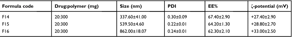
Table 2Characterization of chitosan-coated PLGA nanoparticles
Formula code Drug:polymer (mg) Size (nm) PDI EE% ζ-potential (mV)
F14 20:300 337.60±41.00 0.30±0.09 67.40±2.90 +27.40±2.90
F15 20:300 539.50±4.60 0.22±0.01 64.20±1.30 +28.80±2.70
F16 20:300 862.00±18.07 0.24±0.01 62.30±2.10 +33.00±2.50
Notes:Data are expressed as mean±SD (n=3). F14, F15, and F16 were the selected PLGA nanoparticles (F5) coated with 0.10%, 0.15%, and 0.30% w/v chitosan, respectively.
Abbreviations:PLGA, poly(d,l-lactide-co-glycolide); PDI, poly dispersity index; EE%, entrapment efficiency%.
International Journal of Nanomedicine downloaded from https://www.dovepress.com/ by 118.70.13.36 on 22-Aug-2020
size of the majority of nanoparticles prepared using methylene chloride (F3–F7) rather than ethyl acetate (F8–F13). At high polymer concentration (≥200 mg), the effect of the enhanced viscosity of methylene chloride (F3–F7) predominated due to the higher solubility of PLGA in this solvent relative to ethyl acetate. Thus, the slower drug diffusion may have counteracted Ostwald ripening, and hence smaller nanoparticles were formed.42
As illustrated in Table 2, a distinct increase in the
mean diameter of the selected uncoated PLGA nanopar-ticles (F5) was observed on coating with chitosan at the
three concentrations employed (F14–F16). This may be
explained on the basis that the protonated amino groups of chitosan may enable intermolecular hydrogen bond-ing with carboxylic groups of PLGA allowbond-ing chitosan
adsorption on PLGA surface.28 The smallest particle
size was seen with the lowest chitosan concentration (0.10% w/v, F14). Consequently, this formulation was further studied in comparison with that based on PLGA only (F5).
SEM examination
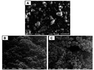
The free drug appeared as irregular microparticles with
wide size distribution (Figure 1A). On the other hand,
uncoated and coated nanoparticles were nanometric with narrower size distribution being nearly spherical with
smooth surface (Figure 1B and C, respectively). Spherical
particles may offer the maximum volume for drug incorporation.44 Selective accumulation in inflamed ulcera-tive tissues, improved cell uptake, and hence reduced dose
and subsequent cost-effectiveness can be expected.28
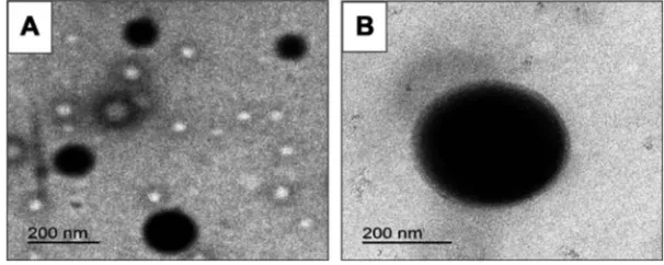
TEM examination
In agreement with SEM results, TEM images exhibited spherical nanoscopic uncoated (F5) and chitosan-coated (F14) nanoparticles (Figure 2). In case of the coated nano-particles, a solid dense polymer core surrounded by evenly distributed coat of chitosan appeared. In accordance with particle size measurements, uncoated nanoparticles pos-sessed smaller size than those coated with chitosan. Possibility of chitosan adsorption and binding to PLGA surface via hydrogen bonding between the protonated amino groups of chitosan and carboxylic groups of
PLGA may still explain these results.28
ζ- potential
The selected uncoated PLGA nanoparticles (F5) showed a relatively low negativeζ-potential equal to−10.50±0.20
Figure 1Scanning electron microscopy.
Notes:(A) Free diosmin, (B) uncoated diosmin-PLGA nanoparticles (F5), and (C) chitosan-coated PLGA nanoparticles (F14).
Abbreviation:PLGA, poly(d,l-lactide-co-glycolide).
International Journal of Nanomedicine downloaded from https://www.dovepress.com/ by 118.70.13.36 on 22-Aug-2020
which could be referred to the carboxylic groups on the nanoparticles surface that were partially shielded by
polyvinyl alcohol.45 Successful coating of PLGA
nano-particles with chitosan was reflected by the higher
posi-tive ζ-potential due to the amino groups in chitosan
(Table 2). This can be attributed to the intermolecular hydrogen bonding of amino groups of chitosan with carboxylic groups of PLGA allowing chitosan adsorption on PLGA surface and masking the original negative
charge of PLGA carboxylic groups.28 The increase in
chitosan concentration to 0.30% w/v resulted in a signifi
-cant rise in average ζ-potential possibly due to the
adsorption of the subsequent layers of chitosan on the
first layer.14 The higher ζ-potential values of
chitosan-coated nanoparticles than those of unchitosan-coated PLGA
nanoparticles can lead to a larger repulsive force; thus, more enhanced stability against aggregation can be expected.46
Solid-state characterization FT-IR
Figure 3depicts FT-IR spectra of uncoated (A) and coated (B) nanoparticles in comparison with free diosmin, the poly-mer(s), the corresponding PM, and the plain nanoparticles.
The characteristic peaks of diosmin at 3500 and 950 cm−1
were detected in its FT-IR spectrum (Figure 3AandB,I).9In addition, it showed absorption bands at 1660 cm−1due to the aromatic ketonic carbonyl stretching (C=O vibration), as well as at 1611 and 1502 cm−1assigned to the stretching of C=C bond in the aromatic ring.3
Figure 2Transmission electron microscopy.
Notes:(A) Uncoated diosmin-PLGA nanoparticles (F5) and (B) chitosan-coated PLGA nanoparticles (F14).
Abbreviation:PLGA, poly(d,l-lactide-co-glycolide).
3500
A
B
3000 2500 2000 1000 500
Wavenumber (cm-1) Wavenumber (cm-1)
1500 3500
V
V VI
IV
IV III
III
II
II
I I
% T
ransmittance
% T
ransmittance
3000 2500 2000 1500 1000 500
Figure 3Fourier transform-infrared spectra.
Notes:(A) Uncoated diosmin-PLGA nanoparticles (F5). (I) Diosmin, (II) PLGA, (III) diosmin:PLGA 1:1 PM, (IV) plain uncoated nanoparticles, and (V) medicated uncoated
PLGA nanoparticles. (B) Chitosan-coated PLGA nanoparticles (F14). (I) Diosmin, (II) PLGA, (III) chitosan, (IV) diosmin:chitosan 1:1 PM, (V) plain chitosan-coated nanoparticles, and (VI) medicated chitosan-coated nanoparticles.
Abbreviations:PLGA, poly(d,l-lactide-co-glycolide); PM, physical mixture.
International Journal of Nanomedicine downloaded from https://www.dovepress.com/ by 118.70.13.36 on 22-Aug-2020
PLGA spectrum (Figure 3AandB, II) exhibited stretching peaks of C=O at 1753 cm−1,47C–H bending at 859–1465 cm−1
as well as CH, CH2, and CH3 stretching vibration
between 2885 and 3000 cm−1, and finally OH stretching
around 3455–3500 cm−1.48 PLGA responded at 2955 cm−1 due to the linear CH2stretching and at 1756 cm−1due to the
ester bond.49According toFigure 3B(III), the intense peaks at
1658 and 1600 cm−1 in chitosan spectrum confirmed the
presence of amide I and amide II. Also, the peak of C–H
stretch and C–H bend appeared at 2875–2900 and 1362–
1426 cm−1, respectively. The peak at 3449 cm−1corresponding to N–H stretch was also present in chitosan spectrum. These spectral characteristics were in agreement with those reported.19 The characteristic peaks of the drug and either polymer were present in the spectrum of the corresponding PM (Figure 3A, III and B, IV) with no additional peaks negating their interaction. The spectrum of medicated nano-particles either uncoated (Figure 3A, V) or coated (Figure 3B, VI) was similar to that of the corresponding plain nanoparti-cles (Figure 3A, IV and B, V, respectively); thus, drug encap-sulation within the examined nanoparticles can be suggested.
DSC
Figure 4represents the thermal behavior of uncoated (A) and coated (B) nanoparticles in comparison with free diosmin, the polymer(s), their PM, and plain nanoparticles. DSC ther-mogram of free diosmin showed a sharp endothermic peak at
285.40°C indicating its melting point and its crystalline nat-ure as well as a broad endothermic peak at 112.90°C corre-sponding to its dehydration (Figure 4AandB,I).3
In DSC curve of PLGA (Figure 4A andB, II), there
was only an endothermic peak at 368.90°C reflecting its
decomposition and amorphous nature.14 In chitosan
ther-mogram, an initial endothermic peak at 75.10°C possibly due to dehydration and higher exothermic peak at
298.80°C due to degradation were identified (Figure 4B,
III).50 The presence of characteristic peaks of diosmin
and either polymer in the thermogram of their PM
negated their interaction (Figure 4A, III and B, IV).
PLGA endothermic peak was shifted to a lower tempera-ture in the thermogram of its PM with diosmin possibly
due to the dissolution of PLGA in the molten diosmin.51
The thermograms of uncoated (Figure 4A, V) or coated
(Figure 4B, VI) nanoparticles were similar to those
describing the corresponding plain nanoparticles (Figure
4A, IV and B, V); thus, drug entrapment within the
medicated nanoparticles can be suggested. Chitosan peak could not be recognized in the thermogram of the
coated PLGA nanoparticles (Figure 4B, VI) probably due
to the lower chitosan amount (30 mg) relative to that of PLGA (300 mg).
XRD
Figure 5 illustrates XRD patterns of free diosmin and the
A
B
V
V VI
IV
IV
III
III
II
Endothermic exothermic
Endothermic exothermic
II
I
50 100 150 200 250 300 350 400
Temperature (ºC)
50 100 150 200 250 300 350 400
Temperature (ºC)
I
Figure 4Differential scanning calorimetry.
Notes:(A) Uncoated diosmin-PLGA nanoparticles (F5). (I) Diosmin, (II) PLGA, (III) diosmin:PLGA 1:1 PM, (IV) plain uncoated nanoparticles, and (V) medicated uncoated
PLGA nanoparticles. (B) Chitosan-coated PLGA nanoparticles (F14). (I) diosmin, (II) PLGA, (III) chitosan, (IV) diosmin:chitosan 1:1 PM, (V) plain chitosan-coated nanoparticles, and (VI) medicated chitosan-coated nanoparticles.
Abbreviations:PLGA, poly(d,l-lactide-co-glycolide); PM, physical mixture.
International Journal of Nanomedicine downloaded from https://www.dovepress.com/ by 118.70.13.36 on 22-Aug-2020
polymer(s) as well as the corresponding PM, plain, and medicated nanoparticles. In agreement with DSC results, diffraction pattern of diosmin showed different intense peaks at 12.24◦, 13.7◦, 15.63◦, 19.79◦, 21.43◦, 22.52◦ and 25.00◦reflecting its crystalline nature (Figure 5AandB, I).9 The amorphous nature of both PLGA and chitosan was
confirmed by the absence of diffraction peaks in their XRD
diffractograms (Figure 5A, II andB, III, respectively). The interaction between diosmin and any of these polymers can-not be suggested due to the presence of drug diffraction peaks in XRD patterns of its PM with either PLGA (Figure 5A, III) or chitosan (Figure 5B, IV). On the other hand, XRD patterns of medicated nanoparticles either uncoated (Figure 5A, V) or coated (Figure 5B, VI) were similar to those of the corre-sponding plain nanoparticles (Figure 5A, IVand B, V, respec-tively) and the drug diffraction peaks were not seen. In accordance with FT-IR and DSC results, this can still indicate the drug encapsulation in the examined nanoparticles.
In vitro release study
In vitro release profiles of diosmin from uncoated (F5) and chitosan-coated nanoparticles (F14) in comparison with free drug are illustrated in Figure 6. The slow dissolution of free diosmin could be attributed to its poor wettability
and consequent agglomeration.9 The release pattern of
diosmin from both types of nanoparticles was biphasic.
An initial rapid release up to 8 h was followed by a sustained release phase till 72 h. Similar results were
reported for PLGA nanoparticles.52The rapid drug release
could be due to the fast dissolution of the drug adsorbed on nanoparticles surface and the enhanced surface diffu-sion of the release medium due to the large surface area of
the nanoparticles.53 The sustained release phase can be
referred to the drug encapsulated within PLGA polymeric matrix that could be released by the slow diffusion. Coating of nanoparticles with chitosan reduced the drug release during the sustained release phase possibly due to the protection against desorption and diffusion of the drug
by chitosan coat on PLGA nanoparticles.15 The rapid
release may provide the therapeutic drug concentrations
that could be maintained by the sustained drug release.28
As well, the drug sustained release may enable the reduc-tion in the dose frequency promoting the patient satisfaction.
Release kinetics
Table 3 shows the results of kinetic analysis of diosmin release results from the uncoated and coated nanoparticles. In vitro release of diosmin from both types of nanoparti-cles during rapid and sustained release phases can be explained by Higuchi model suggesting diffusion-con-trolled drug release. Higuchi diffusion has described
V
V VI
IV
Lin (counts)
Lin (counts)
IV
III
III
II
II
I
I
3 10 20 30 40 50
2-Theta-scale
3 10 20 30 40 50
2-Theta-scale
A
B
Figure 5X-ray diffractometry.
Notes:(A) Uncoated diosmin-PLGA nanoparticles (F5). (I) Diosmin, (II) PLGA, (III) diosmin:PLGA 1:1 PM, (IV) plain uncoated nanoparticles, and (V) medicated uncoated
PLGA nanoparticles. (B) Chitosan-coated PLGA nanoparticles (F14). (I) Diosmin, (II) PLGA, (III) chitosan, (IV) diosmin:chitosan 1:1 PM, (V) plain chitosan-coated nanoparticles, and (VI) medicated chitosan-coated nanoparticles.
Abbreviations:PLGA, poly (d,l-lactide-co-glycolide); PM, physical mixture.
International Journal of Nanomedicine downloaded from https://www.dovepress.com/ by 118.70.13.36 on 22-Aug-2020
Apremilast from PLGA nanoparticles.53 Non-Fickian mechanism (n<0.45) was found to describe the drug release during the rapid release phase from both types of nanoparticles indicating that both diffusion and erosion controlled the drug release during this phase. Fickian diffusion could be suggested to control the drug release during the sustained release phase since 0.45>n>0.87.33
Stability study
Tables 4and5represent the results of the stability study at refrigerator and room temperatures over a period of 3 months, respectively. At refrigerator temperature, there
was an insignificant change in average EE%, drug
retention%, and ζ-potential of both types of nanoparticles
over the 3 months when compared to those initially deter-mined at the beginning of the storage period. Similar results were observed at room temperature except that a significant (P<0.05) decrease in EE% and drug retention% of the uncoated nanoparticles was recorded at the third month. Uniform size distribution of both types of
nano-particles was indicated by mean PDI values of≤0.52±0.01
over the storage period at both temperatures. Compared to the initially determined mean particles size, there was a significant increase (P<0.05) in average size of uncoated nanoparticles only at both temperatures over the 3 months;
yet, the average particles diameter was≤233.20±4.50 nm.
The higher ζ-potential recorded for coated nanoparticles
may explain their higher stability against size enlargement on storage for 3 months at both temperatures. These results collectively can suggest further investigation of both uncoated and coated nanoparticles (F5 and F14, respec-tively) regarding mucoadhesion and AA.
Mucoadhesion study
Figure 7 shows SEM microphotographs of stomach and duodenum of the different groups orally treated with the selected uncoated (F5) or coated (F14) nanoparticles in com-parison with the free drug suspension in 1% w/v CMC. SEM images of stomach and duodenum were recorded at 2 and 8 h post oral treatment (Figure 7AandB, respectively).
After 2 or 8 h, no free diosmin could be detected in stomach (I) or duodenum (II). Uncoated PLGA nanoparticles appeared in both stomach (III) and duodenum (IV) at 2-h post-dosing, while their presence after 8 h could be detected only in duodenum (IV). These results may be attributed to the lack of the bioadhesive capability of PLGA, hence the uncoated nanoparticles did not exhibit gastric retention and
60
60 70 80
Time (h)
Uncoated PLGA nanoparticles (F5)
Chitosan-coated PLGA nanoparticles (F14)
Diosmin
Cumulative diosmin released (%)
50
50 40
40 30
30 20
20 10
10 0
0
Figure 6In vitro diosmin release in borate buffer (pH 10.5) from the selected
nanoparticles compared to the drug alone.
Abbreviation:PLGA, poly(d,l-lactide-co-glycolide).
Table 3Kinetic analysis of drug release data
Formula code Correlation coefficient (r2) Release order
Korsmeyer Main transport mechanism Zero
order
First order
Higuchi model
n r2
Uncoated PLGA nanoparticles (F5)
Rapid release phase 0.887 0.889 0.937 Higuchi 0.485 0.957 Non-Fickian
Sustained release phase 0.865 0. 885 0.904 Higuchi 0.170 0.901 Fickian
Coated nanoparticles (F14)
Rapid release phase 0.915 0.922 0.942 Higuchi 0.599 0.902 Non-Fickian
Sustained release phase 0.939 0.943 0.950 Higuchi 0.107 0.930 Fickian
Abbreviations:PLGA, poly (d,l-lactide-co-glycolide); n, diffusional exponent.
International Journal of Nanomedicine downloaded from https://www.dovepress.com/ by 118.70.13.36 on 22-Aug-2020
passed down to duodenum. In contrast, chitosan-coated nanoparticles were observed in stomach (V) in a massive amount even at 8-h post-dosing, while minor amount were noticed in the duodenum (VI). This confirms the high gastric retention potential of chitosan-coated nanoparticles (F14). As
revealed by ζ-potential measurements, chitosan imparted
positive charge on the coated nanoparticles, and hence mucoadhesion can be promoted through their electrical inter-action with the negatively charged mucus.54
In vivo evaluation
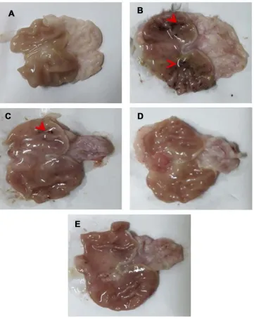
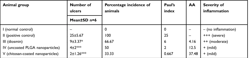
Macroscopic examinationFigure 8represents gross appearance of freshly excised gas-tric tissue of different groups orally pretreated with either free drug suspension in 1% w/v CMC or the selected
nano-particles. Parameters describing the AA through
macroscopic examination are illustrated in Table 6.
Apparently normal gastric mucosa was seen in normal
con-trol (group I) (Figure 8A). On the other hand, glandular
mucosal ulcerations as well as glandular and non-glandular mucosal congestion and edema were extensive in gastric tissues of rats of positive control group (II) (Figure 8B). Accordingly, an incidence of ulcer equal to 100% and the highest Paul’s index of 25 were observed in case of positive control rats. Oral pretreatment with free diosmin (100 mg/kg) resulted in moderate glandular mucosal ulcerations,
glandu-lar and non-glanduglandu-lar mucosal congestion and edema (Figure
8C). Mild congestion and edema appeared in glandular and
non-glandular gastric mucosa in group IV rats that orally
received uncoated nanoparticles (Figure 8D). Apparently
normal gastric mucosa was seen in rats pretreated with
coated nanoparticles (Figure 8E). In agreement, the three
Table 4Storage stability of the lyophilized selected uncoated and coated nanoparticles at refrigerator temperature (4±1°C)
Month Size (nm) PDI EE% Drug retention (%) ζ-potential (mV)
Uncoated nanoparticles (F5)
0 140.30±1.50 0.20±0.15 75.30±2.60 – −10.50±0.20
1 155.90±3.10** 0.30±0.05 73.50±7.30 97.39±6.20 −11.70±0.30
2 197.80±4.40*** 0.26±0.13 68.00±2.80 95.40±2.90 −9.70±0.49
3 233.20±4.50*** 0.40±0.030 71.90±4.70 90.20±0.58 −10.70±0.30
Coated nanoparticles (F14)
0 337.60±41.00 0.30±0.09 67.40±2.90 – 27.40±2.90
1 345.40±15.90 0.30±0.10 64.60±2.10 95.70±1.10 29.40±1.70
2 378.50±6.60 0.49±0.01 65.30±5.10 96.70±3.50 29.40±1.00
3 379.60±20.90 0.49±0.01 65.20±4.70 96.50±3.30 24.60±0.80
Notes:Data are expressed as mean±SD (n=3). **P<0.01 and ***P<0.001 vs initially determined size at the beginning of the storage period.
Abbreviations:PDI, polydispersity index; EE%, entrapment efficiency percentage.
Table 5Storage stability of the lyophilized selected uncoated and coated nanoparticles at room temperature (25±1°C)
Month Size (nm) PDI EE% Drug retention (%) ζ-potential (mV)
Uncoated nanoparticles (F5)
0 155.9±3.1 0.2±0.15 75.3±2.6 – −10.5±0.2
1 175.5±6.3* 0.4±0.02 72.4±5.5 96.25±8.5 −10.9±0.9
2 192.2±8.6*** 0.48±0.006 69.2±4.7 91.75±3 −10.6±0.8
3 230.9±1*** 0.42±0.08 60.6±6.1* 80.3±5.6* −10.6±0.4
Coated nanoparticles (F14)
0 337.6±41 0.3±0.09 67.4±2.9 – 27.4±2.9
1 386.7±15.9 0.5±0.03 64.2±3 95.2±0.4 26.6±1.3
2 367.8±12.4 0.47±0.08 65±4.6 96.3±3.1 23.3±1.1
3 385.7±10.1 0.52±0.01 64.4±4.6 95.4±1.1 21.4±1.3
Notes:Data are expressed as mean±SD (n=3). *P<0.05 vs initially determined size at the beginning of the storage period or drug retention % at thefirst month, ***P<0.001
vs initially determined size at the beginning of the storage period.
Abbreviations:PDI, polydispersity index; EE%, entrapment efficiency percent.
International Journal of Nanomedicine downloaded from https://www.dovepress.com/ by 118.70.13.36 on 22-Aug-2020
pretreatment groups (III, IV, and V) showed significantly (P<0.05) lower mean ulcers number when compared to posi-tive control rats (II). Hence, highly diminished ulcer
inci-dence and Paul’s index values were estimated for these
groups relative to positive control. In comparison with rats orally pretreated with free diosmin, a greater AA was obtained on oral pretreatment with uncoated PLGA nanopar-ticles. Coating of PLGA nanoparticles with chitosan resulted in highly potentiated AA of diosmin.
More prolonged residence time of nanoparticles in ulcerative tissues can be expected due to the greater accu-mulation and the facilitated uptake by immune cells, such
as macrophages.55
The greater AA of the coated nanoparticles than the uncoated ones can be attributed to the adherence of the positively charged chitosan-coated nanoparticles to the
negatively charged cellular membranes56 as well as their
escape from the acidic solution of endosomes–
lysosomes,57 thus inducing their intracellular uptake into
the cytoplasm. The interaction with the cellular membrane results in a structural reorganization of junction proteins that is reversed when the contact with chitosan is
terminated.58 Moreover, mucoadhesion enhanced through
the electrical interaction of positively charged chitosan-coated nanoparticles with the negatively charged mucin
can still account for the potentiated AA.54 In spite of
these biological interactions due to the positively charged surface of chitosan-coated nanoparticles, chitosan cyto-toxicity was negated as clarified by cell viability close to 100% after contacting with chitosan or systems based on it.59 In addition, no significant toxicity was reported fol-lowing repeated oral administration of microparticles or nanoparticles incorporating chitosan at a dose range of
100–125 mg/kg as revealed by the absence of
abnormal-ities in hepatic and renal functions or pathological changes in liver, kidney, and intestinal segments.60,61
Figure 7Scanning electron microscopy of stomach and duodenum of the different groups orally treated with diosmin (100 mg/kg) or an equivalent dose of either uncoated
or chitosan-coated nanoparticles.
Notes:(A) Two-hour post-dosing and (B) eight-hour post-dosing. (I) and (II) stomach and duodenum of rats treated with diosmin, respectively, (III) and (IV) stomach and
duodenum of rats that administered uncoated PLGA nanoparticles, respectively, (V) and (VI) stomach and duodenum of rats given chitosan-coated nanoparticles, respectively. Arrows point to the attached nanoparticles.
Abbreviation:PLGA, poly(d,l-lactide-co-glycolide).
International Journal of Nanomedicine downloaded from https://www.dovepress.com/ by 118.70.13.36 on 22-Aug-2020
Histopathological examination
Figure 9 displays microphotographs of histopathological examination (HE, 100×) of non-glandular (A) and gland-ular (B) gastric tissues of the different groups. Normal control (I) displayed an intact architecture of non-gland-ular and glandnon-gland-ular gastric wall (I). Administration of ethanol to rats of positive control (II) triggered a severe gastric injury with high scores of microscopic damage including extensive congestion and edema in
non-glandular portion, mucosal ulceration and submucosal
inflammation in glandular portion. Submucosal
conges-tion and edema in non-glandular and glandular porconges-tions
besides submucosal inflammation in glandular portion
were moderate in rats pretreated with free diosmin (III), while mild in rats that administered uncoated nanoparti-cles (IV). Non-glandular and glandular gastric walls retained their normal histological pictures in rats that received chitosan-coated nanoparticles (V).
Figure 8Gross appearance of freshly excised stomachs.
Notes:(A) Normal control with normal gastric mucosa. (B) Positive control showing glandular mucosal ulcerations (red arrowheads) as well as extensive glandular and
glandular mucosal congestion and edema. (C) Rats pretreated with diosmin (100 mg/kg) displaying moderate glandular mucosal ulcerations (red arrowheads), glandular and non-glandular mucosal congestion and edema were moderate. (D) Rats pretreated with an equivalent dose of uncoated PLGA nanoparticles exhibiting mild congestion and edema appeared in glandular and non-glandular gastric mucosa. (E) Apparently normal gastric mucosa of rats pretreated with an equivalent dose of chitosan-coated nanoparticles.
Abbreviation:PLGA, poly(d,l-lactide-co-glycolide).
International Journal of Nanomedicine downloaded from https://www.dovepress.com/ by 118.70.13.36 on 22-Aug-2020
In general, oral pretreatment with diosmin either free or as nanoparticles lowered the pathologic scores and attenuated the gastric damage. Results of statistical
analysis of histopathological examination in glandular
portion of gastric tissues are depicted inFigure 10A–D.
The results revealed that there was a significant
Table 6Parameters of macroscopical evaluation of ethanol-induced ulcers in rats
Animal group Number of ulcers
Percentage incidence of animals
Paul’s index
AA Severity of inflammation
Mean±SD n=6
I (normal control) – 0 0 – –(no inflammation)
II (positive control) 25±5.67 100 25 – +++ (severe)
III (diosmin) 9±3.37* 66.67 6 4.16 ++ (moderate)
IV (uncoated PLGA nanoparticles) 4±2*** 50 2 12.5 + (mild)
V (chitosan-coated nanoparticles) 2±1.26*** 33.33 0.667 37.48 + (mild)
Notes:100 mg/kg diosmin or an equivalent dose of the nanoparticles was used; *P<0.05 and***
P<0.001 vs positive control group (II).
Abbreviations:PLGA, poly(lactic-co-glycolic) acid; AA, anti-ulcer activity.
Figure 9Histological examination (HE, 100x) of gastric tissues.
Notes:(A) Non-glandular and (B) glandular gastric mucosa. (I) Normal control displaying an intact architecture of non-glandular and glandular gastric wall. (II) Positive
control showing extensive congestion (red arrows) and edema (black asterisk) in non-glandular portion, mucosal ulceration (thick black arrow), and submucosal inflammation (thin black arrow) in glandular portion. (III)Rrats pretreated with diosmin (100 mg/kg) displaying moderate submucosal congestion (red arrows) and edema (black asterisk) in non-glandular and glandular portions besides submucosal inflammation (thin black arrow) in glandular portion. (IV) Rats pretreated with uncoated PLGA nanoparticles exhibiting mild submucosal congestion (red arrows) and edema (black asterisk) in non-glandular and glandular portions besides very mild submucosal inflammatory cells infiltration in glandular portion (thin black arrow). (V) Non-glandular and glandular gastric walls retained their normal histological pictures in rats pretreated with chitosan-coated nanoparticles.
Abbreviation:PLGA, poly(d,l-lactide-co-glycolide).
International Journal of Nanomedicine downloaded from https://www.dovepress.com/ by 118.70.13.36 on 22-Aug-2020
(P<0.05) elevation in epithelial cells loss, edema,
con-gestion, and inflammation in glandular portions of
positive control and rats orally pretreated with free diosmin or uncoated nanoparticles when compared to normal control. In contrast to free diosmin, oral pre-treatment with either uncoated or coated nanoparticles
significantly (P<0.05) lowered epithelial cells loss,
edema, congestion, and inflammation in glandular
por-tions relative to positive control. In comparison with free diosmin, there was a significant (P<0.05) reduction in these pathological changes only following oral administration of coated nanoparticles.
Significantly (P<0.05) elevated edema in non-glandu-lar portions of gastric tissues was observed in positive control and rats orally pretreated with either free diosmin
or uncoated nanoparticles than normal rats (Figure 10E).
Rats administered coated nanoparticles encountered a sig-nificantly (P<0.05) lower edema in non-glandular portions than positive control, free diosmin, and uncoated nanopar-ticles. Regarding congestion in non-glandular gastric tis-sues, there was a significant (P<0.05) increase in positive control and rats pretreated with free diosmin relative to normal control (Figure 10F). Significantly (P<0.05) dimin-ished congestion was obtained in the three pretreatment
25
A
B
C
D
E
F
Scores of epithelial cell loss
Scores of edema
(0–4)
Scores of edema
(0–4)
Scores of inflammation
(0–3)
Scores of congestion
(0–4)
Scores of congestion
(0–4) 3 3 4 3 4 2 2 2 1 1 1 0 0 3 2 1 0 3 2 1 0 0 20 1.5 1.0 0.5 0.0
Normal controlPositive controlFree diosmin
Uncoated nanoparticlesCoated nanoparticles
Normal controlPositive controlFree diosmin
Uncoated nanoparticlesCoated nanoparticles
Normal controlPositive controlFree diosmin
Uncoated nanoparticlesCoated nanoparticles
Normal controlPositive controlFree diosmin
Uncoated nanoparticlesCoated nanoparticles Normal controlPositive controlFree diosmin
Uncoated nanoparticlesCoated nanoparticles Normal controlPositive controlFree diosmin
Uncoated nanoparticlesCoated nanoparticles
* * * * * * * * * * * * * # *# * # * # * # # # $ # $ # $ @ # $ @ # $ @ # $ @
Figure 10Statistical analysis of microscopic histopathological scores in gastric mucosa.
Notes:(A–D) Glandular and (EandF) non-glandular mucosa. Data are mean±SD, n=6; statistical differences atP<0.05 considered significant; *vs normal control group;#
vs positive control group;$vs diosmin (100 mg/kg) pretreated group;@vs group pretreated with an equivalent dose of uncoated PLGA nanoparticles.
Abbreviation:PLGA, poly(d,l-lactide-co-glycolide).
International Journal of Nanomedicine downloaded from https://www.dovepress.com/ by 118.70.13.36 on 22-Aug-2020
groups when compared to positive control. In contrast to uncoated nanoparticles, oral administration of coated nanoparticles resulted in a significantly (P<0.05) lower congestion in non-glandular gastric tissue than rats given
free diosmin. There was insignificant difference between
rats orally pretreated with uncoated and coated nanoparti-cles regarding congestion scores in non-glandular portion.
There was insignificant difference between normal
control and rats received coated nanoparticles regarding severity of all pathological changes describing ulceration in both glandular and non-glandular gastric tissues. Thus, it can be said that coating of PLGA nanoparticles with chitosan significantly potentiated the cytoprotective activ-ity of diosmin against ethanol-induced ulcer in rats. These results may be due to the increased interaction with the negatively charged cellular membranes and the improved
intracellular uptake into the cytoplasm,56 as well as the
enhanced escape from the acidic solution of endosomes–
lysosomes into the cytoplasm.57 Moreover, the increased
mucoadhesion of positively charged chitosan-coated nano-particles with the negatively charged mucin and the
result-ing gastric retention (Figure 7) can still explain the
superiority of coated nanoparticles.54
Immunohistochemical loclization of NF-κB
Figure 11 illustrates immunohistochemical evaluation of
NF-κB expression in non-glandular (A) and glandular (B)
gastric tissues. Mild positive expression was recognized in both tissues in normal control group (I). Strong immunor-eactivity was observed in both mucosae particularly
around area of mucosal damage and stained inflammatory
cells infiltrating submucosa of glandular portion in
posi-tive control (II). Oral pretreatment with free diosmin sus-pension in 1%w/v CMC (100 mg/kg, III) or an equivalent
Figure 11Microscopic pictures of rats stomach immunostained against NF-κB.
Notes:(A) Non-glandular and (B) glandular gastric mucosa. (I) Normal control showing mild positive expression in glandular and non-glandular gastric mucosa. (II) Positive control
rats displaying strong positive expression in glandular and non-glandular gastric mucosa particularly around area of mucosal damage (thick black arrow) and stained inflammatory cells infiltrating submucosa of glandular portion. (III) Rats pretreated with diosmin (100 mg/kg) or (IV) an equivalent dose of uncoated PLGA nanoparticles exhibiting mild positive expression in non-glandular mucosa, as well as moderate positive expression in gastric mucosa and the inflammatory cells infiltrating submucosa of glandular portion. (V) Rats that received chitosan-coated nanoparticles showing mild positive expression in glandular and non-glandular gastric mucosa. Thin black arrows point to positive signal in mucosa and thin yellow arrows point to positively stained leukocytes infiltrating submucosa. IHC counterstained with Mayer’s hematoxylin (100×), insert (S, 200×).
Abbreviations:NF-κB, nuclear factor kappa-light-chain-enhancer of activated B cells; PLGA, poly (d,l-lactide-co-glycolide); IHC, immunohistochemistry.
International Journal of Nanomedicine downloaded from https://www.dovepress.com/ by 118.70.13.36 on 22-Aug-2020
dose of uncoated nanoparticles (IV) resulted in mild immunostaining in non-glandular mucosa, meanwhile,
moderate positive expression and the inflammatory cells
infiltrating submucosa of glandular portion were recorded
in gastric mucosa (III and IV, respectively). Mild positive expression was detected in both mucosae in rats that received chitosan-coated nanoparticles (V).
Results of statistical analysis of
immunohistochem-ical localization of gastric NF-κB in glandular (A) and
non-glandular portions (B) are depicted in Figure 12.
According to this figure, there was a significant
(P<0.05) increase in gastric NF-κB expression in both portions in positive control when compared to normal control. Oral pretreatment with free diosmin suspension
insignificantly affect the immunoreactivity. On the other
hand, there was a significant (P<0.05) reduction in
glandular and non-glandular NF-κB expression
follow-ing oral administration of diosmin loaded nanoparticles either coated or uncoated in comparison with the posi-tive control. In agreement with histopathological exam-ination, the superiority of coated nanoparticles over the
uncoated ones can be suggested by the significantly
(P<0.05) lower glandular and non-glandular NF-κB
expression than that produced by either free diosmin
or uncoated nanoparticles as well as the insignificantly
different glandular and non-glandular NF-κB expression
from that recorded in normal group. Such superiority can still be attributed to the increased intracellular
uptake into the cytoplasm57 as well as the improved
mucoadhesion and gastric retention of these positively
charged nanoparticles.54
TEM examination of the gastric mucosa ultrastructure
TEM examination of the mucosal surface of normal con-trol rats revealed well-arranged microvilli in neat rows with no loss, normal nucleus, high density of mitochon-dria, regular pattern of rough endoplasmic reticulum, and
dispersed gastric secretion (Figure 13A). Meanwhile, the
mucosal surface of positive control rats showed swollen mitochondria, deleted rough endoplasmic reticulum, abnormal nucleus, cytoplasmic vacuoles, cells with dilated reticulum, complete loss of microvilli, and several
non-homogenated cytoplasmic inclusions (Figure 13B).
Following oral treatment with free diosmin suspension, there was a slight amelioration in the mucosal cells, yet wide junctions between cells were observed and some cytoplasmic organelles were still deleterious, including swollen mitochondria, pyknotic nucleus, and irregular
microvilli (Figure 13C). Regarding rats that received
uncoated PLGA nanoparticles, a slightly ameliorated
Normal controlPositive controlFree diosmin
Uncoated nanoparticlesCoated nanoparticles
Normal controlPositive controlFree diosmin
Uncoated nanoparticlesCoated nanoparticles
3 4
2
1
0 3
4
*** ***
***
* ***
***
### ##
### $$$ @@@
### $$$ @ 2
1
0 IHC scores of positive staining
(0–3)
IHC scores of positive staining
(0–3)
A
B
Figure 12Statistical analysis of IHC scores of NF-κB in gastric mucosa.
Notes:(A) Glandular and (B) non-glandular mucosa. Data are mean±SD, n=6;
*P<0.05 and***P<0.001 vs normal control;##P<0.01 and###P<0.001 vs positive control;$$$P<0.001 vs diosmin (100 mg/kg) pretreated group;@P<0.05 and@@@P<0.001 vs group pretreated with an equivalent dose of uncoated PLGA nanoparticles.
Abbreviations:IHC, immunohistochemistry; NF-κB, nuclear factor kappa-light-chain-enhancer of activated B cells; PLGA, poly(d,l-lactide-co-glycolide).
International Journal of Nanomedicine downloaded from https://www.dovepress.com/ by 118.70.13.36 on 22-Aug-2020
mitochondria, normal rough endoplasmic reticulum, intact cell membrane, and regular microvilli were
observed in the mucosal surface (Figure 13D). Oral
pre-treatment with chitosan-coated nanoparticles of diosmin resulted in obvious amelioration in the mucosal cells, disappearance of vacuoles, intact cell membrane with tight junctions and regularly arranged microvilli with high density as well as normal mitochondria,
endoplas-mic reticulum, and nucleus (Figure 13E).
Conclusion
The selected uncoated nanoparticles consisted of 1:15 drug-PLGA weight ratio using 20 mg diosmin and methy-lene chloride as an organic phase because they showed the highest EE% (75.30±2.60%) and particle size <200 nm (155.90±3.10 nm). Coating of these nanoparticles with
chitosan (0.10%, 0.15%, and 0.30% w/v) significantly
enlarged the size on the increase of chitosan concentration
but EE% did not significantly differ; thus, those coated
Figure 13TEM examination of the gastric mucosa ultrastructure.
Notes:(A) Normal control rats showing well-arranged microvilli in neat rows with no loss, normal nucleus, high density of mitochondria, regular pattern of rough endoplasmic
reticulum, and dispersed gastric secretion. (B) Positive control rats displaying swollen mitochondria, deleted rough endoplasmic reticulum, abnormal nucleus, cytoplasmic vacuoles, cells with dilated reticulum, complete loss of microvilli, and several non-homogenated cytoplasmic inclusions. (C) Rats pretreated with free diosmin (100 mg/kg) exhibited a slight amelioration in the mucosal cells, wide junctions between cells and some deleterious cytoplasmic organelles including swollen mitochondria, pyknotic nucleus, and irregular microvilli. (D) Rats that received uncoated PLGA nanoparticles showing a slightly ameliorated mitochondria, normal rough endoplasmic reticulum, intact cell membrane, and regular microvilli. (E) Rats pretreated with chitosan-coated nanoparticles of diosmin showing obvious amelioration in the mucosal cells, disappearance of vacuoles, intact cell membrane with tight junctions and regularly arranged microvilli with high density as well as normal mitochondria, endoplasmic reticulum, and nucleus.
Abbreviation:TEM, transmission electron microscopy.
International Journal of Nanomedicine downloaded from https://www.dovepress.com/ by 118.70.13.36 on 22-Aug-2020