DOI: 10.1534/genetics.106.064428
The Sensitivity of Yeast Mutants to Oleic Acid Implicates the
Peroxisome and Other Processes in Membrane Function
Daniel Lockshon,* Lauren E. Surface,* Emily O. Kerr,* Matt Kaeberlein
†,1and
Brian K. Kennedy*
,2*Department of Biochemistry, University of Washington, Seattle, Washington 98195 and†Department of Genome Sciences, University of Washington, Seattle, Washington 98195
Manuscript received August 4, 2006 Accepted for publication October 22, 2006
ABSTRACT
The peroxisome, sole site ofb-oxidation inSaccharomyces cerevisiae, is known to be required for optimal growth in the presence of fatty acid. Screening of the haploid yeast deletion collection identified130 genes, 23 encoding peroxisomal proteins, necessary for normal growth on oleic acid. Oleate slightly en-hances growth of wild-type yeast and inhibits growth of all strains identified by the screen. Nonperoxisomal processes, among them chromatin modification by H2AZ, Pol II mediator function, and cell-wall-associated activities, also prevent oleate toxicity. The most oleate-inhibited strains lack Sap190, a putative adaptor for the PP2A-type protein phosphatase Sit4 (which is also required for normal growth on oleate) and Ilm1, a protein of unknown function. Palmitoleate, the other main unsaturated fatty acid of Saccharomyces, fails to inhibit growth of thesap190D,sit4D, andilm1Dstrains. Data that suggest that oleate inhibition of the growth of a peroxisomal mutant is due to an increase in plasma membrane porosity are presented. We propose that yeast deficient in peroxisomal and other functions are sensitive to oleate perhaps because of an inability to effectively control the fatty acid composition of membrane phospholipids.
b
-OXIDATIVE catabolism of fatty acids occurs in peroxisomes of all eukaryotes, yet there is tre-mendous species variation in other functions carried out by this organelle, also called the microbody, the glycosome, and the glyoxisome (van denBosch et al.1992). While b-oxidation occurs in all peroxisomes, only in some species, such as the yeasts, is it a uniquely peroxisomal function (Hiltunenet al.2003). Animals,
by contrast, also performb-oxidation in the mitochon-drion using a separate set of enzymes (Bartlett and
Eaton 2004). A variety of elegant methods using
hu-man cells,Saccharomyces cerevisiae, and other yeast spe-cies have identified 33 Pex proteins, or peroxins, that play a wide range of roles in peroxisome biogenesis and function (Purdue and Lazarow 2001; Eckert
and Erdmann2003; Veenhuiset al.2003; Moyersoen
et al. 2004). Most Pex proteins are highly conserved throughout eukaryotes; human disorders that result from mutations in any one of a number ofpex genes have been studied extensively (Eckertand Erdmann
2003). Only 2 of the 27 known Saccharomyces per-oxins, Pex3 and Pex19, are required to maintain the organelle (Hohfeld et al. 1991; Gotte et al. 1998;
Hettemaet al.2000).
Despite the extensive knowledge of the roles of many of these proteins, the specific biological functions of the peroxisome, as well as the molecular basis of patholo-gies that result from peroxisomal dysfunction, remain poorly understood. Most pointedly, the cellular role of peroxisomalb-oxidation has not been established. Mito-chondrialb-oxidation, found only in animals, is thought to be responsible for catabolism of dietary fatty acid (Bartlettand Eaton2004). Peroxisomalb-oxidation,
found in every eukaryote examined (Kunauet al.1988),
is likely to play a different cellular role. InS. cerevisiae,b -oxidation appears to be the only complete biochemical pathway carried out in the peroxisome (Hiltunenet al.
2003). Such relative simplicity, as well as the unsur-passed genetic utility of this yeast, makes it an attractive system for helping to elucidate the organelle’s cellular function. Soon after it was established that Saccharomy-ces could be used to study the peroxisome (Veenhuis
et al.1987), a genetic screen for mutants that grow slowly in the presence of oleic acid (oleate), an 18-carbon,cis -monounsaturated fatty acid, led to the identification of
PEX1, PEX3, and PEX4 (Erdmann et al. 1989). Slow
growth was attributed to the inability of yeast with defec-tive peroxisomes to carry outb-oxidation and to there-fore be incapable of utilizing fatty acids as a source of carbon.
As part of an effort to identify proteins required for peroxisome inheritance, we sought to identify addi-tional proteins that are required for its maintenance. A set of yeast deletion strains constructed by a consortium 1Present address:Department of Pathology, University of Washington,
Seattle, WA 98195.
2Corresponding author: Department of Biochemistry, Box 357350, University of Washington, Seattle, WA 98195.
E-mail: [email protected]
of laboratories (Winzeleret al. 1999) has been screened
extensively for a broad range of phenotypes. In this work, haploid deletion strains that grow poorly in the presence of oleate were identified. Most Pex proteins previously shown to be required for optimal growth in the presence of oleate were identified by the screen, yet the screen also identified genes whose products have nonperoxisomal functions. Contrary to previous inter-pretations, strains identified in the screen grew poorly in the presence of oleate because of the inhibition of their growth by oleate rather than because they were incapable of using oleate as a carbon source. We present evidence suggesting that the toxicity of oleate topexand other mutants may be the result of changes in the plasma membrane. These findings implicate the perox-isome, an organelle whose biological function has re-mained obscure, in membrane function.
MATERIALS AND METHODS
Yeast media, strains, and growth:Synthetic media with ter-gitol and yeast extract (STY) contains 0.67% yeast nitrogen base (Difco 291940); 0.05% yeast extract (Difco 212750); 1% tergitol [Sigma (St. Louis) NP40S]; a mixture of amino acids, uracil, and adenine at one-tenth the concentration used pre-viously (Sherman 1991); 0.01% ampicillin and 0.01% G418 (both ampicillin and G418 are omitted from liquid media). Oleate and palmitoleate from Nu-Chek Prep were dissolved in warm 10% tergitol before being added to the other compo-nents. Plates contained 2% bactoagar (Difco 214010). Nour-seothricin (clonNAT) was provided by Werner BioAgents.
All yeast strains were derived from BY4741 and BY4742 (Brachmannet al.1998). The oleate screen was carried out using the set ofMATa haploid deletion strains (Open Bio-systems YSC1049) maintained in the supplier’s 96-well format. Frozen stocks were thawed and inoculated into 0.1 ml of YPD 1G418 medium in 50, 96-well dishes using a pinning device mounted on the Beckman Biomek 2000, also used for all fur-ther deletion set manipulations. After growth for 2 days at 30°
without shaking, cultures were diluted100-fold in water and 1ml was pinned onto rectangular agar plates containing STY1 oleate (0.1%) and STY1glycerol (3%) medium for growth at 25°. STY1glycerol plates were grown for 4 days and stored at 4°until sufficient growth of the STY1oleate plates (13 days) allowed growth data to be visually recorded. Strain data and growth properties were managed using Filemaker software. To combine a gene deletion with Pex11-GFP, theMATadeletion strain from the commercial set was mated to the Pex11-GFP fusion derivative of BY4741 constructed by the Yeast GFP Fu-sion Localization Database (Huhet al.2003). The diploid was then sporulated, and His1G418Rspore colonies were
iden-tified from tetrads in which these two markers each segregated 2:2. Flow cytometric analysis of cell cycle distribution was performed as described (Foss2001).
Microscopy, Western blotting, and Sytox green staining: Fluorescence microscopy was carried out using a Zeiss Axiovert 200M with FITC filters to detect GFP. Western blots of yeast protein prepared (Kornitzer 2002) from midlog cultures were probed with mouse monoclonal antibody to GFP (Roche 1814460). The secondary anti-mouse monoclonal antibody, con-jugated to horseradish peroxidase, was reacted with an acridi-nium ester [Amersham (Buckinghamshire, UK) RPN2132], and the product was detected on the Amersham Storm 860
phosphoimager. Blots were then probed with rabbit polyclonal antibody to yeast actin (kindly provided by Alex Merz), further probed with anti-rabbit monoclonal antibody conjugated to horseradish peroxidase, and reacted and visualized as above.
To assay the influence of oleate on cell permeability, yeast grown in STY liquid medium with or without 0.1% oleate to midlog were resuspended in 0.5mmSytox green (BioVision K201) as suggested by the supplier for 10 min at room tem-perature and visualized immediately on the Zeiss microscope with FITC filters.
RESULTS
A screen for proteins that permit normal growth in the presence of oleate:Two previous studies led to the isolation of mutants ofS. cerevisiaethat grow slowly in the presence of oleate (Erdmannet al. 1989; Marziochet al.
1994). Such growth impairment was attributed to the inability to utilize oleate as a carbon source. Six of these mutants fell into four complementation groups, which eventually came to be known asPEX1(Erdmannet al.
1991),PEX3(Hohfeldet al. 1991),PEX4(Wiebeland
Kunau 1992), and PEX7 (Marzioch et al. 1994). To
carry out a screen of the yeast deletion strains using this strategy, a growth medium, STY, was devised (based on the variety of oleate media formulations used since 1989) that in our study maximized the growth differ-ence betweenPex1
andpexDstrains on STY1oleate rela-tive to growth on STY1glycerol. The rationale for using glycerol, a nonfermentable carbon source, as a control in the current and previous screens was that acetyl-CoA produced byb-oxidation can serve as an energy source only in respiratory-competent strains,i.e., those capable of growing on glycerol. Respiratory-deficient mutants, incapable of growth on both glycerol and oleate, could thereby be distinguished from those that grow slowly only on oleate.
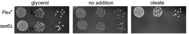
The haploid (MATa) deletion collection (4773 strains) was grown on STY1oleate and STY1glycerol to give 50 pairs of plates, one of which is shown in Figure 1. A total of 136 strains deleted in the genes listed in Table 1 were identified. Twenty-six of these compose 13 pairs of chromosomally adjacent genes. Since five of the six dubious ORFs (predicted to encode proteins that have no orthologs in other fungi) identified by the screen were adjacent in chromosomal location tobona fidegenes listed in Table 1, it is likely that sequence in these dubious ORFs affects expression of adjacent oleate-sensitive dele-tions. These six genes—SPF1,PEX14,OPT1,KNS1,NCS1, andGAL11—were thus in effect identified twice. For the seven other pairs of chromosomally adjacent genes, the identities of the true hits are ambiguous. As has also been observed by others ( J. Aitchisonand P. Lazarow,
Nevertheless, retesting theMATadeletion strains identi-fied in the screen confirmed an oleate phenotype for each strain listed in Table 1. In addition, theMATa ver-sions of most of the strains identified in the screen were also tested and found to correlate well with the pheno-types listed in Table 1.
The screen was judged to be successful because it identified 23 peroxisomal proteins, including 16 Pex proteins that have been reported to be required for optimal growth on oleate. A number of other functional categories are also overrepresented relative to their prominence in the yeast proteome. The Pol II mediator complex is one such category: five of its eight compo-nents (Bjorklundand Gustafsson2005) were
identi-fied (one other of its components is required for respiration and another is essential). Another func-tional category involves H2AZ, the alternative histone whose substitution into chromatin locally influences transcription (Koboret al.2004). Four of nine
compo-nents of SWR-C (four other SWR-C proteins are essen-tial), the complex that substitutes histone H2AZ for H2A in chromatin, were identified, as was H2AZ itself.
Oleate inhibits the growth of peroxisomal mutants and other oleate-sensitive strains: In isolating Saccha-romycespexmutants, Erdmannet al.(1989) attributed
their poor growth in the presence of oleate to an in-ability to use fatty acid as a carbon source. On the contrary, we find that oleate inhibited the growth ofpex
mutants. Figure 2 compares the growth of pex6D and
Pex1
strains on STY containing glycerol, no added car-bon source, or oleate. In contrast to the results of Erdmannet al.(1989), which showed wild-type
Saccha-romyces to be unable to grow unless both 0.05% yeast extract and oleate are present on plates, we found growth to occur without the addition of any carbon source as long as 0.05% yeast extract was included. Tergitol, the detergent in STY medium added for fatty acid solubilization, had no effect on growth (data not shown). Note the slight enhancement in colony size of both strains by the addition of glycerol and of thePex1 strain by the addition of oleate. Growth of the pex6D
strain, however, is severely inhibited by oleate addition. While there is a slight augmentation in growth of the
Pex1
strain by the addition of oleate, the poorer growth on oleate of the strains identified in our screen is mainly due to the inhibition of their growth by oleate rather than to the failure of their growth to be enhanced by oleate. By comparing the growth of all positives ob-tained in the screen on STY, STY1glycerol, and STY1
oleate, oleate inhibition was found to account for the oleate phenotype of all but one of the strains (the ex-ception,pck1D, was capable of growth on STY1glycerol but not on STY and therefore does not appear in Table 1). Because the mutants are oleate inhibited rather than unable to be fed by oleate, the screen in retrospect could have used STY rather than STY 1 glycerol as a control.
STY medium was used to conduct the screen since this minimal medium allows the detection of even weakly oleate-inhibited strains. The possibility therefore arose that the strains appearing to be less strongly inhibited by oleate were exhibiting this phenotype because of starvation. We addressed this possibility with a series of control experiments that examined the effect of oleate on the cell cycle distribution of cells from Pex1 and
pex6Dcultures incubated in STY1oleate. Starvation of yeast is well established to cause cells to accumulate in G1( Johnstonet al.1977). However, at 1, 2, and 4 hr after the shift from glucose- to oleate-containing STY medium, no shift in cell cycle profile of either thePex1 (Figure 3, A, C, E, and G) or thepex6D(Figure 3, B, D, F, and H) strains was detectable. While the viability of the
Pex1
strain is largely unaffected by this 4-hr oleate treat-ment, the progressive decrease in viability of thepex6D
cells during this interval demonstrates the expected toxic effect of oleate on this strain. An accumulation of Figure1.—A genomewide screen for oleate sensitivity. One
TABLE 1
Deletion strains identified in the oleate screen
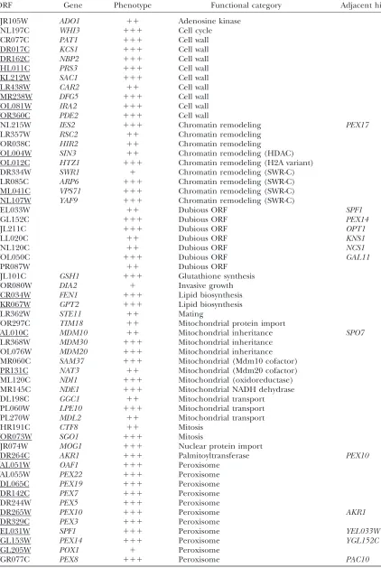
ORF Gene Phenotype Functional category Adjacent hit
YJR105W ADO1 11 Adenosine kinase
YNL197C WHI3 111 Cell cycle
YCR077C PAT1 111 Cell wall
YDR017C KCS1 111 Cell wall
YDR162C NBP2 111 Cell wall
YHL011C PRS3 111 Cell wall
YKL212W SAC1 111 Cell wall
YLR438W CAR2 11 Cell wall
YMR238W DFG5 111 Cell wall
YOL081W IRA2 111 Cell wall
YOR360C PDE2 111 Cell wall
YNL215W IES2 111 Chromatin remodeling PEX17
YLR357W RSC2 11 Chromatin remodeling
YOR038C HIR2 11 Chromatin remodeling
YOL004W SIN3 11 Chromatin remodeling (HDAC)
YOL012C HTZ1 111 Chromatin remodeling (H2A variant)
YDR334W SWR1 1 Chromatin remodeling (SWR-C)
YLR085C ARP6 111 Chromatin remodeling (SWR-C)
YML041C VPS71 111 Chromatin remodeling (SWR-C)
YNL107W YAF9 111 Chromatin remodeling (SWR-C)
YEL033W 11 Dubious ORF SPF1
YGL152C 111 Dubious ORF PEX14
YJL211C 111 Dubious ORF OPT1
YLL020C 11 Dubious ORF KNS1
YNL120C 11 Dubious ORF NCS1
YOL050C 111 Dubious ORF GAL11
YPR087W 11 Dubious ORF
YJL101C GSH1 111 Glutathione synthesis
YOR080W DIA2 1 Invasive growth
YCR034W FEN1 111 Lipid biosynthesis
YKR067W GPT2 111 Lipid biosynthesis
YLR362W STE11 11 Mating
YOR297C TIM18 11 Mitochondrial protein import
YAL010C MDM10 11 Mitochondrial inheritance SPO7
YLR368W MDM30 111 Mitochondrial inheritance YOL076W MDM20 111 Mitochondrial inheritance YMR060C SAM37 111 Mitochondrial (Mdm10 cofactor) YPR131C NAT3 11 Mitochondrial (Mdm20 cofactor) YML120C NDI1 111 Mitochondrial (oxidoreductase)
YMR145C NDE1 111 Mitochondrial NADH dehydrase
YDL198C GGC1 11 Mitochondrial transport
YPL060W LPE10 111 Mitochondrial transport
YPL270W MDL2 11 Mitochondrial transport
YHR191C CTF8 11 Mitosis
YOR073W SGO1 111 Mitosis
YJR074W MOG1 111 Nuclear protein import
YDR264C AKR1 111 Palmitoyltransferase PEX10
YAL051W OAF1 111 Peroxisome
YAL055W PEX22 111 Peroxisome
YDL065C PEX19 111 Peroxisome
YDR142C PEX7 111 Peroxisome
YDR244W PEX5 111 Peroxisome
YDR265W PEX10 111 Peroxisome AKR1
YDR329C PEX3 111 Peroxisome
YEL031W SPF1 111 Peroxisome YEL033W
YGL153W PEX14 111 Peroxisome YGL152C
YGL205W POX1 1 Peroxisome
YGR077C PEX8 111 Peroxisome PAC10
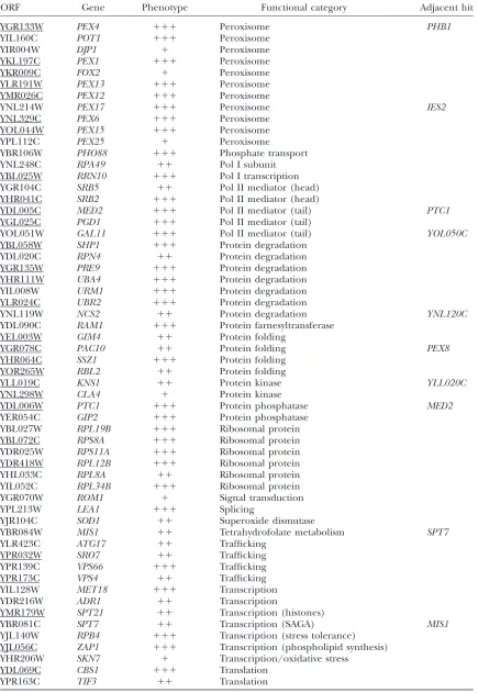
TABLE 1
(Continued)
ORF Gene Phenotype Functional category Adjacent hit
YGR133W PEX4 111 Peroxisome PHB1
YIL160C POT1 111 Peroxisome
YIR004W DJP1 1 Peroxisome
YKL197C PEX1 111 Peroxisome
YKR009C FOX2 1 Peroxisome
YLR191W PEX13 111 Peroxisome
YMR026C PEX12 111 Peroxisome
YNL214W PEX17 111 Peroxisome IES2
YNL329C PEX6 111 Peroxisome
YOL044W PEX15 111 Peroxisome
YPL112C PEX25 1 Peroxisome
YBR106W PHO88 111 Phosphate transport
YNL248C RPA49 11 Pol I subunit
YBL025W RRN10 111 Pol I transcription
YGR104C SRB5 11 Pol II mediator (head)
YHR041C SRB2 111 Pol II mediator (head)
YDL005C MED2 111 Pol II mediator (tail) PTC1
YGL025C PGD1 111 Pol II mediator (tail)
YOL051W GAL11 111 Pol II mediator (tail) YOL050C
YBL058W SHP1 111 Protein degradation
YDL020C RPN4 11 Protein degradation
YGR135W PRE9 111 Protein degradation
YHR111W UBA4 111 Protein degradation
YIL008W URM1 111 Protein degradation
YLR024C UBR2 111 Protein degradation
YNL119W NCS2 11 Protein degradation YNL120C
YDL090C RAM1 111 Protein farnesyltransferase
YEL003W GIM4 11 Protein folding
YGR078C PAC10 11 Protein folding PEX8
YHR064C SSZ1 111 Protein folding
YOR265W RBL2 11 Protein folding
YLL019C KNS1 11 Protein kinase YLL020C
YNL298W CLA4 1 Protein kinase
YDL006W PTC1 111 Protein phosphatase MED2
YER054C GIP2 111 Protein phosphatase
YBL027W RPL19B 111 Ribosomal protein
YBL072C RPS8A 111 Ribosomal protein
YDR025W RPS11A 111 Ribosomal protein
YDR418W RPL12B 111 Ribosomal protein
YHL033C RPL8A 11 Ribosomal protein
YIL052C RPL34B 111 Ribosomal protein
YGR070W ROM1 1 Signal transduction
YPL213W LEA1 111 Splicing
YJR104C SOD1 11 Superoxide dismutase
YBR084W MIS1 11 Tetrahydrofolate metabolism SPT7
YLR423C ATG17 11 Trafficking
YPR032W SRO7 11 Trafficking
YPR139C VPS66 111 Trafficking
YPR173C VPS4 11 Trafficking
YIL128W MET18 111 Transcription
YDR216W ADR1 11 Transcription
YMR179W SPT21 11 Transcription (histones)
YBR081C SPT7 11 Transcription (SAGA) MIS1
YJL140W RPB4 111 Transcription (stress tolerance) YJL056C ZAP1 111 Transcription (phospholipid synthesis) YHR206W SKN7 1 Transcription/oxidative stress
YDL069C CBS1 111 Translation
YPR163C TIF3 11 Translation
G1cells can indeed be observed in stationary cultures of both strains (Figure 3, I and J) or when they are arrested witha-factor (Figure 3, K and L). Since thepex6Dstrain was not starved while oleate was exerting a toxic effect, there is no indication that starvation contributes to the toxic effect of oleate on the pex6D strain. It seems reasonable to extend this conclusion to the other strains that exhibit oleate sensitivity in STY medium but not in rich medium.
Peroxisome structure and function in oleate-sensitive strains: The original goal of the screen was the iden-tification of proteins that, like Pex3 and Pex19, are responsible for the maintenance of the peroxisome. Seventy of the deletions identified by the screen (under-lined in Table 1) were therefore tested for their ability to cause the loss of peroxisomes. Versions of these 70 strains were constructed that express Pex11-GFP in place of Pex11 (this fusion does not influence oleate sensitivity; data not shown). Of these 70 strains, 68 showed punctate GFP fluorescence; the pex3D and
pex19D strains were the only two strains that failed to show punctate GFP fluorescence (data not shown).
Thus, the oleate screen identified no additional pro-teins that are required for maintaining the peroxisome. While many genes identified by the screen encode peroxisomal components, others have no known con-nection to the organelle. The relationship of several of these gene products to the peroxisome was addressed by determining whether deletion influenced oleate-induced enhancement of Pex11-GFP expression. Nu-merous previous studies have shown oleate to cause a substantial induction in the size and/or number of Sac-charomyces peroxisomes (Veenhuiset al.2003).
Oleate-induced expression ofPEX11and the otherPEXgenes depends on Oaf1 and Pip2, transcription factors that bind as a heterodimer to the oleate-responsive element (Luoet al. 1996; Rottensteineret al.1996; Karpichev
and Small 1998). Adr1, the transcription factor first
identified as the inducer of yeast alcohol dehydrogenase (Ciriacy 1975), is also important for PEX gene
in-duction (Simonet al.1991; Gurvitzet al.2001).
To determine the change in Pex11-GFP level in a strain grown under various conditions, total protein was analyzed on GFP-probed Western blots. Figure 4 shows TABLE 1
(Continued)
ORF Gene Phenotype Functional category Adjacent hit
YJL212C OPT1 111 Transporter YJL211C
YAL009W SPO7 111 Unclear MDM10
YAR014C BUD14 11 Unclear
YCR047C BUD23 111 Unclear
YCR063W BUD31 1 Unclear
YFR035C 11 Unclear
YGL127C SOH1 111 Unclear
YGL173C KEM1 111 Unclear
YGR132C PHB1 111 Unclear PEX4
YHR100C 111 Unclear
YJL184W GON7 111 Unclear
YJR118C ILM1 111 Unclear
YKR028W SAP190 111 Unclear
YLR021W 11 Unclear
YLR114C AVL9 1 Unclear
YMR014W BUD22 1 Unclear
YMR032W HOF1 1 Unclear
YMR100W MUB1 111 Unclear
YNR018W 1 Unclear
YOL072W THP1 111 Unclear
A rating of the ratio of growth on STY1glycerol relative to growth on STY1oleate of strains deleted in the indicated ORFs is indicated by ‘‘1’’ (smallest oleate effect) to ‘‘111’’ (largest oleate effect). ‘‘Adjacent hit’’ refers to the appearance in this table of a gene that is adjacent in chromosomal location to any other gene listed in the table. A line under the ORF indicates that the effect of its deletion on the localization of Pex11 was examined (see text).
the results from 12 such strains, grown first in glucose-containing liquid medium, after a subsequent shift to glycerol, and after shifting from glycerol to oleate. The wild-type strain shows a substantial induction in Pex11-GFP expression upon shift to glycerol; a subsequent shift to oleate shows an additional substantial induction. As expected, deletion of eitherOAF1orPIP2permits the initial glycerol induction but blocks the oleate
induc-tion, and deletion of ADR1 blocks the initial glycerol induction but permits the subsequent oleate induction. Deletion ofPEX3,PEX6, orPEX15, as anticipated, allows little if any Pex11-GFP to accumulate even after oleate treatment. Spf1, identified in the oleate screen, is re-ported to be involved in ER function, calcium ho-meostasis (Croninet al.2002), and the orientation of
protein insertion into membranes (Tipperand Harley
2002). It was also shown by mass spectrometry to be associated with the peroxisome (Marelliet al. 2004).
Figure 4 shows that neither glycerol nor oleate cause Pex11-GFP levels to increase in a spf1D background. Spf1 is thus grouped into the ‘‘peroxisome’’ functional category of Table 1.
In contrast, other deletions show glycerol and oleate induction of Pex11-GFP that is indistinguishable from wild type. Figure 4 shows this to be the case forsap190D
and ilm1D, genes that encode proteins of unknown function.akr1D,dfg5D,pox1D,ptc1D, andsac1Dstrains are also unaffected in Pex11-GFP induction (data not shown). These results imply that, although these eight proteins participate in preventing oleate inhibition of growth, they are unlikely to have a role in the mainte-nance or induction of the peroxisome. Indeed, aside from Pox1, which is a peroxisomal protein but which has no known role in organelle maintenance, these pro-teins have no known connection to the peroxisome. An intermediate degree of induction is seen in Figure 4 for ubr2D, implicating Ubr2 perhaps tangentially in peroxisome function. In summary, these Pex11-GFP induction data confirm, as shown in Table 1, that both peroxisomal and nonperoxisomal cellular processes are required to protectS. cerevisiaefrom the toxic effects of oleate.
The strains identified as being most inhibited by oleate also showed inhibition when rich, glucose-con-taining medium (YPD) instead of STY medium was used (Figure 5). Gpt2 and Fen1 have roles in lipid metabo-lism but are unlikely to be related to the peroxisome. Gpt2 is one of two acyl transferases that produce lyso-phosphatidic acid, a phospholipid precursor (Zheng
and Zou 2001; Zaremberg and McMaster 2002;
Sorgerand Daum2003). Its deletion causes substantial
growth inhibition by oleate. In contrast, a strain lacking Sct1, the other glycerol-3-phosphate acyl transferase, fails to show oleate sensitivity (data not shown). Fen1 is
one of three yeast fatty acyl elongases that lengthen the C14, C16, and C18 fatty acyl CoA esters that are pro-duced by the fatty acyl synthetase complex (Oh et al.
1997). Figure 5 shows thefen1Dstrain to be among the most strongly inhibited by oleate, yet strains deleted in genes that encode either of the other elongases, Elo1 (Tokeand Martin 1996) and Sur4 (Oh et al. 1997),
were rechecked and showed no oleate phenotype (data not shown).
Proteins that are implicated in cell-wall function are highly represented among the oleate-inhibited deletion strains. Data for two of these,pde2Dandsac1D, are also presented in Figure 5. Deletion ofPDE2, which encodes the yeast high-affinity cAMP phosphodiesterase, has major effects on cell-wall integrity ( Joneset al. 2003).
Sac1, one of several yeast phosphoinositide
phospha-tases, influences the cell wall by controlling chitin syn-thase trafficking (Schorret al. 2001). As indicated by
the following examples, there is a substantial concur-rence among proteins found from previous studies to influence the cell wall and those reported here that prevent oleate inhibition: mutants in PEX6/PAS8and
FOX2have altered sensitivity to calcofluor white, a gauge of cell-wall strength (Lussieret al. 1997);pex12Dcauses
resistance to killer toxin, another measure of cell-wall integrity (Pageet al. 2003); andFEN1, the elongase
dis-cussed above, was first described asGNS1, mutations in which cause severe defects in synthesis of 1,3-b-glucan, a cell-wall component (el-Sherbeiniand Clemas1995).
The connection between oleate sensitivity and the cell wall is discussed in more detail in thediscussion. As is
also shown in Figure 5, the protein required for resis-tance to the lowest level of oleate (0.01% in YPD) is Ilm1, a 203-residue protein of unknown function predicted to contain four trans-membrane domains. The sensitivity of a number of deletion strains to oleate even in the presence of 2% glucose further supports our conclusion that the reason for slow growth of mutants in the pres-ence of oleate is growth inhibition rather than the in-ability to consume oleate as a carbon and energy source.
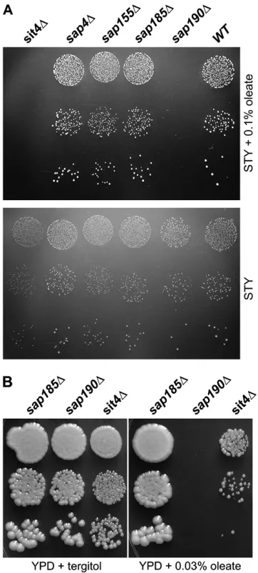
Thesap190Dandilm1Dstrains are highly sensitive to oleate yet insensitive to palmitoleate: The sap190D
strain was also particularly sensitive to oleate; Figure 6B shows it to be inhibited by 0.03% oleate on YPD. Sap190 was first identified as one of two large proteins that co-immunoprecipitate in a cell-cycle-specific man-ner with Sit4 (Suttonet al. 1991), one of five yeast
PP2A-like protein phosphatases (Duveland Broach2004).
Sap190 and Sap155, the other Sit4-associated protein that was initially identified, were subsequently shown by a combination of homology, immunoprecipitation, and genetic criteria, to be two of three (possibly four) Sap proteins of Saccharomyces. Additional genetic evidence from that study suggested that Sap4, Sap155, Sap185, and Sap190 all have distinct but uncharacterized func-tions related in some way to Sit4 (Luke et al. 1996).
Strains lackingSAP4,SAP155, orSAP185were not iden-tified in the oleate screen and their reexamination confirmed them all to be oleate insensitive (Figure 6A). Figure5.—Oleate inhibition of growth on YPD medium.
The indicatedMATastrains from the deletion collection were suspended in water at 10-fold dilutions, spotted onto YPD1 1% tergitol plates containing the indicated amounts of oleate, and grown at 30°for 40 hr. Thelys5Dstrain served as a wild-type control.
As expected on the basis of the relationship of Sap190 and Sit4, however, deletion of SIT4did indeed cause sensitivity to oleate (Figures 5 and 6). Our screen failed to identify thesit4Dstrain probably because of its slow growth even in the absence of oleate.
In addition to oleate, the other main unsaturated fatty acid found in S. cerevisiae is palmitoleate (Daum et al.
1999), which is two carbons shorter but otherwise iden-tical to oleate. A comparison of the ability of the two fatty acids to inhibit growth of several strains is shown in Figure 7. Palmitoleate, instead of severely inhibiting growth, promotes the same slight stimulation of growth of thesap190Dstrain as seen when the wild-type strain is supplemented with oleate. The ilm1D strain is also selectively inhibited by oleate but not by palmitoleate. Growth of thepex6Dstrain, in contrast, is inhibited by palmitoleate almost as much as by oleate. The specificity
with which fatty acids inhibit growth of the sap190D
andilm1Dstrains reinforces our assumption that oleate inhibition is a physiologically relevant phenomenon rather than a more generalized detergent-like effect.
Mutations augment the ability of oleate to permea-bilize the plasma membrane:What can account for the toxicity of oleate to strains in which peroxisomal and other functions are impaired? We reasoned that addi-tion of oleate to these mutant strains might cause mem-brane changes that wild-type strains are capable of resisting. The double bond in oleate’s acyl chain intro-duces a kink that disrupts acyl chain packing in the lipid bilayer. An increase in oleate incorporation into phos-pholipids therefore can increase membrane fluidity (Hazel1995). Too great an increase in fluidity
compro-mises membrane integrity which, at least for the plasma membrane, has lethal consequences. One possible ef-fect of removal of a protein that permits yeast to grow normally in the presence of oleate could be the impair-ment of the cell’s ability to properly control the fatty Figure7.—Oleate and palmitoleate differ in ability to in-hibit growth. The indicatedMATastrains from the deletion collection were suspended in water at 10-fold dilutions, spot-ted on the indicaspot-ted plates, and grown for 7 days at 30°. Figure6.—Oleate inhibition ofsap190Dand related strains.
acid content of its membrane phospholipid. Addition of unsaturated fatty acid could therefore lead to an in-crease in porosity of the plasma membrane.
The effect of oleate on the integrity of the plasma membrane was tested by examining the ability of Sytox green, a vital dye, to enter cells. The fluorescence en-hancement of Sytox upon interaction with nucleic acid has previously been used to probe membrane perme-ability of Aspergillus (Theiset al. 2003), Neurospora,
and Saccharomyces (Thevissenet al. 1999). Figure 8A
shows Sytox-stainedPex1
andpex6Dstrains after 0, 2, and 4 hr of exposure to oleate. Prior to oleate treatment, Sytox entered a similar small fraction (,2%) of cells of the two strains. With increased time of exposure to oleate, a significantly greater fraction ofpex6Dcells than ofPex1cells were capable of taking up the dye. Quan-titation of these results in Figure 8B shows that by 4 hr of oleate treatment, three- to fourfold more pex6D cells thanPex1
cells were Sytox positive.
Exposure to oleate allows entry of Sytox into cells either because oleate directly influences processes at the plasma membrane to allow dye to enter the cell or because oleate kills the cells by some other means after which the dead cells become permeable to Sytox. To distinguish between these two possibilities, yeast were killed by poisoning with clonNAT, a drug that is known to kill by mistranslation (Hauptet al. 1978). Although
,5% of eitherPex1
orpex6Dcells were viable after 2 hr of clonNAT treatment, there was no increase in Sytox-positive cells of either thePex1(Figure 8A) or the
pex6D
(data not shown) strain even after 4 hr of clonNAT treatment. Thus oleate-induced but not clonNAT-induced cell death allowed entry of Sytox into cells within the time course of this experiment. In addition to
PEX6, we also found that deletion ofSAP190,ILM1, or
PEX19led to increases in the fraction of Sytox-positive cells comparable to those seen for thepex6Dstrain in Figure 8 (data not shown).
DISCUSSION
We have identified genes whose products enable optimal growth of Saccharomyces in the presence of oleate. This strategy was used by Erdmannet al.(1989)
because of the peroxisomal location of b-oxidation: impairment of peroxisome function could block fatty acid catabolism, thereby preventing oleate from serving as a carbon and energy source. Our results demonstrate, however, that the growth impairment of the yeast dele-tion strains isolated in our screen is mainly caused by the inhibition of their growth by oleate not by their inability to utilize oleate. Erdmann et al.(1989) reported that
wild-type yeast are completely incapable of growth in the absence of oleate or any other added carbon source. It was therefore not possible, presumably because of dif-ferences in either strain background or media compo-nents in the two studies, for the earlier study to observe
oleate inhibition. In addition to inhibition of the growth of mutants, our data also show that oleate slightly stim-ulates growth of wild-type yeast. Oleate thus appears to be a carbon/energy source preferable to the unknown component(s) of STY medium and/or agar that permits growth in the absence of any added carbon source.
This study had its origins in our search for additional proteins that are required for the maintenance of the peroxisome. At the project’s inception, there was evi-dence that the peroxisome might be an autonomous organelle,i.e., that a new peroxisome could form only from a preexisting peroxisome that would serve as a structural template. The mitochondrion, for exam-ple, is undoubtedly such an autonomous organelle (Lockshon2002), yet, unlike the peroxisome, it
per-forms at least one essential function (Kispalet al.2005)
and must therefore continue to be maintained. Pex3 and Pex19 were known to be required for the mainte-nance of the peroxisome (Hohfeldet al.1991; Gotte
et al.1998), yet the ability of peroxisomes to be reformed inpex3Dandpex19Dstrains after being resupplied with
PEX3andPEX19, respectively, argues against the struc-tural templating of this organelle. However, structures termed ‘‘protoperoxisomes’’ were reported in pex19D
(Snyderet al. 1999) andpex3D(Hazraet al. 2002) mutants
of Pichia pastorisand inpex3 strains ofHansenula poly-morpha(Faberet al. 2002) andS. cerevisiae(K. Huang
and P. B. Lazarow, unpublished data cited in Lazarow
2003). Protoperoxisomes, in principle, would be capa-ble of preserving the putative peroxisomal structural information, which could then serve as a template for the reestablishment of peroxisomes upon genetic com-plementation. Our oleate screen was therefore initiated to identify proteins that are necessary for the main-tenance of both the peroxisome and the protoper-oxisome. None of the 68 deletions examined had any effect on peroxisome integrity. In light of more recent evidence, it is clear why this search was unsuccessful: Hoepfneret al.(2005) have clearly shown that
peroxi-somes of S. cerevisiae, rather than being autonomous, can be derived from the endoplasmic reticulum.
While much is known about the activities of the pro-teins that are responsible for peroxisome function and about the biochemical pathways that occur there, the biological role of this organelle is still poorly under-stood. There is extraordinary variation in metabolic processes carried out in the peroxisomes of the differ-ent eukaryotic species, yet all peroxisomes carry outb -oxidation (Moyersoenet al. 2004). The mitochondrial
b-oxidation pathway found in animals, on the other hand, has thus far been found neither in plants nor in fungi (Kunau et al. 1988). Perhaps the catabolism of
Figure8.—Oleate increases the ability of Sytox to enter a peroxisome-deficient strain. (A)Pex1 (BY4741) and pex6D (YD1115) cultures were grown in STY12% glucose to midlog and then either pelleted and resuspended in STY1oleate (0.1%) or supplemented with clonNAT (0.1 mg/ ml). Samples taken at 0, 2, and 4 hr were stained with Sytox and fluorescence microscopic images were acquired. (B) Quantitation of the percent-age of Sytox-positivePex1
This hypothesis is consistent with the requirement of a functional peroxisome to prevent growth inhibition by oleate. We have also presented data that further suggest that oleate exerts its toxic effect on oleate-sensitive mu-tants by permeabilizing the plasma membrane. This is the anticipated effect of a decrease in the packing den-sity of the membrane bilayer, which would result from the over-incorporation of this unsaturated fatty acid into phospholipids. Although confirmation of an effect of supplemental oleate on phospholipid fatty acid content inpexstrains awaits biochemical analysis of membrane fractions from such cultures, previous studies have in-deed demonstrated the incorporation of fed fatty acids into Saccharomyces phospholipids (Bossieand Martin
1989; Stukey et al. 1989). While it is economical to
propose that oleate exerts its toxicity on mutants in all the functional categories listed in Table 1 by its effect on the plasma membrane, this is not necessarily the case since the mitochondrial entries in Table 1, for example, imply a toxic effect of oleate on mitochondrial membrane. The nuclear-localized proteins listed in Table 1 perhaps cause oleate sensitivity indirectly by adversely influencing the ability ofPEXgenes to be induced by oleate.
Two large-scale screens have identified proteins re-quired for proper cell-wall function in yeast (Lussier
et al.1997; Pageet al. 2003). Twenty of these proteins,
among them those listed in Table 1 in the ‘‘cell wall’’ functional category, were found in our screen:Bud14,
Bud22,Dfg5,Fox2,Gal11,Ilm1,Med2,Nbp2,Pex6,Pex12,
Ptc1,Rps11A,Sac1,Sap190,Sod1,Srb2,Srb5,Ste11,Thp1, andTif3. This overlap implies that, for the ‘‘cell wall’’ func-tional category, oleate might exert its toxic effects directly on the plasma membrane. It is possible that this subset of mutants is deficient primarily in the ability to adequately control plasma membrane composition, a defect that in turn could influence the wall. Indeed, the cell wall is emerging as a dynamic structure that interacts in complex ways with the plasma membrane (Fironet al. 2004).
The sap190D and ilm1D mutants are of interest be-cause they are particularly sensitive to oleate (C18:1) yet unaffected by palmitoleate (C16:1). We hypothesize that Sap190 and Ilm1 function in the control of the ratio of the palmitoleate-to-oleate content of phospho-lipids. The ratio of these two phospholipid components, the predominant unsaturated fatty acids inS. cerevisiae
(Daumet al. 1999), decreases1.7-fold when the growth
temperature of S. cerevisiae increases from 10° to 35°
(Suutariet al. 1990), implying an effect of the C16/C18
ratio on membrane fluidity. Moreover, acyl chain length is known to influence bacterial membrane fluidity (Cronan 1996). Although the mechanisms by which
several prokaryotic species control membrane fluidity is understood in some detail (Mansillaet al. 2004), the
mechanisms by which eukaryotes control this process are completely unknown.
No Sit4-associated proteins other than Sap190 are needed to prevent oleate sensitivity. To date, the Saps of
Saccharomyces have been shown to be involved in sensitivity toKluyveromyces lactiszymocin (Jablonowski
et al.2001, 2004), regulating the level of the Npr1 kinase (Jacinto et al. 2001), influencing the toxic effects of
rapamycin (Rohdeet al.2004), and modulating K1
ef-flux (Manlandro et al. 2005). These previously
re-ported functions, and perhaps oleate sensitivity as well, all concern events at the plasma membrane. None of these previous studies, however, have convincingly dem-onstrated that Sap190 plays a unique role. The sensitiv-ity of the sit4D strain to oleate also suggests that a Sap190/Sit4 complex is the entity that prevents oleate inhibition. The ability of 0.03% oleate in YPD to inhibit thesap190Dstrain but not thesit4Dstrain (Figure 6B), however, suggests that Sap190 may be capable of func-tioning in concert with (an) additional PP2A-type phos-phatase(s) (Duvel and Broach 2004) to form an
alternative functional unit with Sap190.
Deletion of Ilm1, a 203-residue membrane protein of unknown function that resides in the ER, was first re-ported toincrease theloss ofmitochondrial (ilm) DNA (Entian et al. 1999) although we are unable to
re-produce this phenotype (data not shown). Our screen showedilm1Dto cause oleate sensitivity; further exam-ination showed that this deletion, likesap190D, has no effect on sensitivity to palmitoleate. This suggests that Ilm1 may also participate in the control of the C16/C18 ratio. A yeast two-hybrid screen carried out by The Yeast Resource Center (http://www.yeastrc.org/pdr/pages/ front.jsp) has identified Mga2 as one of seven yeast pro-teins that strongly interact with Ilm1. Mga2p is a key transcription factor that controls expression of Ole1, the sole fatty acyl desaturase inS. cerevisiaeresponsible for conversion of the saturated fatty acids stearate (C18) and palmitate (C16) to oleate and palmitoleate, respec-tively (Zhanget al.1999; Chellappaet al. 2001). The
ratio of saturated to unsaturated fatty acids (UFA/SFA) is known from studies ofBacillus subtilis, for example, to be a key determinant of membrane fluidity (Grauand deMendoza 1993; Weber et al.2001). Suutari et al.
(1990), however, showed that the UFA/SFA ratio of Saccharomyces is unaffected by growth temperature. On the other hand, the strong induction of OLE1 ex-pression upon shifting yeast from 30°to 10°(Nakagawa
et al. 2002) strongly implicates the UFA/SFA ratio in yeast membrane fluidity control. Although yeast are poikilotherms, as are all microbes, an understanding of the contributions of C16/C18 and UFA/SFA ratios to yeast membrane fluidity is likely to be relevant to ho-meotherms as well.
phospholipid fatty acid content by oleate. This model is consistent with the role of the peroxisome in fatty acid catabolism, one strategy by which yeast can remove excess oleate. However, the complete absence of b -oxidation in the pox1D and fox2D strains caused only slight oleate sensitivity; deletion of POT1, which enc-odes the third enzyme essential forb-oxidation, caused a more severe oleate phenotype. Thus, prevention of oleate toxicity by the peroxisome is apparently more complex than merely the organelle’s role in fatty acid catabolism. Yeast can also sequester added fatty acid in the lipid particle, a membrane-bound organelle (Athenstaedt et al. 1999). Indeed, yeast that lack a
lipid particle (Sandageret al.2002) are also highly
sen-sitive to oleate (D. Lockshon, unpublished results). An
intimate association between the lipid particle and the peroxisome of Saccharomyces has recently been re-ported (Binnset al.2006), suggesting that perhaps the
peroxisome may also participate in lipid storage. Other possible models, which implicate the peroxisome in membrane resealing (Jedd and Chua 2000; McNeil
and Steinhardt 2003) or in multi-drug efflux pump
function (Ernstet al.2005), have not been ruled out.
A cellular role for the peroxisome has yet to be determined. This work suggests that it could have an important role in governing yeast fatty acid levels and perhaps in governing the composition of membrane phospholipids. The relative simplicity of the metabolic processes that occur in the yeast peroxisome does not readily suggest a cellular function for the organelle. The human peroxisome, on the other hand, houses multiple pathways, perhaps all of which involve membrane com-ponent metabolism (Wandersand Tager1998), thus
implying a close relationship between the organelle and cellular membrane composition.
Stan Fields and members of his lab graciously allowed us to use their Biomek 2000. Thanks to Alex Merz and Margo Murphy for a critical reading of the manuscript. The work was supported by a Congressio-nally Directed Medical Research Program Grant (DAMD17-03-0497) to B.K.K.
LITERATURE CITED
Athenstaedt, K., D. Zweytick, A. Jandrositz, S. D. Kohlweinand
G. Daum, 1999 Identification and characterization of major
lipid particle proteins of the yeast Saccharomyces cerevisiae. J. Bacteriol.181:6441–6448.
Bartlett, K., and S. Eaton, 2004 Mitochondrial beta-oxidation.
Eur. J. Biochem.271:462–469.
Binns, D., T. Januszewski, Y. Chen, J. Hill, V. S. Markin et al.,
2006 An intimate collaboration between peroxisomes and lipid bodies. J. Cell Biol.173:719–731.
Bjorklund, S., and C. M. Gustafsson, 2005 The yeast Mediator
complex and its regulation. Trends Biochem. Sci.30:240–244. Bossie, M. A., and C. E. Martin, 1989 Nutritional regulation of
yeast delta-9 fatty acid desaturase activity. J. Bacteriol.171:6409– 6413.
Brachmann, C. B., A. Davies, G. J. Cost, E. Caputo, J. Liet al.,
1998 Designer deletion strains derived from Saccharomyces cerevisiae S288C: a useful set of strains and plasmids for PCR-mediated gene disruption and other applications. Yeast 14:
115–132.
Chellappa, R., P. Kandasamy, C. S. Oh, Y. Jiang, M. Vemulaet al.,
2001 The membrane proteins, Spt23p and Mga2p, play distinct roles in the activation of Saccharomyces cerevisiae OLE1 gene expression. Fatty acid-mediated regulation of Mga2p activity is independent of its proteolytic processing into a soluble transcrip-tion activator. J. Biol. Chem.276:43548–43556.
Ciriacy, M., 1975 Genetics of alcohol dehydrogenase in
Saccharo-myces cerevisiae. II. Two loci controlling synthesis of the glucose-repressible ADH II. Mol. Gen. Genet.138:157–164.
Cronan, J. E., and C. O. Rock, 1996 Biosynthesis of membrane
lipids, pp. 612–636 in Escherichia coli and Salmonella: Cellular and Molecular Biology, edited by F. C. Neidhardt. American Society
of Microbiology, Washington, DC.
Cronin, S. R., R. Raoand R. Y. Hampton, 2002 Cod1p/Spf1p is a
P-type ATPase involved in ER function and Ca21homeostasis. J. Cell Biol.157:1017–1028.
Daum, G., G. Tuller, T. Nemec, C. Hrastnik, G. Ballianoet al.,
1999 Systematic analysis of yeast strains with possible defects in lipid metabolism. Yeast15:601–614.
Duvel, K., and J. R. Broach, 2004 The role of phosphatases in TOR
signaling in yeast. Curr. Top. Microbiol. Immunol.279:19–38. Eckert, J. H., and R. Erdmann, 2003 Peroxisome biogenesis. Rev.
Physiol. Biochem. Pharmacol.147:75–121.
el-Sherbeini, M., and J. A. Clemas, 1995 Cloning and
characteriza-tion of GNS1: a Saccharomyces cerevisiae gene involved in syn-thesis of 1,3-beta-glucan in vitro. J. Bacteriol.177:3227–3234. Entian, K. D., T. Schuster, J. H. Hegemann, D. Becher, H.
Feldmannet al., 1999 Functional analysis of 150 deletion
mu-tants in Saccharomyces cerevisiae by a systematic approach. Mol. Gen. Genet.262:683–702.
Erdmann, R., M. Veenhuis, D. Mertensand W. H. Kunau, 1989
Iso-lation of peroxisome-deficient mutants of Saccharomyces cerevi-siae. Proc. Natl. Acad. Sci. USA86:5419–5423.
Erdmann, R., F. F. Wiebel, A. Flessau, J. Rytka, A. Beyer et al.,
1991 PAS1, a yeast gene required for peroxisome biogenesis, encodes a member of a novel family of putative ATPases. Cell
64:499–510.
Ernst, R., R. Klemm, L. Schmittand K. Kuchler, 2005 Yeast
ATP-binding cassette transporters: cellular cleaning pumps. Methods Enzymol.400:460–484.
Faber, K. N., G. J. Haan, R. J. Baerends, A. M. Kram and M.
Veenhuis, 2002 Normal peroxisome development from vesicles
induced by truncated Hansenula polymorpha Pex3p. J. Biol. Chem.277:11026–11033.
Firon, A., G. Lesageand H. Bussey, 2004 Integrative studies put
cell wall synthesis on the yeast functional map. Curr. Opin. Micro-biol.7:617–623.
Foss, E. J., 2001 Tof1p regulates DNA damage responses during S
phase inSaccharomyces cerevisiae. Genetics157:567–577. Gotte, K., W. Girzalsky, M. Linkert, E. Baumgart, S. Kammerer
et al., 1998 Pex19p, a farnesylated protein essential for peroxi-some biogenesis. Mol. Cell. Biol.18:616–628.
Grau, R., and D.deMendoza, 1993 Regulation of the synthesis of
unsaturated fatty acids by growth temperature in Bacillus subtilis. Mol. Microbiol.8:535–542.
Gurvitz, A., J. K. Hiltunen, R. Erdmann, B. Hamilton, A. Hartig
et al., 2001 Saccharomyces cerevisiaeAdr1p governs fatty acid beta-oxidation and peroxisome proliferation by regulatingPOX1and PEX11. J. Biol. Chem.276:31825–31830.
Haupt, I., R. Hubenerand H. Thrum, 1978 Streptothricin F, an
in-hibitor of protein synthesis with miscoding activity. J. Antibiot.
31:1137–1142.
Hazel, J. R., 1995 Thermal adaptation in biological membranes: Is
homeoviscous adaptation the explanation? Annu. Rev. Physiol.
57:19–42.
Hazra, P. P., I. Suriapranata, W. B. Snyder and S. Subramani,
2002 Peroxisome remnants in pex3delta cells and the require-ment of Pex3p for interactions between the peroxisomal docking and translocation subcomplexes. Traffic3:560–574.
Hettema, E. H., W. Girzalsky, M.vanDenBerg, R. Erdmannand
B. Distel, 2000 Saccharomyces cerevisiae Pex3p and Pex19p
are required for proper localization and stability of peroxisomal membrane proteins. EMBO J.19:223–233.
Hiltunen, J. K., A. M. Mursula, H. Rottensteiner, R. K. Wieranga,
b-oxidation in the yeastSaccharomyces cerevisiae. FEMS Microbiol. Rev.27:35–64.
Hoepfner, D., D. Schildknegt, I. Braakman, P. Philippsenand
H. F. Tabak, 2005 Contribution of the endoplasmic reticulum
to peroxisome formation. Cell122:85–95.
Hohfeld, J., M. Veenhuisand W. H. Kunau, 1991 PAS3, a
Saccharo-myces cerevisiae gene encoding a peroxisomal integral membrane protein essential for peroxisome biogenesis. J. Cell Biol.114:1167– 1178.
Huh, W. K., J. V. Falvo, L. C. Gerke, A. S. Carroll, R. W. Howson
et al., 2003 Global analysis of protein localization in budding yeast. Nature425:686–691.
Jablonowski, D., A. R. Butler, L. Fichtner, D. Gardiner, R.
Schaffrathet al., 2001 Sit4p protein phosphatase is required
for sensitivity of Saccharomyces cerevisiae to Kluyveromyces lactis zymocin. Genetics159:1479–1489.
Jablonowski, D., L. Fichtner, M. J. Stark and R. Schaffrath,
2004 The yeast elongator histone acetylase requires Sit4-dependent dephosphorylation for toxin-target capacity. Mol. Biol. Cell15:1459–1469.
Jacinto, E., B. Guo, K. T. Arndt, T. Schmelzleand M. N. Hall,
2001 TIP41 interacts with TAP42 and negatively regulates the TOR signaling pathway. Mol. Cell8:1017–1026.
Jedd, G., and N. H. Chua, 2000 A new self-assembled peroxisomal
vesicle required for efficient resealing of the plasma membrane. Nat. Cell Biol.2:226–231.
Johnston, G. C., J. R. Pringle and L. H. Hartwell, 1977
Co-ordination of growth with cell division in the yeast Saccharomy-ces cerevisiae. Exp. Cell Res.105:79–98.
Jones, D. L., J. Petty, D. C. Hoyle, A. Hayes, E. Ragni et al.,
2003 Transcriptome profiling of a Saccharomyces cerevisiae mutant with a constitutively activated Ras/cAMP pathway. Physi-ol. Genomics16:107–118.
Karpichev, I. V., and G. M. Small, 1998 Global regulatory functions
of Oaf1p and Pip2p (Oaf2p), transcription factors that regulate genes encoding peroxisomal proteins in Saccharomyces cerevi-siae. Mol. Cell. Biol.18:6560–6570.
Kispal, G., K. Sipos, H. Lange, Z. Fekete, T. Bedekovics et al.,
2005 Biogenesis of cytosolic ribosomes requires the essential iron-sulphur protein Rli1p and mitochondria. EMBO J.24:589– 598.
Kobor, M. S., S. Venkatasubrahmanyam, M. D. Meneghini, J. W.
Gin, J. L. Jenningset al., 2004 A protein complex containing
the conserved Swi2/Snf2-related ATPase Swr1p deposits histone variant H2A.Z into euchromatin. PLoS Biol.2:E131.
Kornitzer, D., 2002 Monitoring protein degradation. Methods
Enzymol.351:639–647.
Kunau, W. H., S. Buhne, M.de laGarza, C. Kionka, M. Mateblowski
et al., 1988 Comparative enzymology of beta-oxidation. Bio-chem. Soc. Trans.16:418–420.
Lazarow, P. B., 2003 Peroxisome biogenesis: advances and
conun-drums. Curr. Opin. Cell Biol.15:489–497.
Lockshon, D., 2002 A heritable structural alteration of the yeast
mitochondrion. Genetics161:1425–1435.
Luke, M. M., F. DellaSeta, C. J. DiComo, H. Sugimoto, R. Kobayashi
et al., 1996 The SAPs, a new family of proteins, associate and function positively with the SIT4 phosphatase. Mol. Cell. Biol.
16:2744–2755.
Luo, Y., I. V. Karpichev, R. A. Kohanski and G. M. Small,
1996 Purification, identification, and properties of a Saccharo-myces cerevisiae oleate-activated upstream activating sequence-binding protein that is involved in the activation of POX1. J. Biol. Chem.271:12068–12075.
Lussier, M., A. M. White, J. Sheraton, T.diPaolo, J. Treadwell
et al., 1997 Large scale identification of genes involved in cell surface biosynthesis and architecture inSaccharomyces cerevisiae. Genetics147:435–450.
Manlandro, C. M., D. H. Haydonand A. G. Rosenwald, 2005
Abil-ity of Sit4p to promote K1efflux via Nha1p is modulated by Sap155p and Sap185p. Eukaryot. Cell4:1041–1049.
Mansilla, M. C., L. E. Cybulski, D. Albanesiand D.deMendoza,
2004 Control of membrane lipid fluidity by molecular thermo-sensors. J. Bacteriol.2004:6681–6688.
Marelli, M., J. J. Smith, S. Jung, E. Yi, A. I. Nesvizhskiiet al.,
2004 Quantitative mass spectrometry reveals a role for the
GTPase Rho1p in actin organization on the peroxisome mem-brane. J. Cell Biol.167:1099–1112.
Marzioch, M., R. Erdmann, M. Veenhuis and W. H. Kunau,
1994 PAS7 encodes a novel yeast member of the WD-40 protein family essential for import of 3-oxoacyl-CoA thiolase, a PTS2-containing protein, into peroxisomes. EMBO J.13:4908–4918. McNeil, P. L., and R. A. Steinhardt, 2003 Plasma membrane
disruption: repair, prevention, adaptation. Annu. Rev. Cell Dev. Biol.19:697–731.
Moyersoen, J., J. Choe, E. Fan, W. G. Hol and P. A. Michaels,
2004 Biogenesis of peroxisomes and glycosomes: trypanosoma-tid glycosome assembly is a promising new drug target. FEMS Microbiol. Rev.28:603–643.
Nakagawa, Y., N. Sakumoto, Y. Kaneko and S. Harashima,
2002 Mga2p is a putative sensor for low temperature and oxy-gen to induce OLE1 transcription in Saccharomyces cerevisiae. Biochem. Biophys. Res. Commun.291:707–713.
Oh, C. S., D. A. Toke, S. Mandalaand C. E. Martin, 1997 ELO2
and ELO3, homologues of the Saccharomyces cerevisiae ELO1 gene, function in fatty acid elongation and are required for sphingolipid formation. J. Biol. Chem.272:17376–17384. Page, N., M. Gerard-Vincent, P. Menard, M. Beaulieu, M. Azuma
et al., 2003 ASaccharomyces cerevisiaegenome-wide mutant screen for altered sensitivity to K1 killer toxin. Genetics163:875–894. Purdue, P. E., and P. B. Lazarow, 2001 Peroxisome biogenesis.
Annu. Rev. Cell Dev. Biol.17:701–752.
Rohde, J. R., S. Campbell, S. A. Zurita-Martinez, N. S. Cutler, M.
Asheet al., 2004 TOR controls transcriptional and translational
programs via Sap-Sit4 protein phosphatase signaling effectors. Mol. Cell. Biol.24:8332–8341.
Rottensteiner, H., A. J. Kal, M. Filipits, M. Binder, B. Hamilton
et al., 1996 Pip2p: a transcriptional regulator of peroxisome proliferation in the yeast Saccharomyces cerevisiae. EMBO J.
15:2924–2934.
Sandager, L., M. H. Gustavsson, U. Stahl, A. Dahlqvist, E.
Wiberget al., 2002 Storage lipid synthesis is non-essential in
yeast. J. Biol. Chem.277:6478–6482.
Schorr, M., A. Then, S. Tahirovic, N. Hug and P. Mayinger,
2001 The phosphoinositide phosphatase Sac1p controls traf-ficking of the yeast Chs3p chitin synthase. Curr. Biol.11:1421– 1426.
Sherman, F., 1991 Getting started with yeast. Methods Enzymol.
194:3–21.
Simon, M., G. Adam, W. Rapatz, W. Spevakand H. Ruis, 1991 The
Saccharomyces cerevisiae ADR1 gene is a positive regulator of transcription of genes encoding peroxisomal proteins. Mol. Cell. Biol.11:699–704.
Snyder, W. B., K. N. Faber, T. J. Wenzel, A. Koller, G. H. Luers
et al., 1999 Pex19p interacts with Pex3p and Pex10p and is es-sential for peroxisome biogenesis in Pichia pastoris. Mol. Biol. Cell10:1745–1761.
Sorger, D., and G. Daum, 2003 Triacylglycerol biosynthesis in yeast.
Appl. Microbiol. Biotechnol.61:289–299.
Stukey, J. E., V. M. McDonoughand C. E. Martin, 1989 Isolation
and characterization of OLE1, a gene affecting fatty acid desatu-ration from Saccharomyces cerevisiae. J. Biol. Chem.264:16537– 16544.
Sutton, A., D. Immanueland K. T. Arndt, 1991 The SIT4 protein
phosphatase functions in late G1 for progression into S phase. Mol. Cell. Biol.11:2133–2148.
Suutari, M., K. Liukkonenand S. Laakso, 1990 Temperature
ad-aptation in yeasts: the role of fatty acids. J. Gen. Microbiol.136:
1469–1474.
Theis, T., M. Wedde, V. Meyerand U. Stahl, 2003 The antifungal
protein from Aspergillus giganteus causes membrane permeabi-lization. Antimicrob. Agents Chemother.47:588–593.
Thevissen, K., F. R. Terrasand W. F. Broekaert, 1999
Perme-abilization of fungal membranes by plant defensins inhibits fungal growth. Appl. Environ. Microbiol.65:5451–5458. Tipper, D. J., and C. A. Harley, 2002 Yeast genes controlling
responses to topogenic signals in a model transmembrane pro-tein. Mol. Biol. Cell13:1158–1174.
Toke, D. A., and C. E. Martin, 1996 Isolation and characterization
van denBosch, H., R. B. Schutgens, R. J. Wandersand J. M. Tager,
1992 Biochemistry of peroxisomes. Annu. Rev. Biochem.61:
157–197.
Veenhuis, M., M. Mateblowski, W. H. Kunau and W. Harder,
1987 Proliferation of microbodies in Saccharomyces cerevisiae. Yeast3:77–84.
Veenhuis, M., J. A. Kieland I. J. VanDerKlei, 2003 Peroxisome
assembly in yeast. Microsc. Res. Tech.61:139–150.
Wanders, R. J., and J. M. Tager, 1998 Lipid metabolism in
perox-isomes in relation to human disease. Mol. Aspects Med.19:69– 154.
Weber, M. H., W. Klein, L. Muller, U. M. Niess and M. A.
Marahiel, 2001 Role of the Bacillus subtilis fatty acid
desatur-ase in membrane adaptation during cold shock. Mol. Microbiol.
39:1321–1329.
Wiebel, F. F., and W. H. Kunau, 1992 The Pas2 protein essential
for peroxisome biogenesis is related to ubiquitin-conjugating enzymes. Nature359:73–76.
Winzeler, E. A., D. D. Shoemaker, A. Astromoff, H. Liang, K. Anderson
et al., 1999 Functional characterization of the S. cerevisiae ge-nome by gene deletion and parallel analysis. Science285:901–906. Zaremberg, V., and C. R. McMaster, 2002 Differential partitioning
of lipids metabolized by separate yeast glycerol-3-phosphate acyl-transferases reveals that phospholipase D generation of phospha-tidic acid mediates sensitivity to choline-containing lysolipids and drugs. J. Biol. Chem.277:39035–39044.
Zhang, S., Y. Skalskyand D. J. Garfinkel, 1999 MGA2 or SPT23 is
required for transcription of theD9 fatty acid desaturase gene, OLE1, and nuclear membrane integrity inSaccharomyces cerevisiae. Genetics151:473–483.
Zheng, Z., and J. Zou, 2001 The initial step of the glycerolipid
path-way: identification of glycerol 3-phosphate/dihydroxyacetone phosphate dual substrate acyltransferases in Saccharomyces cer-evisiae. J. Biol. Chem.276:41710–41716.