ABSTRACT
MOORE, MATTHEW DOUGLAS. Novel Alternative Methods for the Detection and Study of Human Noroviruses. (Under the direction of Dr. Lee-Ann Jaykus.)
Human norovirus is the leading cause of viral gastroenteritis and foodborne illness worldwide. Because it is highly transmissible, rapid detection of human norovirus is critical for control. In the absence of an in vitro cultivation system, genome amplification is
commonly used for detection (specifically RT-qPCR), but this method does not discriminate infectious from non-infectious particles. The purpose of this work was to develop and evaluate novel methods to improve detection and study of human norovirus.
While quite accurate, RT-qPCR it is not amenable to true real-time (near
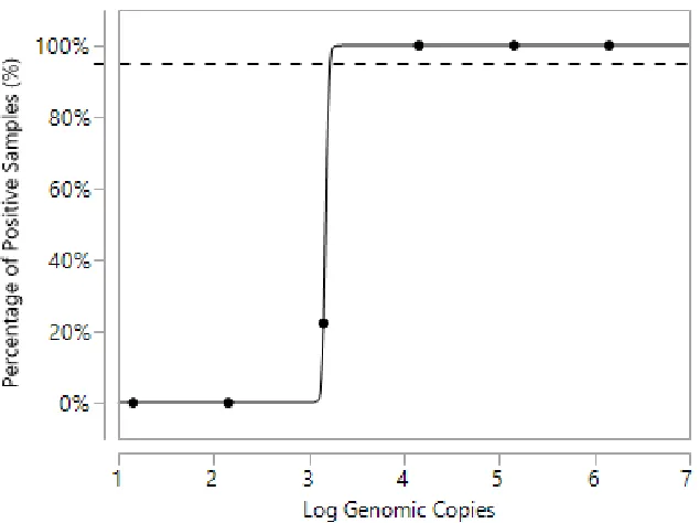
instantaneous) detection in the field. Recombinase polymerase amplification (RPA) is a portable, rapid isothermal genome amplification method with time-to-result of only 20 minutes. An RT-RPA method was developed that was able to amplify and detect human norovirus from patient outbreak stool that was either subject to RNA purification or directly boiled to release viral RNA. The assay demonstrated specificity, reacting only with the two most recent pandemic norovirus strains (GII.4), and had a limit of detection of 3.2±0.01 log10
genomic copies. It is promising for application in clinical point-of-care or field settings in which rapid detection is required.
to an established (longer) assay when evaluating the efficacy of a physical (heat) and chemical (copper) virus inactivation method. The streamlined approach provides a rapid, high throughput means of obtaining mechanistic insight for further norovirus study.
HBGAs are expensive and difficult to synthesize, purify, and modify, as are antibodies. Nucleic acid aptamers are emerging ligands that are easily synthesized and modified, inexpensive, and stable. Aptamers targeting a region of the outermost capsid domain of a pandemic norovirus strain were produced using SELEX (Systematic Evolution of Ligands by EXponential enrichment). Two candidates, aptamers M1 and M6-2, showed broad reactivity with a panel of VLPs corresponding to 14 different norovirus strains; they were also capable of binding and concentrating infectious virus from stool. These aptamers are among the most broadly reactive norovirus ligands reported to date.
Utilization of aptamers for discrimination of infectious from non-infectious norovirus particles has not been investigated. Thus, aptamer M6-2 binding behavior was compared to HBGA and antibody NS14 as applied to SYV exposed to various heat treatments. Aptamer M6-2 behaved similarly to HBGAs, as both displayed a high degree of target conformation-dependent binding (98.0 ± 1.3% and 99.5 ± 1.2% signal, respectively), while NS14 had significantly less (73.6 ± 3.9%). Dynamic light scattering (DLS) and transmission electron microscopy (TEM) confirmed binding results. Ligand docking simulations placed M6-2 in similar capsid binding regions as HBGA, with NS14 in a different region. This is the first instance in which aptamers have been utilized for estimation of infectivity of a virus.
and GII.2 Snow Mountain (SMV)], were used for the simulations, and their heat
Novel Alternative Methods for the Detection and Study of Human Noroviruses
by
Matthew Douglas Moore
A dissertation submitted to the Graduate Faculty of North Carolina State University
in partial fulfillment of the requirements for the degree of
Doctor of Philosophy
Food Science
Raleigh, North Carolina 2016
APPROVED BY:
_______________________________ _______________________________
Dr. Lee-Ann Jaykus Dr. John Cavanagh
Committee Chair
_______________________________ _______________________________
DEDICATION
BIOGRAPHY
ACKNOWLEDGMENTS
First and foremost, I would like to acknowledge and thank Dr. Lee-Ann Jaykus for being such an amazing advisor, person, and role model. I would also like to thank my
committee members Dr. Barbara Sherry, Dr. John Cavanagh, and Dr. Todd Klaenhammer for providing a lot of good guidance, thought, and help in this process. I would like to thank Dr. Benjamin Bobay and Brittany Mertens for all of their help and the work they have done with us in this manuscript. I would like to thank Dr. Rebecca Goulter, Dr. Clyde Manuel, Dr. Blanca Escudero-Abarca, Erin Almand, Dr. Soo Hwan Suh, and the rest of the Jaykus Lab members who are all awesome friends and co-workers. I would like to thank Dr. Joshua Gurtler, Dr. Haley Oliver, Dr. Kathryn Boor, Dr. Hari Dwivedi, Dr. Frank N. Berry, Jack Handey, and Dr. David Levitsky for helping me so much in my career. I would like to thank Julibeth Briseno, Fred Jimenez, and all of the other awesome staff members of our
department for helping me with all the forms and adminstrative requirements needed for this. I would like to thank my parents for all of the support and guidance they have
provided me to get me to this point. I‘d like to thank my brother and friends. Finally, I‘d like
TABLE OF CONTENTS
LIST OF TABLES……….vii
LIST OF FIGURES………..viii
CHAPTER ONE- Human Norovirus as a Foodborne Pathogen: Challenges and Developments………1
Introduction………1
In Vitro and In Vivo Cultivation……….5
Understanding the Epidemiology of Human Norovirus………..12
Norovirus Evolution and Epidemic Strain Emergence………17
Detection of Human Norovirus in Environmental and Food Samples………20
Conclusions………..25
References………31
CHAPTER TWO- Development of a Recombinase Polymerase Amplification Assay for Detection of Epidemic Human Noroviruses………44
Abstract………44
Introduction………..45
Materials and Methods……….46
Results………..50
Discussion………53
References………68
CHAPTER THREE- A Rapid Plate-Based Histo-Blood Group Antigen Binding Assay for Estimation of Human Norovirus Functionality………72
Abstract………72
Introduction………..73
Materials and Methods……….75
Results………..78
Discussion………80
CHAPTER FOUR- Generation and Characterization of Nucleic Acid Aptamers
Targeting the Capsid P Domain of a Human Norovirus GII.4 Strain…..………90
Abstract………90
Introduction………..91
Materials and Methods………...93
Results………102
Discussion………..104
References………..117
CHAPTER FIVE- Use of DNA Aptamers as Alternative Ligands for Estimation of Infectious Human Norovirus Particles…….………..123
Abstract………..123
Introduction………124
Materials and Methods………...126
Results………135
Discussion………..140
References………..159
CHAPTER SIX- Differences in Heat Susceptibility of Human Norovirus Strains is Predicted by Ligand Docking and Molecular Dynamics Simulations..………..165
Abstract………..165
Introduction………166
Materials and Methods………...168
Results………175
Discussion………..181
References………..202
LIST OF TABLES
Table 1.1 Studies Highlighting Issues in Positive RT-(q)PCR Interpretation………...27 Table 1.2 Studies Regarding In Vitro Estimation of Human Norovirus Infectivity Using RT-(q)PCR……….28
Table 2.1 Primer Sequences Used for RT-RPA and RT-qPCR Assays………..59 Table 2.2 RT-RPA Performance with Purified RNA and Heated Stool Samples………...…61 Table 2.3 Exclusivity (Specificity) Analysis………...62 Table 2.4 Sequence Alignment of GII.4 Strains and GII.3………..63 Table 4.1 Oligonucleotides Used in the Selection and Characterization of Aptamers with Binding Affinity to Human NoV………...111
Table 4.2 Aptamer Sequences Obtained After SELEX Against NoV GII.4 P Domain……112
Table 4.3 Binding Affinity of Selected Aptamers (M1 and M6-2) to a Broad Panel of VLPs Based on ELASA………...113
Table 5.1 Exponential Decay Rates of SYV Calculated Using Different Ligands……...…148
Table 6.1 Exponential Decay Rates and Melting Temperatures for HOV, SMV, and
LIST OF FIGURES
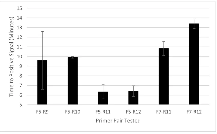
Figure 1.1 Sample Phylogenetic Tree of Selected GII Strains………....30 Figure 2.1 Time to Signal Detection of Different RT-RPA Primer Pairs………64 Figure 2.2 Sensitivity of RT-RPA Assay for Purified GII.4 New Orleans RNA as Predicted by Probit Regression………....65
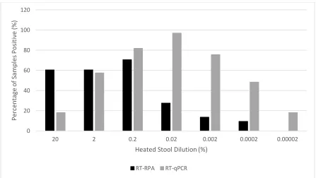
Figure 2.3 Comparison of RT-RPA and RT-qPCR Assays for Detection of Heated Stool Isolates……….66
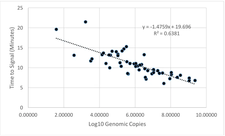
Figure 2.4 Linear Regression of RT-RPA Assay with Purified RNA……….67 Figure 3.1 Optimization of Antibody Buffer and Concentration for Streamlined HBGA Binding Assay………..83
Figure 3.2 Streamlined HBGA Capture Assay Performance with Different Concentrations of SYV VLPs………...84
Figure 3.3 Comparison of Long Duration and Streamlined HBGA Binding Assays as Applied to Heat Treated SYV VLPs………..85
Figure 3.4 Comparison of Long Duration and Streamlined HBGA Binding Assays as Applied to SYV VLPs on Stainless Steel and Copper Surfaces………86
Figure 4.1 Predicted Secondary Structures of the M1 and M6-2 ssDNA Aptamers Generated Against the GII.4 P Domain with Common Aptamer Pool Motifs Circled………...114 Figure 4.2 Binding Ratios of Partially Purified, Serially Diluted 20% GII.4 and Human NoV-Negative Stool Samples to Selected Aptamers by ELASA………...115 Figure 4.3 Capture of GII.4 New Orleans in Stool Using Aptamer Magnetic Capture
Figure 5.1 Conformation-Dependent Binding Behavior of Three Different Ligands to SYV VLPs Treated at Select Time-Temperature Combinations………149 Figure 5.2 Apparent Percentage of Sequence-Dependent Binding of Three Human Norovirus Ligands………...151
Figure 5.3 Dynamic Light Scattering Results for Heat-Treated GII.4 Sydney VLPs……...153 Figure 5.4 Ligand Docking of M6-2 to SYV VP1……….154 Figure 5.5 Electron microscopy of SYV VLPs treated at different temperatures for one minute………156
Figure 5.6 Sequence Alignment of 1lHM and SYV Capsid Sequences………157 Figure 5.7 Cluster Analysis of M6-2 and SYV VP1 Binding………158 Figure 6.1 Docked Models of Aptamer M6-2 to Human Norovirus Major Capsid
Proteins………..188 Figure 6.2 M6-2, HBGA, and NS14 Interaction Interface with HOV, SMV, and SYV
VP1………189 Figure 6.3 Most Populated Docking Interactions Between the VP1 Domain and Aptamer M6-2………..190 Figure 6.4 Secondary Structure of the Residue Over the Length of the Simulation……….191 Figure 6.5 Comparison of Heat Stability of Different Human Norovirus Strains Using
CHAPTER 1
Human Norovirus as a Foodborne Pathogen: Challenges and Developments Matthew D. Moore1, Rebecca M. Goulter1, and Lee-Ann Jaykus1
1
Department of Food, Bioprocessing and Nutrition Science, North Carolina State University, Raleigh, NC 27695
Introduction
Human noroviruses (NoV) are the most common cause of acute gastroenteritis and responsible for substantial morbidity and mortality worldwide (Patel et al. 2008; Glass et al., 2009). One important mode of NoV infection is via contaminated food, as these viruses are considered the leading known foodborne agent in the United States (U.S.), accounting for over 50% of foodborne illnesses annually (Scallan et al., 2011b). Human NoV may also be a significant cause of foodborne disease of unknown etiology, which is estimated to be as many as 80% of food-related illness (Scallan et al., 2011a).
Noroviruses are a member of the family Caliciviridae, which derives its name from the Greek word for cup (calyx), in reference to cup-like depressions on the surface of the virus (Glass et al., 2009). The Caliciviridae family contains six genera: Norovirus, Vesivirus, Lagovirus, Recovirus, Sapovirus, and Becovirus. The Norovirus genus is the most common cause of disease in humans relative to the other five. These viruses were previously referred to by other names, such as ―small round structured viruses‖ and ―Norwalk-like viruses‖
ORF 1 codes for a nonstructural polyprotein that is believed to self-cleave into six to seven proteins (NS1-NS7). Some of the nonstructural proteins include an NTPase (Pfister and Wimmer, 2001), a protease (Blakeney et al., 2003; Liu et al., 1996), an RNA-dependent RNA polymerase (Fukushi et al., 2004; Rohayem et al., 2006), and a protein (VPg) that is covalently linked to the 5‘ end of the genome suspected to initiate translation and possibly
transcription (Subba-Reddy et al. 2011, Belliot et al. 2008, Daughenbaugh et al. 2003, Goodfellow et al. 2005). For a more thorough discussion of NoV genomic structure and function, refer to Thorne & Goodfellow (2014).
ORF2 and ORF3 encode major (VP1) and minor (VP2) capsid proteins, respectively (Glass et al. 2000, Prasad et al. 1999). The VP1 protein contains internal N-terminal and shell (S) domains as well as a protruding (P) domain comprised of a P2 subdomain that is the most exposed part of the virus; and the P1 subdomain lies below P2 (closer to the S domain). The viral capsid is a T= 3 icosahedron that contains 180 copies of VP1, which form dimers. When expressed in certain models, the proteins self-assemble in empty capsids called virus-like particles (VLPs, Jiang et al. 1992). Human NoV cannot be cultivated in vitro and there is no known animal model for reliable virus propagation outside of human beings.
Historically, NoV have been classified based on the degree of difference between the amino acid sequences of the VP1 major capsid protein (Zheng et al., 2006). This protein was selected for many reasons, one of which was that it contains the highly variable P2
(2005) and Donaldson et al. (2010). Historically, genogroups have been defined as having 45-61% uncorrected pairwise difference in the VP1 region (Zheng et al. 2006). Accordingly, the Norovirus genus has been divided into six genogroups (GI – GVI), three of which cause disease in humans (GI, GII, and GIV). The recent GVI and a tentatively-proposed GVII genogroups consist of canine NoV (Martella et al., 2008; Tse et al., 2012). Further subdivisions within genogroups, representing genetic clusters, constitute genotypes.
Genotypes have been defined to contain 14-44% VP1 amino acid difference, while strains are defined to have 0-14% difference (Zheng et al., 2006). To date, over 31 genotypes of human NoV have been reported, of which 9 and 22 belong to GI and GII, respectively. At the time of this writing, this constitutes over 150 strains, but genotypic and strain designations are evolving.
Traditional nomenclature has been based on genogroup-genotype combination (e.g., GII/4 or GII.4). With the discovery that recombination can occur in the ORF1-ORF2 junction and other areas, and identification of more genotypes/strains, an alternative international standard for strain classification has been proposed. This assignment is a dual typing system based on specific difference cutoffs of the partial RdRp (ORF1, 1300 nucleotides) and complete VP1 (ORF2) sequences. The new nomenclature of an identified isolate would be written as: ―norovirus Genogroup/Host/Country/Isolation Year/Partial ORF1_ORF2_Strain NameIndex Year/Isolate city name (possibly with a unique isolate number).‖ For example, an
isolate of genogroup II found in Houston, TX (U.S.) that has a GII.P4 partial ORF1 grouping with a GII.4 Hunter ORF2 sequence would be designated as norovirus
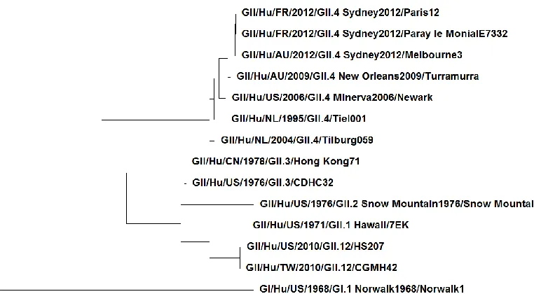
norovirus GII/Hu/US/2005/GII.4_Hunter2004/Houston23 (Kroneman et al. 2013). An example phylogenetic tree of some GII human NoV strains is provided in figure 1.
A number of features of NoV make them highly transmissible through the food supply. First, the infectious dose is low, ranging from 20 to several thousand particles, depending upon the study and corresponding modeling assumptions (Teunis et al. 2008; Atmar et al. 2013). Also, these viruses are antigenically and genetically diverse, resulting in many different strains with limited immunological cross-protection between strains.
Although still debatable, there is evidence that immunity to any one strain may be as short as a few months to two years (Parrino et al., 1977). Human NoV are persistent in the
environment, lasting weeks to years depending on environmental conditions such as temperature and relative humidity. They are also resistant to nearly all of the active ingredients in cleaning products, sanitizers, and disinfectants commonly used in food production and processing, including quaternary ammonium compounds, detergents, alcohols, and even chlorine at regulated concentrations. The same can be said about food processing and preservation methods such as heat, ionizing radiation, organic acids, preservatives, and manipulation of pH or water activity. These issues are discussed in multiple reviews (Hirneisen et al. 2010, Hoelzer et al. 2013, Li et al. 2012b). Some call human NoV the ―near perfect‖ foodborne pathogen, except for the fact that they cannot
multiply (but persist) in foods and the environment.
foodborne transmission route. These topics include efforts in human NoV cultivation; recent epidemiological findings; strain evolution; and key detection conundrums. The reader will come away with a better understanding of why this foodborne pathogen is so difficult to study and control, and how scientists are working to address these issues.
In Vitro and In Vivo Cultivation
The single most important limitation to the ability to study human NoV is the lack of an in vitro cultivation method, despite almost 50 years of attempts. Cultivation using many mammalian cell lines and human NoV strains has been attempted. For example, Duizer et al. (2004) tested A549, AGS, Caco-2, CCD-18, CRFK, CR-PEC, Detroit 551, Detroit 562, FRhK-4, HCT-8, HeLa, HEC, HEp-2, Ht-29, HuTu-80, I-407, IEC-6, IEC-18, Kato-3, L20B, MA104, MDBK, MDCK, RD, TMK, Vero and 293 cell lines along with about 32 different human NoV strains, all leading to no significant findings. Contrary to the case for murine NoV (GV), human macrophages and dendritic cells were unable to support infection by human strains (Lay et al., 2010). Guix et al. (2007) transfected Norwalk virus (the
be at the attachment and uncoating stages of infection. Further, this study implicated the viral VPg as necessary for viral replication (Guix et al., 2007). Some initial success was observed when challenging adult duodenal tissues in ex vivo culture, however, when using the in vitro human glandular epithelial cell line HIEC-6 only low (2 log10 or less) viral RNA production
was observed, as was the absence of a cytopathic effect (Leung et al., 2010).
A three-dimensional (3D) cell culture model was reported almost a decade ago for human NoV cultivation. Human small intestinal cells were grown on collagen I-coated porous microcarrier beads in a rotating wall vessel incubator (Straub et al. 2007). This mimics the fluid-shear environment of the intestine and thus allows the intestinal epithelium to grow and differentiate in three dimensions. Straub et al. (2007) showed replication of two GII and one GI human NoV strains in five passages using this model. The same group utilized the 3D cell culture method in Caco-2 cells and seemed to have some limited success (Straub et al., 2011). However, coordinated international efforts have failed to replicate these findings (Herbst-Kralovetz et al., 2013; Papafragkou et al., 2013; Takanashi et al., 2014). More recent efforts focus on the use of human intestinal enteroids or organoids, derived from human intestinal epithelial stem cells that are created ex vivo into 3D small intestinal
A number of animal models have been proposed for human NoV propagation. One of these is gnotobiotic pigs, which when challenged with a GII.4 strain, appear to exhibit mild diarrhea, shed virus, support some degree of viral replication, and exhibit a specific immune response profile (Cheetham et al. 2006). Additionally, the binding patterns of different human NoV VLPs to HBGA-expressing gnotobiotic pig buccal and duodenal tissues have been observed (Cheetham et al., 2007). Gnotobiotic calves have also been reported to host human NoV GII.4 infection (Souza et al. 2008).
The presence of GI and GII human NoV antibodies has been reported in several non-human primate species (Jiang et al., 2004). Rockx et al. (2005) found some evidence that rhesus macaques had the potential to serve as a model when inoculated with Norwalk virus (GI.1). Chimpanzees were also shown to host infection, harbor viral antigen production, and shed virus when injected intravenously with filtered Norwalk virus (Bok et al., 2011). A recent study reported that some strains of BALB/c mice and BALB/c mice that had been ―humanized‖ (grafted with certain human stem cells) were able to host replication of a
In the absence of cell culture or animal models, scientists have resorted to human challenge studies. Perhaps the first example of a human NoV challenge study occurred shortly after the original 1968 Norwalk virus outbreak, in which case fecal specimens derived from secondary cases were diluted and fed to volunteers (Dolin et al., 1972). Some important fundamental questions about human NoV infection have been addressed using human
feeding studies. For instance, the aforementioned information on NoV infectious dose were derived from human challenge data (Atmar et al., 2013; Teunis et al., 2008). Other
information was also elucidated from these studies, as Atmar et al. (2013) found that the incubation period of the virus ranged from 20-50 hours, with some association of a shorter incubation period for higher inoculation doses. Further, the range of symptom duration was found to be from 8-60 hours; however, viral shedding of infected patients ranged from 6-55 days. Viral shedding in vomitus was also reported, with a median of 41,000 genomic equivalents of NoV per ml of vomitus. This is compared to medians of 160 billion genomic equivalents/ml and 10 billion genomic equivalents/ml in the stool of people who exhibited gastroenteritis symptoms and those who did not, respectively (Atmar et al., 2013).
expensive, time consuming, and require rigorous regulatory approvals. They must therefore be carefully designed to optimize data collection but are, nonetheless, only rarely done.
In the absence of a cultivable human NoV strain, cultivable surrogate viruses have been widely used. These include feline calicivirus (FCV; Doultree et al. 1999); murine norovirus (MNV; Wobus et al. 2006); Tulane Virus (Farkas et al., 2008); rabbit hemorrhagic disease virus (Meyers et al., 1991); porcine enteric calicivirus (Flynn & Saif 1988); poliovirus; hepatitis A virus; and MS2 bacteriophage (Bae & Schwab 2008, Shin & Sobsey 2003). To date, FCV propagated in Crandell-Reese feline kidney cells and MNV propagated in mouse macrophage RAW 264.7 cells are the most widely used surrogates. Feline calicivirus is a member of the Vesivirus genus in the Caliciviridae family and causes a respiratory disease in cats, but is considered appropriate because it was a member of the same family as Norovirus and of similar genome structure, at least by the standards of the late 1990‘s, when it was first used. FCV is still considered the ―gold standard‖ by the U.S. Environmental Protection
Agency for products seeking to make anti-noroviral claims. MNV was reported by Karst et al. (2003) about a decade ago and was rapidly adopted because it is a member of the Norovirus genus (categorized as a GV NoV) and hence has more similar genetic and
structural characteristics to human NoV than other surrogates. However, it causes meningitis, not gastroenteritis, in mice (Kahan et al., 2011).
the surrogates to human NoV, in part because the latter cannot be cultivated in vitro. A full accounting of all these studies is beyond the scope of this paper, but they have been reviewed elsewhere (Baert et al., 2009a; Hirneisen et al., 2010; Hoelzer et al., 2013). Differences in susceptibility of the surrogates to environmental conditions and inactivation treatments are now well documented. Richards (2012) provides a critical review of these issues. Many of the surrogates differ in terms of resistance to elevated temperature, extremes of pH, and susceptibility to organic solvents as well as various chemical sanitizers and disinfectants. Generally, FCV appears to be more susceptible to pH and chlorine than is MNV, while MNV is more susceptible to alcohols (Cannon et al., 2006; Park et al., 2010; Tung et al., 2013). A recent systematic review and meta-analysis of studies using two or more surrogates
concluded that MNV, hepatitis A virus (HAV), and bacteriophage MS2 were generally more stable than FCV to chemical disinfectants, but by an average of less than or equal to 1.5 log10
PFU (Hoelzer et al., 2013).
Tulane virus (TV) is a newer surrogate, first reported by Farkas et al. (2008). Tulane virus was isolated from the stool of captive rhesus macques, and was phylogenetically classified into a new Caliciviridae genus called Recovirus. The TV genome has as general genetic similarity to Norovirus, and the capsid has similar size (~36 nm) and buoyant density as other members of the Caliciviridae family (Farkas et al., 2008). Unlike MNV and FCV, TV has been reported to bind HBGA types A and B (Farkas et al., 2010). This is significant in that this is the same putative host receptor as human NoV, suggesting that TV may be a more relevant surrogate in cases where capsid functionality and behavior (disinfection
Recent studies have sought to characterize the susceptibility of TV to different inactivation strategies. One such study compared TV and MNV sensitivity to pH, chlorine, heat, and survival in tap water at room and refrigeration temperatures. MNV was found to be more stable in refrigerated tap water and at various pH values compared to TV. However, MNV and TV behaved similarly in 20C tap water, under different heat treatments (50-75C
for 2 minutes) and at most concentrations of chlorine (0.2, 20, 200, and 2,000 ppm)
(Hirneisen and Kniel, 2013a). In another study, MNV and TV showed inactivation profiles similar to human NoV (assessed by RT-qPCR) when inoculated on spinach for up to a week (Hirneisen and Kniel, 2013b). Similarly, no statistically significant differences in the survival of MNV, TV, and hepatitis A virus on alfalfa seeds and sprouts held at 22C for 20 days was
observed by Wang et al. (2013). Certainly, further work must be done to identify the appropriate, most conservative (e.g., resistant) surrogate to be used for a given condition or treatment. It is likely that the choice of appropriate surrogate will be a function of the
Understanding the Epidemiology of Norovirus Disease
As stated previously, human NoV are the leading cause of acute viral gastroenteritis and of foodborne disease in most--if not all--of the world. But food is only one means by which the virus is spread, and person-to-person (P2P) transmission remains the most
common route. Recently, P2P was reported to account for 62-84% of all reported outbreaks (Vega et al., 2014, 2011). The overall burden of disease from all transmission routes
combined is staggering. A compilation of different studies of incidence/outcome rates produced estimates that NoV are responsible for 19-21 million illnesses, 1.7-1.9 million outpatient visits, 400,000 emergency department visits, 56,000-71,000 hospitalizations, and 570-800 deaths annually in the U.S. alone. When disease rates are applied to a U.S. resident who lives to the age of 79 years, he/she would experience five incidents of human NoV disease in a lifetime, have a 1 in 2 chance of a disease related outpatient visit, a 1 in 50-70 chance of hospitalization, and 1 in 5,000-7,000 chance of death due to NoV infection (Hall et al., 2013; Scallan et al., 2011b). Long-term healthcare facilities are particularly important, constituting 62.5% (2,475/3,960) of NoV cases reported to the U.S. Centers for Disease Control and Prevention (CDC) from 2009-2013 (although most NoV cases are not reported to the CDC) (Vega et al., 2014). European statistics are generally similar to those from the U.S. (Phillips et al. 2010; Baert et al. 2009b; Tam et al. 2012).
transmission (Matthews et al. 2012, Vega et al. 2014). For example, 72% of long-term healthcare facility outbreaks reported from 2009-2013 were caused by GII.4 strains, but non-GII.4 strains also caused considerable morbidity, especially in association with restaurant and food-related settings (Vega et al., 2014). Of late, there appears to have been an increase in human NoV disease incidence in multiple countries, probably due to the emergence of a new GII.4 strain (the Sydney strain which appeared first in 2012) (van Beek et al., 2013). In fact, human NoV mortality can spike by as much as 50% during seasons in which an emergent epidemic (GII.4) strain is circulating (Hall et al., 2012a).
There are limited statistics relative to the amount of NoV disease attributable to foods. Obtaining these estimates is extremely challenging because multiple factors must be considered and assumptions made in order to generate population-level estimations of the burden of foodborne illness. For laboratory confirmed surveillance, for example, the infected patient must seek medical attention; a clinical sample must be obtained; a laboratory must identify the pathogen; and the illness must be correctly reported up the chain of health authorities (Scallan et al., 2011b). Unfortunately, disease associated with human NoV is widespread, frequently goes undiagnosed, is not normally life threatening, and is not reported to public health authorities in the U.S. Consequently, foodborne disease incidence estimates for NoV are largely determined based on population-level data and mathematical modeling. The most commonly cited U.S. statistic is that of Scallan et al. (2011b), which estimates that viruses accounted for 59% of all foodborne disease, and NoV accounted for 99% (5.5
(26%, 2nd in rank) and 150 deaths (11%, 4th in rank) annually (Scallan et al., 2011b). In Canada, NoV is also considered the leading cause of foodborne illness, accounting for 65% of the known illnesses (Thomas et al., 2013).
However, estimates of the comparative importance of food relative to other
transmission routes vary widely. Based on outbreaks published from 1983-2011, Matthews et al. (2012) suggested that the majority of NoV infections were transmitted by foodborne routes (54%), with person-to-person ranking second (26%). However, this was a
meta-analysis of published outbreaks and not necessarily based upon population-based surveillance data. On the other hand, using U.S. CaliciNet surveillance system data from 2009-2013, Vega et al. (2014) concluded that 83.7% of NoV outbreaks were person-to-person while only 16.1% were foodborne. In another study focused only on foodborne outbreaks, 46% (3,000) of approximately 6,300 outbreaks of known cause reported to the U.S. CDC between 2001-2008 were attributed to NoV (Hall et al. 2012b).
Human NoV outbreaks are difficult to identify and even more difficult to trace back to a common food source. In addition to the general problems described above, most foodborne NoV outbreaks are small, associated with a retail setting (and hence investigated locally or regionally), and secondary P2P spread (propagated outbreaks) is very common. Even in the case of a foodborne source being identified, many factors hinder detection of the virus in the food (discussed later).
conjunction with epidemiological data to retrospectively identify common source outbreaks occurring in Europe from 1999-2008. The method found a notable increase in common source and international outbreaks compared to the numbers identified originally by the Food-Borne Viruses in Europe (FBVE) network (Verhoef et al., 2011). In a later study, the same team analyzed 500 publicly-available full-length NoV capsid sequences for outbreak identification. Not surprisingly, a region containing most of the P2 subdomain (nucleotide positions 900-1,400) was found to be the best for assigning genotypes while also recognizing variants and identifying outbreak events. Within this region, the minimum fragment of 950-1,350 nucleotides of the capsid gene could be used to identify greater than 80% of outbreak events (Verhoef et al., 2012). A similar approach has been taken in tracing and identifying nosocomial infections (Sukhrie et al., 2011). With a distinctly foodborne focus, Verhoef et al. (2010) used genotype frequency distributions from NoV isolated from European bivalve mollusks in conjunction with genotype frequency derived from human illness surveillance to differentiate outbreaks due to contamination early in the food chain (i.e., during production or processing) vs. those occurring later in the chain (i.e., food-handler-associated). In a completely different approach, Rha et al. (2013) utilized BioSense, a national electronic U.S. healthcare surveillance system, to statistically correlate emergency department chief
Another epidemiological challenge related to NoV—as well as other foodborne pathogens—is attribution of cases or outbreaks to specific food commodities. Because surveillance data are so sparse, many attribution studies are based largely on outbreak data. Over the last several years, the U.S. CDC has embarked on more comprehensive human NoV food attribution studies. Using data from 1998-2008 obtained from the Foodborne Disease Outbreak Surveillance System, they have been able to make some general conclusions. First of all, the majority of food-associated NoV outbreaks in their database could not be
attributable to one or more specific foods. When they could be attributed to a food, such foods could be subdivided into multicomponent or ―complex‖ foods and single component or ―simple‖ foods (Painter et al., 2013). Human NoV outbreaks were more often caused by
complex foods relative to simple foods. Further breakdown of the simple foods suggested that fresh vegetables were responsible for about 30-40% of simple food outbreaks, followed by fruits and nuts (10-20%), mollusks (10-15%), and dairy (5-15%) (Gould et al., 2013; Hall et al., 2014, 2012b; Painter et al., 2013). Very similar results were obtained using 2009-2012 data (Hall et al., 2014) and by others not affiliated with CDC (Batz et al., 2012; Greig and Ravel, 2009).
source (pre- or post-harvest) could not be resolved. Not unexpectedly, the only food for which pre-harvest contamination was a notable proportion was mollusks (Hall et al., 2012b). Of the 528 foodborne NoV outbreaks reported during 2009-2012 for which specific
contamination factors were identified, infectious workers were responsible for 70% of these outbreaks, and bare hand contact with ready-to-eat foods was identified in 54% of the outbreaks (Hall et al., 2014). Clearly, the food handler appears to be responsible for the majority of foodborne outbreaks.
In conclusion, it appears that the majority of NoV outbreaks appear to be transmitted between individuals, although foodborne transmission is significant. Estimates of the relative proportion of transmission that is food-associated vary widely and more refined data are needed. Complex, prepared, ready-to-eat foods are over-represented in outbreaks, and the most common setting for these is restaurants/delicatessens and catering facilities, implicating food handling as the likely contamination source. In the case of simple foods, vegetables, fruits and nuts are the most likely culprits, but it is difficult to discern how important pre-harvest contamination is for these products. Even in the absence of comprehensive data, it appears that targeting the food preparation sector would be an efficacious way to address the foodborne NoV problem.
Norovirus Evolution and Epidemic Strain Emergence
program from 2009-2013 found that GII.4 was implicated in 72% of nearly 4,000 reported NoV outbreaks in the U.S. (Vega et al., 2014). This genotype has been found to circulate and evolve rapidly after periods of stasis (a phenomenon called epochal evolution) such that different pandemic/epidemic strains emerge with unique properties every few years. In the last two decades, these have included: Camberwell (1994); US 95/96 (1995-1996);
Farmington Hills (2002); GII.4b (2002); Hunter (2004); Sakai (2006); Minerva (2006); New Orleans (2009); and Sydney (2012). Interested readers are referred to White et al. (2014) and Vinje et al. (2014) for additional discussion of this subject.
changes in antigenic profiles pose challenges to creation of vaccines and universal recognition ligands for development of detection assays.
Another aspect of human NoV evolution is the occurrence of recombinant strains, as these viruses have been found to undergo spontaneous recombination at different genome locations including ORF1 (Waters et al., 2007); the ORF1/2 junction (Bull et al., 2007, 2005; Eden et al., 2013); the ORF2/3 junction (Eden et al., 2013); and ORF2 (Eden et al., 2013; Rohayem et al., 2005). Novel recombinants of multiple genotypes have been recently
reported. For example, an emerging GII.12 strain is a likely recombinant (with a GII.g ORF1 and GII.12 ORF2) (Giammanco et al. 2012, Takanashi et al. 2011) that interestingly did not bind synthetic HBGAs yet infected humans (Takanashi et al., 2011). This same strain had a rate of evolution comparable to a previously reported rate for GII.4 (Giammanco et al., 2012).
maintain itself even in the face of widespread population immunity (Bull et al., 2010; Donaldson et al., 2008; White, 2014).
Detection of Human Norovirus in Environmental and Food Samples
Despite extensive efforts, development of robust methods to detect viral contamination in foods and environmental samples remains challenging. Because the
numbers of virus particles present in contaminated foods are usually low and no universal or rapid culture-based methods are available, enrichment is not possible. Consequently, the viruses must be concentrated and purified from complex sample matrices prior to detection, with molecular amplification (reverse transcription qPCR or RT-qPCR) being the detection method of choice. Sequence-based determination of amplicon identity is ideally used for amplicon confirmation. The major steps required for the detection of viruses in foods can be designated as follows: (1) virus concentration and purification; (2) nucleic acid extraction; (3) detection; and (4) confirmation. A full description of these methods is well beyond the scope of this document, as entire monographs are written about this. The interested reader is referred to these more comprehensive reviews (Bosch et al. 2008, Butot et al. 2014, Knight et al. 2013, Maurer 2011, Vinjé 2014). Here we discuss two recalcitrant problems associated with the detection of NoV in complex sample matrices, those being interpretation of presumptively positive samples and the ―infectivity dilemma.‖
human NoV in food and environmental samples where the concentration of viruses is usually quite low. In earlier days, sample concentrates were processed for viral RNA isolation, followed by detection by RT-PCR and confirmation of amplicon identify using nucleic acid hybridization. In the middle of the last decade, RT-PCR was replaced almost exclusively by real-time or reverse transcription-quantitative PCR (RT-qPCR), a method that incorporates a fluorescently labeled probe or specifically intercalating fluorescent dye in the reaction mix, theoretically allowing one to by-pass time consuming DNA hybridization steps. This method remains the ―gold standard‖ at the current time.
But incorporation of a hybridization step during amplification can be problematic when low template concentrations are present. First of all, matrix-associated inhibition persists in many food and environmental samples, and can impact the efficiency of nucleic acid extraction and RT-qPCR (Rutjes et al., 2006; Schrader et al., 2012). Such inhibition is usually identified using internal amplification controls, but these can interfere with the efficiency of amplification of the target (Espy et al., 2006; Hoorfar et al., 2004). When inhibition occurs, the usual solution is sample dilution. Unfortunately, this then reduces the analytical sensitivity of the assay. Another issue is interpretation of results for samples producing high cycle threshold (Ct) values by RT-qPCR. There have been a number of studies that have employed RT-qPCR to detect viruses in various types of naturally
contaminated sample matrices, and a snapshot of these studies is provided in Table 1. Note that in most cases, some sort of amplicon confirmation step was used, ranging from cloning and sequencing to ―double‖ amplifications to hybridization. The most reliable of these is
samples to successfully acquire sequences, especially when Cycle threshold (Ct) values exceed 35. Unfortunately, low levels of viral contamination (and associated high Ct values) are the rule rather than the exception for these sample types.
Thus, in many cases definitive confirmation of amplicon identity may not be possible. This means that the analyst is faced with the conundrum of wrongly interpreting sample positivity, leading to false positive or false negative reporting. At this point in time, it is probably prudent to interpret such samples as ―presumptively‖ positive. But also keep in mind that sample negativity is impacted by residual RT-qPCR inhibition (and potential sample dilution), competition with internal amplification controls, and sampling. Even a negative result does not assure the analyst of the absence of viral contamination.
Unfortunately, these interpretation dilemmas remain unaddressed. As more food and environmental samples are screened, it is increasingly important to come to consensus on these data interpretation issues, or there is the risk of under- or over-estimating the prevalence of virus contamination and/or wrongly predicting its risk.
demonstrated that the RNA associated with inactivated viruses remains detectable by nucleic acid amplification long after viral infectivity has been lost (Richards 1999). In short, in the absence of a culture method or a suitable marker for infectivity, it is impossible to confirm that the detection of human NoV by RT-qPCR is indicative of infectious virus, as RT-qPCR tends to overestimate the actual amount of infectious virus. There are two major reasons why food scientists need reliable means to discriminate human NoV infectivity status: (1) to identify products that actually pose a risk to human health; and (2) to support studies of virus inactivation, including characterization of the efficacy of both traditional and novel control measures.
Alternative methods for predicting virus infectivity based on molecular amplification approaches have been investigated. These methods fall into two major classifications: (1) those based on examining the stability of the virus genome; and (2) those based on
examining capsid integrity. Examples of these approaches are provided in Table 2. The latter approach is the most popular, and one commonly used method is to precede RT-qPCR with enzymatic (RNase with or without prior proteinase K) treatment. Sometimes called
―virolysis,‖ the idea is that destruction of the viral capsid will result in release of viral RNA,
infectivity discrimination assumes that if the viral capsid is non-intact, it will not be able to bind to the receptors.
There are inactivation methods that target the viral nucleic acid. In this case, detection of small genomic target fragments using RT-qPCR may not indicate the presence of
infectious virus for two reasons. The first reason is that naked viral RNA can persist long after a viral capsid has been destroyed. The second reason is that even though a single strand break occurring anywhere in the viral genome will render a virus particle non-infectious, the same break cannot be detected by RT-PCR if the target region for amplification remains intact. To account for this problem, it has been suggested that measurement of the overall integrity of genomic RNA could provide a useful marker for infective particles. This has been accomplished by techniques using multiple amplifications (Pecson et al. 2011), amplification of near full length genomes (Kostela et al., 2008), or a combination in which long range reverse transcription is followed by more efficient small fragment RT-qPCR (Wolf et al., 2009).
Note that each of the methods to discriminate infectivity status of human NoV using nucleic acid amplification have their own advantages and disadvantages, and the interested reader is referred to the comprehensive review of Knight et al. (2013) for more details. There is great debate amongst scientists working in this field as to which is the ―best‖ method.
When looking at the data collectively as applied to surrogate viruses for which infectivity and nucleic acid amplification are compared, preceding RT-qPCR with enzymatic pre-treatments or ligand binding (capsid integrity methods) provide better but still not complete agreement with virus infectivity. Methods to assess genome integrity and the use of nucleic acid
intercalating agents are difficult to optimize and frequently inefficient. Also, data on method performance frequently differs as a function of inactivation strategy. To date, no one method has been demonstrated to accurately discern infectious from non-infectious viral particles in the absence of an in vitro human NoV cultivation method.
Conclusions
In all, human NoV remain a significant challenge. Elements of NoV‘s basic biology
challenges, particularly in vaccine development and detection of emerging strains. Although there is better understanding of disease burden and attribution, outbreaks are still vastly under-reported, and adequately addressing the important role of the food handler remains a recalcitrant issue. In the absence of reliable detection methods for viruses in foods, it is difficult to attribute specific foods to outbreaks, or to better understand the prevalence of naturally occurring contamination. In addition to its existing features as a ―near perfect‖
Table 1.1 Studies Highlighting Issues in Positive RT-(q)PCR Interpretation
Sample type Controls/ Confirmation Issues Citation
Leafy greens Second PCR targeting a different region and attempted sequencing of
amplicon.
Of 275 samples total, 148 (54%) were found to be positive for HuNoV by RT-qPCR. Only 40 samples (15%) produced a band of expected size for the second
amplification. Of these 40 amplicons, only 16 (6%) could be sequenced to confirm HuNoV RNA.
Mattison et al. 2010
Raspberries, cherry tomatoes, strawberries
and fruit salad
IAC included; attempted sequencing of amplicon.
‗Positive‘ Ct values were >37. Not a single ‗positive‘ could be confirmed by sequencing.
Stals et al. 2011
Environmental (catering kitchens and
restrooms)
Nested PCR; attempted sequencing amplicon.
Samples with Ct‘s >40 could not always be confirmed by sequencing.
Boxman et al. 2011
Raw and treated sewage water
Southern hybridization. Not always correlation between positive RT-qPCR and hybridization results.
van den Berg et al.
2005 Shellfish Dilution, hybridization
and attempted sequencing amplicon.
Sequencing not possible. Differences in sensitivities of hybridization method compared to RT-qPCR between GI
and GII genogroups of NoV. Inhibition seen in 27% of positive samples.
Loisy et al. 2005
Shellfish IAC and sequencing. Most ‗positive‘ virus samples had Ct‘s >45 (of 50 cycles). Of 15 ‗positive‘ samples, only 5 could be sequenced.
DePaola et al. 2010 N/A N/A A broad review focused on molecular methods in food
microbiology for interested readers.
Table 1.2 Studies Regarding In Vitro Estimation of Human Norovirus Infectivity Using RT-(q)PCR
Method Viruses Tested Treatment(s) Results Citation(s)
Long range genome amplification
Bacteriophage MS2; human NoV GI.1 and GII.4; murine
norovirus (MNV); poliovirus I (PV1) Ultraviolet (UV) light; heat;
Method generally overestimated viral load by a range of 0.5 to as much as 4 log10, but usually
performed better than traditional RT-qPCR. For UV, method design had to account for
different genomic region susceptibility. Amplification tended to be inefficient. Does
not account for capsid integrity.
Kostela et al. 2008, Pecson et al. 2011, Shin & Sobsey 2003, Wolf et al. 2009
RNase and Proteinase K pre-treatment followed by RT-PCR MS2; MNV; HAV, PV1, feline calicivirus (FCV); human NoV GII UV; hypochlorite; heat; singlet oxygen; high hydrostatic pressure (HPP);
Method generally overestimated viral load by a range of 2->3 log10, sometimes performing better than traditional RT-qPCR. One study gave negative endpoint results, but low initial loads of virus were used (~103 logs). Generally
does not account for genome integrity.
Diez-Valcarce et al. 2011, Nuanualsuwan & Cliver 2002, Pecson et al. 2009, Tang et
al. 2010 RNase pre-treatment
with RT-qPCR
FCV, human HuNoV , MNV,
human adenovirus, human NoV GI.1 Hypochlorite; heat; alcohols; quarternary ammonium compounds; chlorine dioxide; oregano extract
Method correlates to infectivity over a narrow disinfection range for hypochlorite (low available chlorine concentrations) and heat initially, but overestimates viral load in most
cases, by 1 to >3 log10. A human feeding
study found this method overestimated viral persistence in groundwater by nearly 3 years.
Mathematical modeling was applied to make estimation fairly accurate in one case. Generally does not account for genome
integrity.
de Abreu Corrêa et al. 2012, Gilling et al. 2014, Nowak et
al. 2011a,b; Seitz et al. 2011, Topping
Table 1.2 Studies Regarding In Vitro Estimation of Human Norovirus Infectivity Using RT-(q)PCR (Continued). Cell-binding (Raw
264.7 or Caco-2) or receptor binding (Ganglioside GD1a
or porcine gastric mucin, PGM) followed by
RT-qPCR
MNV; human NoV GI.1, GI.8
and GII.4
Heat; HPP; UV; hydrogen peroxide; grape
seed extract;
Some evidence that PGM binds preferentially to intact particles. Collectively, the method
overestimated viral load by 1-4.5 log10 but
usually performed better than traditional RT-qPCR. Generally does not account for genome
integrity.
Dancho et al. 2012, Li et al. 2011, 2012a; Tang et al. 2010
Pre-treatment with nucleic acid intercalating agents
(propidium monoazide, PMA
followed by RT-qPCR
HAV; human NoV GII.2; MS2; MNV
Heat Two studies compared PMA-RTqPCR to RNase-RT-qPCR. The former method appeared to perform equivalently or better than
the latter, and certainly better than RT-qPCR alone. However, the method is difficult to optimize and results appear to vary by virus.
Escudero-Abarca et al. 2014, Kim & Ko 2012, Parshionikar et
al. 2010b, Sánchez et al.
2012 Hydrazide binding
followed by RT-qPCR
Astrovirus serotype 4;
human NoVGII.4
Hypochlorite Method overestimated astrovirus load by about 1-2 log10 compared to plaque assay. Only
relevant for disinfection approaches in which oxidative damage occurs.
Sano et al. 2010
Proteinase K and RNase pretreatment followed by NASBA
FCV, human NoV GII
Heat Enzymatic treatment removed all positive signal for human NoV that had been seen without treatment. No comparison to surrogate
References
Atmar, R.L., Opekun, A.R., Gilger, M.A., Estes, M.K., Crawford, S.E., Neill, F.H., Ramani, S., Hill, H., Ferreira, J., Graham, D.Y., 2013. Determination of the Human Infectious Dose-50% for Norwalk Virus. J. Infect. Dis. jit620–. doi:10.1093/infdis/jit620 Bae, J., Schwab, K.J., 2008. Evaluation of murine norovirus, feline calicivirus, poliovirus,
and MS2 as surrogates for human norovirus in a model of viral persistence in surface water and groundwater. Appl. Environ. Microbiol. 74, 477–84.
doi:10.1128/AEM.02095-06
Baert, L., Debevere, J., Uyttendaele, M., 2009a. The efficacy of preservation methods to inactivate foodborne viruses. Int. J. Food Microbiol. 131, 83–94.
doi:10.1016/j.ijfoodmicro.2009.03.007
Baert, L., Uyttendaele, M., Stals, a, VAN Coillie, E., Dierick, K., Debevere, J., Botteldoorn, N., 2009b. Reported foodborne outbreaks due to noroviruses in Belgium: the link between food and patient investigations in an international context. Epidemiol. Infect. 137, 316–25. doi:10.1017/S0950268808001830
Batz, M.B., Hoffmann, S., Morris, J.G., 2012. Ranking the disease burden of 14 pathogens in food sources in the United States using attribution data from outbreak investigations and expert elicitation. J. Food Prot. 75, 1278–91. doi:10.4315/0362-028X.JFP-11-418 Belliot, G., Sosnovtsev, S. V, Chang, K.-O., McPhie, P., Green, K.Y., 2008.
Nucleotidylylation of the VPg protein of a human norovirus by its
proteinase-polymerase precursor protein. Virology 374, 33–49. doi:10.1016/j.virol.2007.12.028 Blakeney, S.J., Cahill, A., Reilly, P. a, 2003. Processing of Norwalk virus nonstructural
proteins by a 3C-like cysteine proteinase. Virology 308, 216–224. doi:10.1016/S0042-6822(03)00004-7
Bok, K., Parra, G.I., Mitra, T., Abente, E., Shaver, C.K., Boon, D., Engle, R., Yu, C.,
Kapikian, A.Z., Sosnovtsev, S. V, Purcell, R.H., Green, K.Y., 2011. Chimpanzees as an animal model for human norovirus infection and vaccine development. Proc. Natl. Acad. Sci. U. S. A. 108, 325–30. doi:10.1073/pnas.1014577107
Bosch, A., Guix, S., Sano, D., Pintó, R.M., 2008. New tools for the study and direct surveillance of viral pathogens in water. Curr. Opin. Biotechnol. 19, 295–301. doi:10.1016/j.copbio.2008.04.006
Bull, R. a, Eden, J.-S., Luciani, F., McElroy, K., Rawlinson, W.D., White, P. a, 2012.
Contribution of intra- and interhost dynamics to norovirus evolution. J. Virol. 86, 3219– 29. doi:10.1128/JVI.06712-11
Bull, R. a, Eden, J.-S., Rawlinson, W.D., White, P. a, 2010. Rapid evolution of pandemic noroviruses of the GII.4 lineage. PLoS Pathog. 6, e1000831.
Bull, R. a, Tanaka, M.M., White, P. a, 2007. Norovirus recombination. J. Gen. Virol. 88, 3347–59. doi:10.1099/vir.0.83321-0
Bull, R.A., Hansman, G.S., Clancy, L.E., Tanaka, M.M., Rawlinson, W.D., White, P.A., 2005. Norovirus Recombination in ORF1/ORF2 Overlap. Emerg. Infect. Dis. 11, 1079– 1085.
Butot, S., Zuber, S., Baert, L., 2014. Sample preparation prior to molecular amplification: Complexities and opportunities. Curr. Opin. Virol. 4, 66–70.
doi:10.1016/j.coviro.2013.12.004
Cannon, J.L., Lindesmith, L.C., Donaldson, E.F., Saxe, L., Baric, R.S., Vinjé, J., 2009. Herd immunity to GII.4 noroviruses is supported by outbreak patient sera. J. Virol. 83, 5363– 74. doi:10.1128/JVI.02518-08
Cannon, J.L., Papafragkou, E., Park, G.W., Osborne, J., Jaykus, L.-A., Vinjé, J., 2006. Surrogates for the study of norovirus stability and inactivation in the environment: aA comparison of murine norovirus and feline calicivirus. J. Food Prot. 69, 2761–5. Cheetham, S., Souza, M., McGregor, R., Meulia, T., Wang, Q., Saif, L.J., 2007. Binding
patterns of human norovirus-like particles to buccal and intestinal tissues of gnotobiotic pigs in relation to A/H histo-blood group antigen expression. J. Virol. 81, 3535–44. doi:10.1128/JVI.01306-06
Cheetham, S., Souza, M., Meulia, T., Grimes, S., Han, M.G., Saif, L.J., 2006. Pathogenesis of a genogroup II human norovirus in gnotobiotic pigs. J. Virol. 80, 10372–81.
doi:10.1128/JVI.00809-06
Choi, J.-M., Hutson, A.M., Estes, M.K., Prasad, B.V.V., 2008. Atomic resolution structural characterization of recognition of histo-blood group antigens by Norwalk virus. Proc. Natl. Acad. Sci. U. S. A. 105, 9175–80. doi:10.1073/pnas.0803275105
D‘Souza, D.H., Su, X., 2010. Efficacy of Chemical Treatments Against Murine Norovirus, Feline Calicivirus, and MS2 Bacteriophage. Foodborne Pathog. Dis. 7, 319–326. Daughenbaugh, K.F., Fraser, C.S., Hershey, J.W.B., Hardy, M.E., 2003. The genome-linked
protein VPg of the Norwalk virus binds eIF3, suggesting its role in translation initiation complex recruitment. EMBO J. 22, 2852–9. doi:10.1093/emboj/cdg251
Daughenbaugh, K.F., Wobus, C.E., Hardy, M.E., 2006. VPg of murine norovirus binds translation initiation factors in infected cells. Virol. J. 3, 33. doi:10.1186/1743-422X-3-33
Dawson, D.J., Paish, a, Staffell, L.M., Seymour, I.J., Appleton, H., 2005. Survival of viruses on fresh produce, using MS2 as a surrogate for norovirus. J. Appl. Microbiol. 98, 203–9. doi:10.1111/j.1365-2672.2004.02439.x
Pothier, P., Le Pendu, J., Boireau, W., Belliot, G., 2011. Qualitative and quantitative analysis of the binding of GII.4 norovirus variants onto human blood group antigens. J. Virol. 85, 4057–70. doi:10.1128/JVI.02077-10
Debbink, K., Donaldson, E.F., Lindesmith, L.C., Baric, R.S., 2012. Genetic mapping of a highly variable norovirus GII.4 blockade epitope: potential role in escape from human herd immunity. J. Virol. 86, 1214–26. doi:10.1128/JVI.06189-11
Debbink, K., Lindesmith, L.C., Donaldson, E.F., Costantini, V., Beltramello, M., Corti, D., Swanstrom, J., Lanzavecchia, A., Vinjé, J., Baric, R.S., 2013. Emergence of New Pandemic GII.4 Sydney Norovirus Strain Correlates With Escape From Herd Immunity. J. Infect. Dis. 208, 1877–87. doi:10.1093/infdis/jit370
Dolin, R., Blacklow, N.R., DuPont, H., Buscho, R.F., Wyatt, R.G., Kasel, J. a., Hornick, R., Chanock, R.M., 1972. Biological Properties of Norwalk Agent of Acute Infectious Nonbacterial Gastroenteritis. Exp. Biol. Med. 140, 578–583. doi:10.3181/00379727-140-36508
Donaldson, E.F., Lindesmith, L.C., Lobue, A.D., Baric, R.S., 2008. Norovirus pathogenesis: mechanisms of persistence and immune evasion in human populations. Immunol. Rev. 225, 190–211. doi:10.1111/j.1600-065X.2008.00680.x
Donaldson, E.F., Lindesmith, L.C., Lobue, A.D., Baric, R.S., 2010. Viral shape-shifting: norovirus evasion of the human immune system. Nat. Rev. Microbiol. 8, 231–41. doi:10.1038/nrmicro2296
Doultree, J.C., Druce, J.D., Birch, C.J., Bowden, D.S., Marshall, J. a, 1999. Inactivation of feline calicivirus, a Norwalk virus surrogate. J. Hosp. Infect. 41, 51–7.
Duizer, E., Schwab, K.J., Neill, F.H., Atmar, R.L., Koopmans, M.P., Estes, M.K., 2004. Laboratory efforts to cultivate noroviruses. J. Gen. Virol. 85, 79–87.
doi:10.1099/vir.0.19478-0
Eden, J.-S., Tanaka, M.M., Boni, M.F., Rawlinson, W.D., White, P. a, 2013. Recombination within the pandemic norovirus GII.4 lineage. J. Virol. 87, 6270–82.
doi:10.1128/JVI.03464-12
Escudero-Abarca, B.I., Rawsthorne, H., Goulter, R.M., Suh, S.H., Jaykus, L. a, 2014. Molecular methods used to estimate thermal inactivation of a prototype human norovirus: more heat resistant than previously believed? Food Microbiol. 41, 91–5. doi:10.1016/j.fm.2014.01.009
Espy, M., JR, U., LM, S., SP, B., Jones, M., Vetter, E.A., Yao, J.D.C., Wengenack, N., Rosenblatt, J., Cockerill III, F., Smith, T.F., 2006. Real-Time PCR in Clinical
Microbiology : Applications for Routine Laboratory Testing. Clin. Microbiol. Rev. 19, 165. doi:10.1128/CMR.19.1.165
diversity and histo-blood group antigen interactions of rhesus enteric caliciviruses. J. Virol. 84, 8617–25. doi:10.1128/JVI.00630-10
Farkas, T., Sestak, K., Wei, C., Jiang, X., 2008. Characterization of a rhesus monkey calicivirus representing a new genus of Caliciviridae. J. Virol. 82, 5408–16. doi:10.1128/JVI.00070-08
Flynn, W.T., Saif, L.J., 1988. Serial propagation of porcine enteric calicivirus-like virus in primary porcine kidney cell cultures. J. Clin. Microbiol. 26, 206–12.
Fukushi, S., Kojima, S., Takai, R., Hoshino, F.B., Oka, T., Takeda, N., Katayama, K., Kageyama, T., 2004. Poly ( A ) - and Primer-Independent RNA Polymerase of Norovirus. J. Virol. 78, 3889–3896. doi:10.1128/JVI.78.8.3889
Giammanco, G.M., Rotolo, V., Medici, M.C., Tummolo, F., Bonura, F., Chezzi, C., Martella, V., De Grazia, S., 2012. Recombinant norovirus GII.g/GII.12 gastroenteritis in children. Infect. Genet. Evol. 12, 169–74. doi:10.1016/j.meegid.2011.10.021
Gill, O.N., Cubitt, W.D., McSwiggan, D.A., Watney, B.M., Bartlett, C.L.R., 1983. Epidemic of gastroenteritis caused by with small round structured viruses contaminated with small round structured viruses. Br. Med. J. 287, 1532–1534.
Glass, P.J., White, L.J., Ball, J.M., Leparc-Goffart, I., Hardy, M.E., Estes, M.K., 2000. Norwalk Virus Open Reading Frame 3 Encodes a Minor Structural Protein. J. Virol. 74, 6581–6591. doi:10.1128/JVI.74.14.6581-6591.2000.Updated
Glass, R.I., Parashar, U.D., Estes, M.K., 2009. Norovirus Gastroenteritis. N. Engl. J. Med. 361, 1776–1785.
Goodfellow, I., Chaudhry, Y., Gioldasi, I., Gerondopoulos, A., Natoni, A., Labrie, L., Laliberté, J.-F., Roberts, L., 2005. Calicivirus translation initiation requires an interaction between VPg and eIF4E. EMBO Rep. 6, 968–72.
doi:10.1038/sj.embor.7400510
Gould, L.H., Walsh, K. a, Vieira, A.R., Herman, K., Williams, I.T., Hall, A.J., Cole, D., 2013. Surveillance for foodborne disease outbreaks - United States, 1998-2008. MMWR. Surveill. Summ. 62, 1–34.
Greig, J.D., Ravel, a, 2009. Analysis of foodborne outbreak data reported internationally for source attribution. Int. J. Food Microbiol. 130, 77–87.
doi:10.1016/j.ijfoodmicro.2008.12.031
Guix, S., Asanaka, M., Katayama, K., Crawford, S.E., Neill, F.H., Atmar, R.L., Estes, M.K., 2007. Norwalk virus RNA is infectious in mammalian cells. J. Virol. 81, 12238–48. doi:10.1128/JVI.01489-07
United States, 1999-2007. Clin. Infect. Dis. 55, 216–23. doi:10.1093/cid/cis386 Hall, A.J., Eisenbart, V.G., Etingüe, A.L., Gould, L.H., Lopman, B.A., Parashar, U.D.,
2012b. Epidemiology of Foodborne Norovirus Outbreaks, United States, 2001-2008. Emerg. Infect. Dis. 18, 2001–2008.
Hall, A.J., Lopman, B. a, Payne, D.C., Patel, M.M., Gastañaduy, P. a, Vinjé, J., Parashar, U.D., 2013. Norovirus disease in the United States. Emerg. Infect. Dis. 19, 1198–205. doi:10.3201/eid1908.130465
Hall, A.J., Wikswo, M.E., Pringle, K., Gould, L.H., Parashar, U.D., 2014. Vital signs: foodborne norovirus outbreaks - United States, 2009-2012. MMWR. Morb. Mortal. Wkly. Rep. 63, 491–5.
Han, M.G., Cheetham, S., Azevedo, M., Thomas, C., Saif, L.J., 2006. Immune responses to bovine norovirus-like particles with various adjuvants and analysis of protection in gnotobiotic calves. Vaccine 24, 317–26. doi:10.1016/j.vaccine.2005.07.071
Herbst-Kralovetz, M.M., Radtke, A.L., Lay, M.K., Hjelm, B.E., Bolick, A.N., Sarker, S.S., Atmar, R.L., Kingsley, D.H., Arntzen, C.J., Estes, M.K., Nickerson, C. a, 2013. Lack of norovirus replication and histo-blood group antigen expression in 3-dimensional
intestinal epithelial cells. Emerg. Infect. Dis. 19, 431–8. doi:10.3201/eid1903.121029 Hirneisen, K. a, Kniel, K.E., 2013a. Comparing human norovirus surrogates: murine
norovirus and Tulane virus. J. Food Prot. 76, 139–43. doi:10.4315/0362-028X.JFP-12-216
Hirneisen, K. a, Kniel, K.E., 2013b. Norovirus surrogate survival on spinach during
preharvest growth. Phytopathology 103, 389–94. doi:10.1094/PHYTO-09-12-0231-FI Hirneisen, K. a., Black, E.P., Cascarino, J.L., Fino, V.R., Hoover, D.G., Kniel, K.E., 2010.
Viral Inactivation in Foods: A Review of Traditional and Novel Food-Processing Technologies. Compr. Rev. Food Sci. Food Saf. 9, 3–20. doi:10.1111/j.1541-4337.2009.00092.x
Hoelzer, K., Fanaselle, W., Pouillot, R., Van Doren, J.M., Dennis, S., 2013. Virus inactivation on hard surfaces or in suspension by chemical disinfectants: systematic review and meta-analysis of norovirus surrogates. J. Food Prot. 76, 1006–16. doi:10.4315/0362-028X.JFP-12-438
Hoorfar, J., Malorny, B., Abdulmawjood, A., Cook, N., Fach, P., Wagner, M., 2004. Practical Considerations in Design of Internal Amplification Controls for Diagnostic PCR Assays MINIREVIEW Practical Considerations in Design of Internal
Amplification Controls for Diagnostic PCR Assays. J. Clin. Microbiol. 42, 1863–8. doi:10.1128/JCM.42.5.1863
bind to human ABO, Lewis, and secretor histo-blood group antigens: identification of 4 distinct strain-specific patterns. J. Infect. Dis. 188, 19–31. doi:10.1086/375742
Huang, P., Farkas, T., Zhong, W., Tan, M., Thornton, S., Morrow, A.L., Jiang, X., Harbor, P., 2005. Norovirus and Histo-Blood Group Antigens : Demonstration of a Wide Spectrum of Strain Specificities and Classification of Two Major Binding Groups among Multiple Binding Patterns. J. Virol. 79, 6714–6722. doi:10.1128/JVI.79.11.6714 Jiang, B., McClure, H.M., Fankhauser, R.L., Monroe, S.S., Glass, R.I., 2004. Prevalence of
rotavirus and norovirus antibodies in non-human primates. J. Med. Primatol. 33, 30–3. doi:10.1111/j.1600-0684.2003.00051.x
Jiang, X., Wang, M., Graham, D.Y., Estes, M.K., 1992. Expression, self-assembly, and antigenicity of the Norwalk virus capsid protein. J. Virol. 66, 6527–32.
Jiang, X., Wang, M., Wang, K., Estes, M.K., 1993. Sequence and Genomic Organization of Norwalk Virus. Virology 195, 51–61.
Kahan, S.M., Liu, G., Reinhard, M.K., Hsu, C.C., Livingston, R.S., Karst, S.M., 2011. Comparative murine norovirus studies reveal a lack of correlation between intestinal virus titers and enteric pathology. Virology 421, 202–10.
doi:10.1016/j.virol.2011.09.030
Karst, S.M., Wobus, C.E., Goodfellow, I.G., Green, K.Y., Virgin, H.W., 2014. Advances in Norovirus Biology. Cell Host Microbe 15, 668–680. doi:10.1016/j.chom.2014.05.015 Karst, S.M., Wobus, C.E., Lay, M., Davidson, J., Virgin, H.W., 2003. STAT1-dependent
innate immunity to a Norwalk-like virus. Science 299, 1575–8. doi:10.1126/science.1077905
Kim, S.Y., Ko, G., 2012. Using propidium monoazide to distinguish between viable and nonviable bacteria, MS2 and murine norovirus. Lett. Appl. Microbiol. 55, 182–8. doi:10.1111/j.1472-765X.2012.03276.x
Knight, A., Li, D., Uyttendaele, M., Jaykus, L.-A., 2013. A critical review of methods for detecting human noroviruses and predicting their infectivity. Crit. Rev. Microbiol. 39, 295–309. doi:10.3109/1040841X.2012.709820
Kroneman, A., Vega, E., Vennema, H., Vinjé, J., White, P. a, Hansman, G., Green, K., Martella, V., Katayama, K., Koopmans, M., 2013. Proposal for a unified norovirus nomenclature and genotyping. Arch. Virol. 158, 2059–68. doi:10.1007/s00705-013-1708-5
Lambden, P.R., Caul, E.O., Ashley, C.R., Clarke, I.N., 1993. Sequence and Genome
Organization of a Human Small Round-Structured Virus (Norwalk-Like) Virus. Science (80-. ). 259, 516–519.
Estes, M.K., 2010. Norwalk virus does not replicate in human macrophages or dendritic cells derived from the peripheral blood of susceptible humans. Virology 406, 1–11. doi:10.1016/j.virol.2010.07.001
Leon, J.S., Kingsley, D.H., Montes, J.S., Richards, G.P., Lyon, G.M., Abdulhafid, G.M., Seitz, S.R., Fernandez, M.L., Teunis, P.F., Flick, G.J., Moe, C.L., 2011. Randomized, double-blinded clinical trial for human norovirus inactivation in oysters by high hydrostatic pressure processing. Appl. Environ. Microbiol. 77, 5476–82.
doi:10.1128/AEM.02801-10
Leung, W.K., Chan, P.K.S., Lee, N.L.S., Sung, J.J.Y., 2010. Development of an in vitro cell culture model for human noroviruses and its clinical application. Hong Kong Med. J. 16, S18–21.
Li, J., Predmore, A., Divers, E., Lou, F., 2012. New interventions against human norovirus: progress, opportunities, and challenges. Annu. Rev. Food Sci. Technol. 3, 331–52. doi:10.1146/annurev-food-022811-101234
Lindesmith, L., Moe, C., Lependu, J., Frelinger, A., Treanor, J., Baric, R.S., 2005. Cellular and Humoral Immunity following Snow Mountain Virus Challenge Cellular and Humoral Immunity following Snow Mountain Virus Challenge. J. Virol. 79, 2900. doi:10.1128/JVI.79.5.2900
Lindesmith, L.C., Beltramello, M., Donaldson, E.F., Corti, D., Swanstrom, J., Debbink, K., Lanzavecchia, A., Baric, R.S., 2012a. Immunogenetic mechanisms driving norovirus GII.4 antigenic variation. PLoS Pathog. 8, e1002705. doi:10.1371/journal.ppat.1002705 Lindesmith, L.C., Costantini, V., Swanstrom, J., Debbink, K., Donaldson, E.F., Vinjé, J.,
Baric, R.S., 2013. Emergence of a norovirus GII.4 strain correlates with changes in evolving blockade epitopes. J. Virol. 87, 2803–13. doi:10.1128/JVI.03106-12 Lindesmith, L.C., Debbink, K., Swanstrom, J., Vinjé, J., Costantini, V., Baric, R.S.,
Donaldson, E.F., 2012b. Monoclonal antibody-based antigenic mapping of norovirus GII.4-2002. J. Virol. 86, 873–83. doi:10.1128/JVI.06200-11
Lindesmith, L.C., Donaldson, E.F., Baric, R.S., 2011. Norovirus GII.4 strain antigenic variation. J. Virol. 85, 231–42. doi:10.1128/JVI.01364-10
Lindesmith, L.C., Donaldson, E.F., Lobue, A.D., Cannon, J.L., Zheng, D.-P., Vinje, J., Baric, R.S., 2008. Mechanisms of GII.4 norovirus persistence in human populations. PLoS Med. 5, e31. doi:10.1371/journal.pmed.0050031
Liu, B., Clarke, I.N., Lambden, P.R., 1996. Polyprotein processing in Southampton virus: identification of 3C-like protease cleavage sites by in vitro mutagenesis. J. Virol. 70, 2605–10.