1
Predictive factors of response to Sunitinib in Imatinib-Resistant Gastrointestinal Stromal
1
Tumors (GISTs): A multi-institutional study
2
Giuseppe Badalamenti1*, Lorena Incorvaia1*, Bruno Vincenzi2*, Antonio Galvano1*, Giovanni 3
Grignani3, Maria Pantaleo4, Daniele Fanale1, Gianni Pantuso5, Oronzo Brunetti6, Ida De Luca1, 4
Nadia Barraco1, Margherita Nannini4, Lidia Gatto4, Lorenzo D'Ambrosio3, Nello Grassi5, Nicola 5
Silvestris7, Viviana Bazan8ᶲ and Antonio Russo1ᶲ. 6
7
1 Section of Medical Oncology, Department of Surgical, Oncological and Oral Sciences,University 8
of Palermo, Italy 9
2 Department of Oncology, University Campus Bio-Medico, Rome, Italy, 10
3 Sarcoma Unit, Candiolo CancerInstitute - FPO, IRCCS, Candiolo, Italy 11
4 Department of Specialized, Experimental and Diagnostic Medicine, S.Orsola-Malpighi Hospital, 12
University of Bologna, Bologna, Italy. 13
5 Department of Surgical, Oncological and Oral Sciences, Section of Surgical Oncology, University 14
of Palermo 15
6 Medical Oncology Unit, IRCCS Istituto Tumori “Giovanni Paolo II”, Bari, Italy 16
7 Scientific Directorate, IRCCS Istituto Tumori “Giovanni Paolo II”, Bari, Italy 17
8 Department of Biomedicine, Neuroscience and Advanced Diagnostics, University of Palermo, Italy.
*Giuseppe Badalamenti, Lorena Incorvaia, Bruno Vincenzi and Antonio Galvano should be considered equally first authors.
ᶲViviana Bazan and Antonio Russo should be considered equally co-last authors. 18
19
Corresponding author:Nicola Silvestris, MD 20
Medical Oncology Unit and Scientific Directorate 21
National Cancer Institute “Giovanni Paolo II” 22
Viale O. Flacco, 65 - 70124 Bari, ITALY 23
Phone/Fax: +39-0805555419 24
E-mail: n.silvestris@oncologico.bari.it 25
26
2 27
Abstract
28 29
Imatinib 400 mg is the standard of care for medical treatment of advanced GISTs. In the majority of 30
cases, however, GISTs eventually develop resistance to imatinib. The optimal second line treatment 31
has not been established yet and imatinib dose escalation (800 mg) or sunitinib represent two 32
feasible options. The objective of this retrospective, multi-institutional, study is to analyze the 33
validity of several parameters as possible predictive factors of response to sunitinib after imatinib 34
failure. 35
We reviewed 128 metastatic GISTs treated with sunitinib between January 2007 to June 2017. 36
Primary tumour site, metastatic site, c-KIT/PDGFR-α mutational status, PET-FDG status and type 37
of disease progression to sunitinib were assessed as possible predictive factors of response. 38
This study identifies the gastric site of primary tumor as a predictive factor to sunitinib efficacy in 39
second line setting. The mutational status (GIST WT), the site of metastasis (peritoneum) and the 40
FDG-PET status (negative), although not statistically significant, seem to be elements of increased 41
activity for sunitinib treatment. 42
These results provide the rationale to drive physician for sunitinib choice in second line setting for 43
metastatic GISTs, to improve patients selection and to maximize the benefit from the treatment, on 44
the basis of possible predictive factors of response. 45
46
Keywords: Gastrointestinal Stromal Tumors, GIST, Sunitinib, Imatinib, Predictive Factors 47
3 62
63
1. Introduction
64
Gastro intestinal stromal tumours (GISTs) are the most common mesenchymal neoplasm of gastro 65
intestinal (GI) tract, nevertheless are considered as rare tumors accounting for 1% of all primary GI 66
cancers[1]. The incidence of these tumors differs profoundly among regions; in USA there are 7 to 67
29 new cases per million population annually whereas in North Europe there are 14.5 per million 68
population [2,3]. The mean age at diagnosis is 63, as reported by the National Cancer Institute's 69
Surveillance, Epidemiology and End Results (SEER) but diagnosis under the age of 40 is not 70
uncommon[2]. Despite the GISTs can arise in any portion of the GI tract as they originate from the 71
Cajal cells, some district are more affected by this disease. Indeed the majority of GISTs originate 72
in the stomach (50%) and in the jejunum or ileum (30%), only 5% are located in the duodenum and 73
the rectum and less than 1% arise in the esophagus [1,4]. 74
Over the past 30 years GISTs have emerged from a poorly understood neoplasm to a well-defined 75
tumor entity. The major breakthrough derived from the discovery of the CD117 antigen that 76
enabled a better distinction between GISTs and other group of spindle cell neoplasms arising from 77
the GI tract including lipomas, leiomyomas schwannomas, hemangiomas. Indeed these 78
malignancies are usually CD117 negative while GISTs are typically CD117 positive [5]. CD117 is 79
encoded by the proto-oncogene KIT and its product is a membrane tyrosine kinase receptor (TKR), 80
which activates cell proliferation[5]. and it is over-expressed in 80-90% of GISTs. Moreover in the 81
early 2000s, it was demonstrated that c-KIT gene could harbor driver somatic mutations that were 82
responsible for the onset of the disease. Almost 80% of GISTs harbor activating mutations in c-KIT 83
exon 11, encoding for the juxtamembrane domain of TKR, which results in the constitutive and 84
ligand-independent receptor dimerization and activation. However mutations can occur in exon 9, 85
13 and 17 determining the same activation of receptor signaling[6]. Another gene that is involved in 86
GISTs carcinogenesis is the platelet-derived growth factor receptor alpha (PDGFRα) [7] which is 87
mutated in approximately 5-8% of GISTs. Nevertheless 10% of GISTs does not harbor any 88
mutations in both c-KIT and PDGFRα gene. These tumors are defined as “wild type” (WT) [8] and 89
are characterized by mutations in the succinate dehydrogenase gene, which is also identified in 85 90
% of familial GISTs [9]. 91
GISTs are oncogene-addicted tumors and this finding has profoundly changed treatment strategy 92
and patients management. Indeed before 2000s locally advanced or metastatic GISTs were 93
4
mutation able to enhance GISTs cancer cells growth, led to the introduction of effective treatment 95
targeting the TKRs. The first tyrosin kinase inhibitor (TKI) used in the treatment of locally 96
advanced or metastatic GISTs was Imatinib, which immediately reached impressive results in 97
improving prognosis in this subset of patients. Imatinib was originally approved for chronic 98
myeloid leukaemia and it is able to block KIT and PDGFRα activationby inhibiting ATP binding to 99
the receptor catalytic site required for phosphorylation and signaling activation [12]. Interestingly, 100
Imatinib dosage for GISTs treatment is dependent on c-KIT mutational status. Despite the 101
400mg/die schedule improved median overallsurvival (OS) for metastatic GISTs patients from 18 to 102
57 months, achieving a disease response in more than 50% of patients, a metanalysis of two large 103
randomized phase III trials showed that the imatinib 800mg/die schedule significantly improved 104
progression free survival (PFS) in a small subset of patients harboring exon 9 105
mutation[13,14,15,16]. 106
Nevertheless, although the high efficacy of imatinib, complete response are reached in less than 107
10% of patients, and all metastatic GISTs will incontrovertibly acquire resistance to the treatment 108
due to the onset of secondary mutation in c-KIT [17]. 109
For these metastatic resistant patients,who was started on first line imatinib 400 mg daily and 110
experienced disease progression, there are two feasible options:imatinib dose escalation (800 111
mg/die) and sunitinib, an oral TKI with a greater selectivity for KIT and PDGFRα. 112
Both are valid options to overcome resistance in imatinib-refractory patients treatment, however the 113
optimal treatment has not been established yet:imatinib dose escalation activity was demonstrated in 114
two large dose finding randomized phase III trials[15,16]. For sunitinib, the schedule50 mg 4 weeks 115
on and 2 off, improved significantly PFS over placebo in a randomized controlled trial, in second 116
line setting for those patients who had progression to first line imatinib [18]. However, sunitinib 117
37.5 mg continuously seems to be similarly effective and safety to sunitinibstandard dose[19]. 118
Nevertheless there is still the lack of a direct comparison between these treatment option. 119
With the present study we have retrospectively evaluated the value of several parameters as possible 120
predictive factors of response to sunitinib in metastatic GISTs in order to drive the physician choice 121
and maximize the effectiveness of second line treatment. 122
123
2. Patients and methods
124
Patients were eligible if they had histologically confirmed diagnosis of GISTs, metastatic disease, 125
radiological progression to imatinib,and received second-line treatment with sunitinib. All patients 126
5
toxicity. A written informed consent was obtained from each patient for the acquisition of clinical 128
and molecular data to be included in the study. 129
The following information were recordedfor all patient when they started sunitinib: age, ECOG 130
performance status,site of primary tumour and site of metastasis, c-KIT and PDGFRα mutational 131
status, baseline PET-status.Radiological evaluation of treatment efficacy by CT-scan was performed 132
after 4 months of therapy and responses were evaluated by Response Evaluation Criteria in Solid 133
Tumors (RECIST) version 1.1 together with CHOI criteria on the basis of physicians’ experience. 134
Three types of progression disease by CT scan have been identified: “dimensional PD” (dPD) 135
characterized by the growth of known lesions, “numerical PD” (nPD) characterized by the 136
appearance of new lesions, and “mixed PD” (mPD) characterized by both dimensional and 137
numerical PD. Survival outcomes, including PFS and OS defined as the time between the date of 138
sunitinib starting and the date of disease progression or death, respectively, were assessed. 139
140
3. Statistical analysis
141
Patient’s characteristics were reported using median values and range. The Chi-squared test was 142
used to assess differences between groups. Survival analysis was performed using Kaplan Meyer 143
method, providing HR estimates and their 95% CI, with the use of the logrank test for comparisons. 144
All statistical tests were two-sided. Patients with no evidence of PD were censored at the last 145
tumour assessment. Multi-parametric, Cox proportional hazard models were usedto assess 146
interactions betweenpatients’ characteristics, including tumor site, metastasis site, gene status, 147
baseline PET, type of progression to sunitinib, andsurvival outcomes.We obtained separate models 148
for PFS and OS, adjusting for all the covariates that predicted, afterunivariate analysis, for PFS or 149
OS, as appropriate. A stepwise procedure was used with a significance level of p=0.05 to retain 150
variables inthe model.All thestatistical analysis were performed using “Medcalc” software version 151
11.5.0. 152
153
4. Results
154
One hundred twenty eight patientswith histologically confirmed diagnosis of advanced GISTs 155
refractory to imatinib who received second-line treatment with sunitinib from January 2007 to June 156
2017were included in the study. Sixty-eight were male, 60 were female. Median age was 54.5 years 157
(range: 30-81). The primary tumor wassited in the stomach for 38 patients (29.5%), small bowel for 158
67 (52%), rectum for14 (11%), others for 6 patients (4.5%). Median follow up was 83.4 months 159
6
stomach as primary tumor sites achieved a statistically significant longer median PFS (12and 10 161
months, p<0,0001) as compared to the other sites (Figure 1).
162
The specific subtype of c-KIT and PDGFRα mutations were available for 119 of 128 patients 163
(91%): 78harbored c-KIT exon 11 deletion, 20 patients were c-KIT exon 9 mutant, 8 patients 164
showed PDGFRα mutation, whereas 13 patients were WT. Contrary to previous studies,mutational 165
status of c-KIT and PDGFR α did not resultin a significant association with GISTs PFS (p=0.62). 166
Liver and peritoneumconfirmed to be the most frequent metastatic sites in our cohort: 45 liver, 33 167
peritoneum, 40 both, 8other site. Evaluating the correlation between thesite of metastasis and PFS, 168
amajor efficacy of sunitinib has been observed in patients with peritoneal metastasis, who achieved 169
10 months ofmedian PFS. Although a trend toward better PFS, site of metastasis did not 170
reachstatistical significance at the univariate analysis (p=0,24). 171
Baseline PET status was available in 83 of 128 cases, 63were PET positive, whereas 20 were PET 172
negative. Sunitinib was more effective in terms of medianPFS in PET negative than PET positive 173
metastatic GISTs, although this difference was notstatistically significant (12 versus 10 months, 174
p=0.11). 175
The type of progression was available for 106 of 128 patients (83%). The median PFS for sunitinib 176
was 11 months in the cohort with dPD, 9months for nPD, and 11 months for mPD (p=0.2). 177
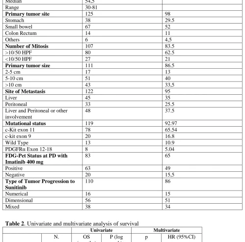
Regarding Overall Survival (OS), the results of multivariable analysis of mortality in the 178
competitive risks model are summarized in the Table 2. Baseline PET status, site of metastasis and 179
mutational status were notsignificantly associated with GISTs OS; tumor site was able to predict for 180
OS at multivariate analysis (p=0,0004): patients with small intestine [HR: 0.22 (0.08-0.57), 181
p=0.002] and rectum as primary tumor sites [HR: 0.19 (0.07-0.561), p=0.005] achieved a 182
statistically significant longer median OS as compared to stomach [HR: 0.51 (0.19-1.35)]. 183
184
5. Discussion
185
A deeper understanding of the molecular alterations underlying the development of GISTs, 186
including mutational activation of KIT or PDGFRα has led to the approval of new effective targeted 187
therapies thatrevolutioned the management and treatment of this disease. Imatinib 400 mgcurrently 188
represents the new backbone of first line treatment in patients with metastatic GISTs leading to a 189
significant improvement in termsof PFS, OS, and quality of life, as reported in a phase II and III 190
trials[12-14]. However, despite the high efficacy of this therapy, the majority of tumors develop 191
acquired resistance to imatinib and experience diseaseprogression. Imatinib 800 mgand sunitinib 192
represent the current standard second-line treatments, representing two feasible and effective 193
7
17, 18]. Imatinib dose escalation could be considered in patients who started on imatinib 400 mg,on 195
the basis of randomized dosefindingtrials revealing theefficacy of this strategy in both American 196
and European populations[12, 13]. In the European study, 247 of the 473 patients were randomly 197
assigned toimatinib low dose arm, and 133 who progressed were crossed over to higher dose of 198
imatinib [13].There were almost 36 patients with a prolonged stable disease and 3 partial 199
responses.Median PFS were 1.7 and 2.0 years for imatinib 400 mg and 800 mg arms respectively 200
(HR: 0.91;p= 0.18), and median OS was 3.9 years in both treatment arms. Similar results were 201
reported inthe American trial. Median PFS was 18 months for patients on imatinib 400 mg, and 20 202
months forthose receiving imatinib 800 mg [14]. Median OS was 55 and 51 months, 203
respectively.Sunitinib is an active TKI that has been approved on the basis of a large phase III trial 204
where 412patients imatinib refractory were randomized 2:1 to receive sunitinib or placebo. Median 205
time totumour progression was 27.3 weeks in sunitinib group and 6.4 weeks in placebo group (HR: 206
0.33;p<0.0001). Although survival was significantly better with sunitinib in the initial report, over 207
timeOS converged in the sunitinib and placebo arms (median 72.7 vs. 64.9 weeks; HR, 0.876; P 208
=0.306), given the cross-over design [19]. 209
Given the uncertainly on the best therapy to adopt in secondline for imatinib refractory GISTs 210
patients, our retrospective study reports a real word series assessingpotential predictive factors of 211
response to sunitinib in order to drive physicians’ choice in thissetting. 212
Mutational status in controlled clinical trials, significantly influences the activity of sunitinib [20]. 213
In a phase I/II trial, PFS and OS were meaningfully longer for patients with a primary KIT 9 exon 214
(58 %) or PDGFRα mutation (56 %) than for those with a KIT exon 11 mutation (34 %). However, 215
our study did not confirm previously data. 216
The present study, according to our best knowledge, represents the largest series of GIST patients 217
after imatinib failure analyzed for mutational status as predictive factors of response to sunitinib 218
treatment, in routine clinical practice outside randomized, controlled, clinical trials. 219
In previous studies, KIT mutation status appears as a predictor of tumor response to sunitinib. 220
The first study (M. C. Heinrich et al, 2008) explore the relationship between GIST kinase 221
mutations, KIT or PDGFRA, and the response to sunitinib in 77 patients with Imatinib-Resistant 222
GIST. PFS and OS were significantly longer in patients with primary KIT exon 9 mutations or a 223
wild-type genotype than in those with KIT exon 11 mutations. 224
Anotehr study on 74 patients tested for KIT and PDGFRA mutations (Dok Hyun Yoon et al, 2010), 225
showed that patients with KIT exon 9 mutant GIST (n=11, 14.9%) have better TTP (median 13.6 226
mo vs 6.9 mo, p=0.631) than those with KIT exon 11 mutant GIST (n=47, 63.5%), although 227
8
Consistent with previous studies, a third studying a group of 89 patients (Rutkowski et al, 2012) 229
confirmed that patients with primary tumors carrying mutations in KIT exon 9 or wild-type had 230
substantially better 1-year PFS (68% and 57%; median 65.5 and 50.5 weeks, respectively) than 231
patients having tumors with KIT exon 11 or PDGFRA mutations (34% and 15%; median 36.8 and 9 232
weeks, respectively). 233
Of note, our results, contrary to previous studies, don’t show that PFS were longer for patients who 234
had mutations in KIT exon 9, than KIT exon 11 mutations. Median PFS for KIT exon 9 was 10 235
months (n=20, 16.8%), similar to PFS for KIT exon 11, that was 10 months (n=78, 65.5%). 236
Median PFS was, instead, significantly longer in patients with wild-type genotype than exon 11 237
patients. (n=13, 10.9%, median PFS 20 mo). 238
Therefore, contrary to previous studies, with a larger sample (119 pts), we have proven that, GISTs 239
harboring KIT exon 9 mutations not appear to be more sensitive to sunitinib than those with 240
primary KIT exon 11 mutations. Consistent with cited studies, instead, wild-type genotype patients 241
have better PFS than KIT exon 11 and exon 9. 242
Similar results were for overall survival, with a median of 76 months for KIT exon 11 mutation and 243
72 months for exon 9. The median OS for wild-type was, instead, lower than Kit mutant GIST (67 244
mo). 245
Our data showed that primary tumor site is predictive for PFS. Patients with small bowel and 246
stomach as primary tumor sites achieved a statistically significant longer median PFS compared to 247
the other sites. 248
Regarding the site of metastasis, the peritoneal localization seems to achieve longer PFS than 249
othermetastatic sites, although not statistically significant. The greater efficacy on peritoneal 250
metastasis might be explained by the role ofangiogenesis in peritoneal progression and the strong 251
anti-angiogenic activity of sunitinib [21]. Not all metastases have similar vascularization. In the 252
liver there can be small or large liver metastases; necrosis is common in larger masses. Peritoneal 253
metastasis are usually small (often < 2 cm) and homogeneously enhancing and vascularization. This 254
can be an explanation for the better activity of sunitinib in patients with peritoneal metastasis. Many 255
studies indicate that dual inhibition of PDGFR and VEGFR from sunitinib produces greater 256
antiangiogenic effects than inhibition of only one such as for imatinib, and the lower dimension and 257
the homogeneous vascularization typical of the peritoneal metastasis, may contribute to better 258
activity from sunitinib[22,23,24]. 259
Moreover, our study showed that thetype of radiologic progression and baseline PET status
260
werenot statistically significant predictors of response to sunitinib, although numeric progression 261
9 263
6. Conclusion
264
This study identifies the gastric site of primarytumor as a predictive factor to sunitinib efficacy in 265
second line setting. The mutational status(GIST WT), the site of metastasis (peritoneum) and the 266
FDG-PET status (negative), although notstatistically significant, seem to be elements of increased 267
activity for sunitinib treatment. These results provide the rationale to drive physician’schoice in 268
second line setting formetastatic GISTs, to improve patients selection and maximize survival 269
benefit onthe basis of possible predictive factors of response. 270
271
Figure 1
272
273
Figure 1: Small bowel and stomach as primary tumor sites achieved a statistically significant longer median PFS (12 274
and 10 months, p<0,0001) compared to the other sites. 275
276
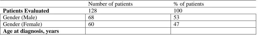
Table 1. Main characteristics of study patients. 277
Number of patients % of patients
Patients Evaluated 128 100
Gender (Male) 68 53
Gender (Female) 60 47
10
Median 54,5
Range 30-81
Primary tumor site 125 98
Stomach 38 29.5
Small bowel 67 52
Colon Rectum 14 11
Others 6 4,5
Number of Mitosis 107 83.5
>10/50 HPF 80 62.5
<10/50 HPF 27 21
Primary tumor size 111 86.5
2-5 cm 17 13
5-10 cm 51 40
>10 cm 43 33,5
Site of Metastasis 122 95
Liver 45 35
Peritoneal 33 25.5
Liver and Peritoneal or other involvement
48 37.5
Mutational status 119 92.97
c-Kit exon 11 78 65.54
c-kit exon 9 20 16.8
Wild Type 13 10.9
PDGFRα Exon 12-18 8 5.04
FDG-Pet Status at PD with Imatinib 400 mg
83 65
Positive 63 49
Negative 20 15,5
Type of Tumor Progression to Sunitinib
110 86
Numerical 16 15
Dimensional 56 51
Mixed 38 34
278
Table 2. Univariate and multivariate analysis of survival 279
Univariate Multivariate
N. OS
(months)
P (log rank)
p HR (95%CI)
Sex 0.14
M 68 79.9
F 60 88.5
N. of mitosis/50
HPF
0.06
≤5 27 107.1
>5 80 82.1
Tumor size 0.05
2-5 cm 17 92.9
5-10cm 51 95.7
>10cm 43 75.5
Tumor Site <0.0001 0.0004
Stomach 38 68.9 0.18 0.51 (0.19-1.35)
11
Rectum 14 105.1 0.005 0.19 (0.07-0.61)
Others 6 46.3
Metastasis site 0.54
Liver 45 81.7
Liver and Peritoneum
40 78.2
Peritoneum 33 92.4
Others 8 99.2
Gene status 0.62
c-kit 11 78 86.3
c-kit 9 20 80.3
PDGFRα 12 2 45.0
PDGFRα 18 6 80.3
WT 13 80.9
PET 0.35
N/A 35
Positive 63 77.2
Negative 20 89.1
12
Compliance with Ethical Standards
303 304
Acknowledgments: The authors thank Dr. Chiara Drago for the English language revision. 305
306
Availability of data and materials
307
All data used or analyzed in this study are included in this published article or are available from the 308
corresponding author on reasonable request. 309
Authors’ contributions
310
All authors have contributed, read and approved the final manuscript. 311
Ethics approval and consent to participate
312
A written informed consent was obtained from each patient for the acquisition of clinical and 313
molecular data to be included in the study. All clinical information for each patient was 314
anonymously recorded and coded after a written informed consent. The study was approved by 315
ethical committee (Comitato Etico Palermo 1) of the university- affiliated hospital AOUP ‘P. 316
Giaccone’ of Palermo (G-Land 2017, approval number: 01-03-2017). 317
Patient consent for publication
318
Not applicable. 319
Competing interests
320
The authors declare no competing interests. 321
13 335
336 337
References
338
1. Miettinen, M.; Lasota, J. Gastrointestinal stromal tumors--definition, clinical, histological, 339
immunohistochemical, and molecular genetic features and differential diagnosis. Virchows Arch. 340
2001,438,1-12. 341
2. Tran, T.; Davila, J.A.; El-Serag, H.B. The epidemiology of malignant gastrointestinal stromal 342
tumors: an analysis of 1,458 cases from 1992 to 2000. Am J Gastroenterol. 2005,100,162-168. 343
3. Nilsson, B.; Bumming, P.; Meis-Kindblom, J.M.; et al. Gastrointestinal stromal tumors: the 344
incidence, prevalence, clinical course, and prognostication in the preimatinib mesylate era—a 345
population-based study in western Sweden. Cancer. 2005,103,821-829. 346
4. Reith, J.D.; Goldblum, J.R.; Lyles, R.H.; Weiss, S.W. Extragastrointestinal (soft tissue) stromal 347
tumors: an analysis of 48 cases with emphasis on histologic predictors of outcome. Mod Pathol. 348
2000,13,577-585. 349
5. Rubin, B.P.; Fletcher, J.A.; Fletcher, C.D. Molecular Insights into the Histogenesis and 350
Pathogenesis of Gastrointestinal Stromal Tumors. Int J Surg Pathol. 2000,8,5-10. 351
6. Medeiros F, Corless CL, Duensing A, et al. KIT-negative gastrointestinal stromal tumors: proof 352
of concept and therapeutic implications. Am J SurgPathol. 2004,28,889-894. 353
7. Andersson, J.; Sjogren, H.; Meis-Kindblom, J.M.; Stenman, G.; Aman, P.; Kindblom, L.G. The 354
complexity of KIT gene mutations and chromosome rearrangements and their clinical correlation in 355
gastrointestinal stromal (pacemaker cell) tumors. Am J Pathol. 2002,160,15-22. 356
8. Mussi, C.; Schildhaus, H.U.; Gronchi, A.; Wardelmann, E.; Hohenberger, P. Therapeutic 357
consequences from molecular biology for gastrointestinal stromal tumor patients affected by 358
neurofibromatosis type 1. Clin Cancer Res. 2008,14,4550-4555. 359
9. Boikos, S.A.; Pappo, A.S.; Killian, J.K.; et al. Molecular Subtypes of KIT/PDGFRA Wild-Type 360
Gastrointestinal Stromal Tumors: A Report From the National Institutes of Health Gastrointestinal 361
Stromal Tumor Clinic. JAMA Oncol. 2016,2,922-928. 362
10. Plaat, B.E.; Hollema, H.; Molenaar, W.M.; et al. Soft tissue leiomyosarcomas and malignant 363
gastrointestinal stromal tumors: differences in clinical outcome and expression of multidrug 364
resistance proteins. J Clin Oncol. 2000,18,3211-3220. 365
11. Badalamenti, G.; Rodolico, V.; Fulfaro, F.; et al. Gastrointestinal stromal tumors (GISTs): focus 366
on histopatological diagnosis and biomolecular features. Ann Oncol. 2007,18(supp 6),vi 136-vi140, 367
14
12. Joensuu, H.; Roberts, P.J.; Sarlomo-Rikala, M.; et al. Effect of the tyrosine kinase inhibitor 369
STI571 in a patient with a metastatic gastrointestinal stromal tumor. N Engl J Med. 2001,344 ,1052-370
1056. 371
13. Blanke, C.D.; Demetri, G.D.; von Mehren, M.; et al. Long-term results from a randomized 372
phase II trial of standard- versus higher-dose imatinib mesylate for patients with unresectable or 373
metastatic gastrointestinal stromal tumors expressing KIT. J Clin Oncol. 2008,26,620-625. 374
14. Verweij, J.; Casali, P.G.; Zalcberg, J.; et al. Progression-free survival in gastrointestinal stromal 375
tumours with high-dose imatinib: randomised trial. Lancet. 2004,364,1127-1134. 376
15. Blanke, C.D.; Rankin, C.; Demetri, G.D.; et al. Phase III randomized, intergroup trial assessing 377
imatinib mesylate at two dose levels in patients with unresectable or metastatic gastrointestinal 378
stromal tumors expressing the kit receptor tyrosine kinase: S0033. J Clin Oncol. 2008,26,626-632. 379
16. Comparison of two doses of imatinib for the treatment of unresectable or metastatic 380
gastrointestinal stromal tumors: a meta-analysis of 1,640 patients. J Clin Oncol. 2010,28 ,1247-381
1253. 382
17. Rutkowski, P.; Nowecki, Z.I.; Debiec-Rychter, M.; et al. Predictive factors for long-term effects 383
of imatinib therapy in patients with inoperable/metastatic CD117(+) gastrointestinal stromal tumors 384
(GISTs). J Cancer Res Clin Oncol. 2007,133,589-597. 385
18. Demetri, G.D.; van Oosterom, A.T.; Garrett, C.R.; et al. Efficacy and safety of sunitinib in 386
patients with advanced gastrointestinal stromal tumour after failure of imatinib: a randomized 387
controlled trial. Lancet. 2006,368,1329-1338. 388
19. George, S.; Blay, J.Y.; Casali, P.G.; et al. Clinical evaluation of continuous daily dosing of 389
sunitinib malate in patients with advanced gastrointestinal stromal tumour after imatinib failure. Eur 390
J Cancer. 2009,45,1959-1968. 391
20. Choi, H.; Charnsangavej, C.; Faria, S.C.; et al. Correlation of computed tomography and 392
positron emission tomography in patients with metastatic gastrointestinal stromal tumor treated at a 393
single institution with imatinib mesylate: proposal of new computed tomography response criteria. J 394
Clin Oncol. 2007,25,1753-1759. 395
21. Demetri, G.D.; Garrett, C.R.; Schoffski, P.; et al. Complete longitudinal analyses of the 396
randomized, placebo-controlled, phase III trial of sunitinib in patients with gastrointestinal stromal 397
tumor following imatinib failure. Clin Cancer Res. 2012,18,3170-3179. 398
22. Demetri, G.D.; Heinrich, M.C.; Fletcher, J.A.; et al. Molecular target modulation, imaging, and 399
clinical evaluation of gastrointestinal stromal tumor patients treated with sunitinib malate after 400
15
23. Heinrich, M.C.; Maki, R.G.; Corless, C.L.; et al. Primary and secondary kinase genotypes 402
correlate with the biological and clinical activity of sunitinib in imatinib-resistant gastrointestinal 403
stromal tumor. J Clin Oncol. 2008,26,5352-5359. 404
24. Vincenzi, B.; Nannini, M.; Badalamenti, G.; et al. Imatinib rechallenge in patients with 405
advanced gastrointestinal stromal tumors following progression with imatinib, sunitinib and 406