Electronic Thesis and Dissertation Repository
8-9-2013 12:00 AM
Ionic and Osmotic Mechanisms Of Insect Chill-Coma And Chilling
Ionic and Osmotic Mechanisms Of Insect Chill-Coma And Chilling
Injury
Injury
Heath A. MacMillan
The University of Western Ontario Supervisor
Dr. Brent J. Sinclair
The University of Western Ontario
Graduate Program in Biology
A thesis submitted in partial fulfillment of the requirements for the degree in Doctor of Philosophy
© Heath A. MacMillan 2013
Follow this and additional works at: https://ir.lib.uwo.ca/etd Part of the Systems and Integrative Physiology Commons
Recommended Citation Recommended Citation
MacMillan, Heath A., "Ionic and Osmotic Mechanisms Of Insect Chill-Coma And Chilling Injury" (2013). Electronic Thesis and Dissertation Repository. 1410.
https://ir.lib.uwo.ca/etd/1410
This Dissertation/Thesis is brought to you for free and open access by Scholarship@Western. It has been accepted for inclusion in Electronic Thesis and Dissertation Repository by an authorized administrator of
i
IONIC AND OSMOTIC MECHANISMS OF INSECT CHILL-COMA AND CHILLING INJURY
(Thesis format: Integrated Article)
by
Heath Andrew MacMillan
Graduate Program in Biology
A thesis submitted in partial fulfillment of the requirements for the degree of
Doctor of Philosophy
The School of Graduate and Postdoctoral Studies The University of Western Ontario
London, Ontario, Canada
ii
Abstract
iii
Keywords
iv
Co-authorship statement
Chapter 2 was published as a review article in Journal of Insect Physiology (reprint permission in Appendix D). I was the first author of this publication, and the co-author was Brent J. Sinclair (BJS), who contributed in the conception of the ideas and helped in writing the manuscript.
Chapter 3 was published in The Journal of Experimental Biology (reprint permission in Appendix D). I was the first author, and the co-authors were Caroline M. Williams (CMW) James F. Staples (JFS) and BJS. CMW helped in the design and implementation of the respirometry experiment, and JFS and BJS contributed to the overall experimental design. All of the authors contributed to the preparation of the manuscript and were involved in the synthesis of many of the ideas.
Chapter 4 was published in The Journal of Experimental Biology (reprint permission in Appendix D). I was the first author, and BJS was the co-author. BJS contributed to the experimental design, was involved in the synthesis of many of the ideas, and helped write the manuscript.
Chapter 5 was published in Proceedings of the National Academy of Sciences of the United States of America (reprint permission in Appendix D). I was the first author, and the co-authors were CMW, JFS and BJS. CMW helped in the design and implementation of the respirometry experiments, and JFS and BJS contributed to the overall experimental design. All of the authors contributed to the preparation of the manuscript and were involved in the synthesis of many of the ideas.
v
Acknowledgments
First, I am thankful for my wonderful wife, whose patience for my academic pursuits is (nearly!) endless. Christine, thank you for your support over the past 10 years. You mean everything to me. I would also like to thank my mother, Victoria MacMillan, for encouraging me to do what I love most in this life, and my father, Rick MacMillan, for introducing me to the beauty of the natural world as a child. I thank my brother, Tavis MacMillan, for his enthusiasm for science and his love of the absurd. While on the topic of family, I thank my imminently expected daughter, whose due date has been an effective motivator for thesis writing. Thank you for your timely (and entirely out of your control) existence.
It is impossible for me to imagine a PhD thesis under the supervision of better mentors than Brent Sinclair and Jim Staples. Both Brent and Jim have had an enormous influence on how I view the world, and on my role in it as a scientist. I thank Brent for his endless enthusiasm, criticism, and advice during my academic development, for recognizing my potential, and for pointing that potential toward an important topic. I thank Jim for his positivity and encouragement throughout both my undergraduate and graduate careers, and for inspiring me to pursue a career in science at a critical point in my life.
Other faculty members at Western have had an important impact on my career. I wish to thank André Lachance, for educating me on the importance of systematics in modern biology, for allowing me to pester him with questions on phylogenetics, and for officiating my marriage to Christine. I would also like to thank Mark Bernards, Jane Bowles, Robert Cumming, Sheila Macfie, Jeremy McNeil, and Amanda Moehring, for their contributions to my undergraduate education and/or useful discussions during my time in the graduate program.
vi
Brown, Dillon Chung, Litza Coello, Alex Cooper, Jill Crosthwaite, Lauren Des Marteaux, Laura Ferguson, Alexander and Meghan Gerson, Ruth Jakobs, Liam McGuire, Kristen Nicholson, Eddy Price, Arun Rajamohan, Golnaz Salehipour-shirazi, Justin Saindon, Stephanie Sobek-Swant, Lauren Strachan, Raymond Thomas, Hiroko Udaka, and Jian Zhang, thank you. I am known for my poor memory among my peers, and I trust that anyone I have missed in this list will forgive me. Whoever you may be, you probably mean a lot to me!
I am grateful for the Ontario Graduate Scholarship program, the Natural Sciences and Engineering Research Council of Canada (NSERC) and the Department of Biology at the University of Western Ontario for financial support over the last 6 years. I strongly believe that receipt of this financial support seeded my success as an undergraduate and graduate student by giving me the financial freedom to focus on my professional development.
Lastly, I would like to thank Brent Sinclair, Jim Staples, Chris Guglielmo, Norman Hüner, and Andrew Watson, for encouraging me to think beyond the scope of my initial research goals and embrace the unexpected. The perspective you fostered has stuck with me, and has led me down an exciting research path I could not have foreseen when I decided to start a PhD on chill-coma.
“Traditional fields of study are going to continue to grow, and in doing so, inevitably they will meet and create new disciplines. In time, all of science will come to be a continuum of description, an explanation of networks, of principles and laws. That’s why
you need not just be training in one specialty, but also acquire breadth in other fields, related to and even distant from your own initial choice.” – Edward O. Wilson, Advice to
vii
Table of contents
Abstract ... ii!
Co-authorship statement ... iv!
Acknowledgments ... v!
Table of contents ... vii!
List of tables ... xiii!
List of figures ... xiv!
List of videos... xvii!
List of symbols and abbreviations ... xviii!
1! General introduction ... 1!
1.1! Biotic impacts of a changing climate ... 1!
1.2! What determines thermal limits to performance? ... 3!
1.2.1! There is little evidence for oxygen- and capacity-limitation in terrestrial ectotherms ... 4!
1.3! Insects at low temperatures ... 5!
1.3.1! The insect CTmin ... 6!
1.3.2! Freeze tolerance and freeze avoidance ... 6!
1.3.3! Chill susceptibility ... 9!
1.3.4! Variation in cold tolerance ... 11!
1.3.5! Does a loss of ion homeostasis underlie insect chill-coma and chilling injury? ... 11!
1.4! Insect ion and water balance ... 12!
1.4.1! Ion homeostasis in excitable tissues ... 12!
1.4.2! The insect renal system ... 14!
viii
2! Physiological mechanisms of insect chill-coma ... 30!
2.1! Introduction ... 30!
2.2! Measuring chill-coma ... 31!
2.3! Plasticity of chill-coma ... 33!
2.4! Possible mechanisms underlying chill-coma in insects ... 34!
2.4.1! Whole organism oxygen limitation ... 34!
2.4.2! Signal transmission failure ... 35!
2.4.3! Disruption of ion regulation ... 37!
2.4.4! Mechanisms underlying chill-coma recovery ... 45!
2.5! Conclusions ... 45!
2.6! Literature cited ... 47!
3! Metabolism and energy supply below the critical thermal minimum of a chill-susceptible insect ... 56!
3.1! Introduction ... 56!
3.2! Materials and methods ... 59!
3.2.1! Respirometry ... 59!
3.2.2! ATP and lactate quantification during cold exposure and recovery ... 60!
3.2.3! Data analysis ... 61!
3.3! Results ... 64!
3.4! Discussion ... 69!
3.5! Literature cited ... 72!
4! The role of the gut in insect chilling-injury: cold-induced disruption of osmoregulation in the fall field cricket, Gryllus pennsylvanicus ... 77!
4.1! Introduction ... 77!
ix
4.2.2! Determination of CTmin and survival of chronic cold exposure ... 81!
4.2.3! Tissue sampling ... 81!
4.2.4! Sample preparation and analysis ... 82!
4.2.5! Data analysis ... 83!
4.3! Results ... 84!
4.3.1! CTmin and survival at 0 °C ... 84!
4.3.2! Hemolymph and muscle ion concentrations ... 84!
4.3.3! Tissue water content ... 87!
4.3.4! Hemolymph and gut ion content ... 91!
4.3.5! Hemolymph color ... 94!
4.4! Discussion ... 94!
4.4.1! Osmotic balance of the hemolymph and muscle ... 95!
4.4.2! Osmotic balance of the alimentary canal ... 97!
4.4.3! Conclusion ... 99!
4.5! Literature cited ... 100!
5! Reestablishment of ion homeostasis during chill-coma recovery in the cricket Gryllus pennsylvanicus ... 105!
5.1! Introduction ... 105!
5.2! Materials and methods ... 107!
5.2.1! Animal husbandry ... 107!
5.2.2! Chill-coma recovery time ... 107!
5.2.3! Measurement of ion and water balance ... 107!
5.2.4! Respirometry ... 110!
5.2.5! Data analysis ... 111!
x
5.3.2! Loss and recovery of ion concentration, ion content and muscle
equilibrium potentials ... 114!
5.3.3! Recovery of osmotic homeostasis after cold exposure is metabolically costly ... 116!
5.3.4! A conceptual model of chill-coma onset and recovery ... 120!
5.4! Literature cited ... 122!
6! Phenotypic plasticity and evolution of Drosophila cold tolerance are associated with modulation of Na+ and K+ homeostasis ... 126!
6.1! Introduction ... 126!
6.1.1! Drosophila as a model for variation in cold tolerance ... 127!
6.1.2! Disruption of ion homeostasis underlies CTmin and chilling injury in insects ... 128!
6.1.3! Na+/K+-ATPase plays important ionoregulatory roles in Drosophila ... 129!
6.1.4! Does modulation of ion homeostasis underlie variation in insect cold tolerance? ... 130!
6.1.5! Objectives and hypotheses ... 131!
6.2! Materials and methods ... 133!
6.2.1! Animal origins and husbandry ... 133!
6.2.2! Measurement of CTmin ... 134!
6.2.3! Hemolymph ion concentration ... 134!
6.2.4! Na+/K+-ATPase activity ... 137!
6.2.5! Data analysis ... 141!
6.2.6! Phylogenetically-independent contrasts ... 144!
6.3! Results ... 147!
6.3.1! Critical thermal minima ... 147!
xi
6.3.4! Na+/K+-ATPase transcript and protein abundance ... 153!
6.4! Discussion ... 156!
6.4.1! Cold acclimation mitigates the effects of low temperature on hemolymph [K+] ... 156!
6.4.2! Improvements in cold tolerance through modulation of resting hemolymph [Na+] and [K+] ... 157!
6.4.3! Do cold tolerant insects decouple Na+ and water balance? ... 158!
6.4.4! Drosophila cold tolerance is associated with low Na+/K+-ATPase activity ... 159!
6.4.5! Conclusion ... 162!
6.5! Literature cited ... 162!
7! General Discussion ... 173!
7.1! Thesis summary ... 173!
7.2! A conceptual model of chill-susceptibility ... 174!
7.3! Limitations and remaining gaps in the model ... 177!
7.3.1! Taxonomic and contextual expansion of the conceptual model ... 178!
7.3.2! Does ion balance disruption cause chill-coma and chilling injury? ... 179!
7.3.3! Elucidating the routes of ion and water migration in the cold ... 181!
7.4! What are the mechanisms of cold tolerance plasticity and evolution? ... 181!
7.4.1! Variation in ion pump thermal sensitivity and the role of the membrane environment ... 184!
7.4.2! Modulation of transcellular and paracellular permeability ... 185!
7.4.3! Modification of cellular and epithelial ion gradients and diet effects on cold tolerance ... 188!
7.4.4! Inhibition of apoptosis – a final stand against chilling injury? ... 190!
xii
Appendix A: Chapter 4 supplementary material ... 199!
Appendix B: Chapter 5 supplementary material ... 202!
Appendix C: Chapter 6 supplementary material ... 208!
Supplementary methods ... 208!
Hemolymph collection ... 208!
Measurement of Na+/K+-ATPase activity ... 208!
Appendix D: Reprint permissions ... 218!
Reprint permission for chapter 2 ... 218!
Reprint permission for chapters 3 and 4 ... 222!
Reprint permission for chapter 5 ... 223!
xiii
List of tables
Table 7.1. Physiological mechanisms hypothesized to underlie variation in the cold tolerance of chill susceptible insects. ... 183!
Table A.1. Results of false discovery rate corrected t-tests and general linear models of hemolymph and muscle ion concentrations and calculated muscle equilibrium potentials of Gryllus pennsylvanicus exposed to 0 °C. ... 199!
Table A.2. Results of false discovery rate corrected t-tests and general linear models of hemolymph and tissue water content of Gryllus pennsylvanicus exposed to 0 °C. ... 200!
Table A.3. Results of false discovery rate corrected t-tests and general linear models of hemolymph, foregut, midgut and hindgut ion content of Gryllus pennsylvanicus exposed to 0 °C. ... 201!
Table B.1. Results of linear and non-linear regression of cricket chill-coma recovery time and cold exposure duration. ... 202!
Table B.2. Results of t-tests of the effects of 24 h of exposure to 0 °C on hemolymph, muscle and gut ion and water content. ... 203!
Table B.3. Results of linear (L) and non-linear (NL) models (M) of G. pennsylvanicus hemolymph, muscle and gut ion and water content during chill-coma recovery following 24 h at 0 °C. ... 204!
Table C.1. Details of Drosophila species stock origins and critical thermal minima
estimates. ... 214!
xiv
List of figures
Figure 1.1. Terminology of cold tolerance measures typically quantified in a
chill-susceptible insect in the laboratory. ... 7!
Figure 1.2. Overview of physiological mechanisms of ion and water homeostasis of the hemolymph (white) gut lumen (light grey) and cell cytoplasm (dark grey) of a typical phytophagous insect. ... 13!
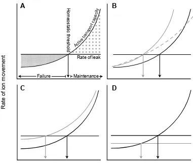
Figure 2.1. Schematics of the relationship between the capacity to actively transport ions across membranes (which has a Q10 between 2 and 3) and the rate of ion diffusion through available pathways (relatively unaffected by temperature; Zachariassen et al., 2004) to illustrate possible mechanisms underlying chill-coma in insects. ... 39!
Figure 2.2: The interactive effects of cold exposure temperature and duration on chill-coma recovery time of female Drosophila melanogaster reared at 25 °C. ... 44!
Figure 2.3. Schematic of physiological mechanisms of neuromuscular transmission
failure thought to underlie insect chill-coma at low temperatures. ... 46!
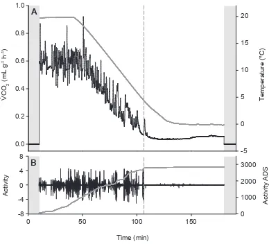
Figure 3.1. An example of respirometry and activity data from one adult female Gryllus pennsylvanicus during a temperature ramp. ... 63!
Figure 3.2. CO2 release and slope of activity absolute difference sum (ADS) of female Gryllus pennsylvanicus during a linear temperature ramp from 20°C to 0°C at 0.25°C min-1. ... 65! Figure 3.3. Concentrations of ATP (black), lactate (white) and alanine (gray) in the hind femur of female adult Gryllus pennsylvanicus (A) during exposure to 0 °C and (B) during recovery at 20 °C from 24 h in chill-coma at 0°C. ... 66!
Figure 3.4. Concentrations of ATP, lactate and alanine in the hind femur of female adult Gryllus pennsylvanicus under normoxic conditions (white bars) and following 1h of
xv
exposure to 0 °C for up to 120 h. ... 85!
Figure 4.2. Concentration (mean ± sem) of Na+ (A), K+ (B), Mg2+ (C) and Ca2+ (D) in hemolymph (filled) and femur muscle tissue (open) of G. pennsylvanicus during exposure to 0 °C for up to 120 h. ... 86!
Figure 4.3. Calculated equilibrium potentials (mean ± sem) of Na+ (filled circles), K+ (open circles), Mg2+ (filled triangles) and Ca2+ (open triangles) of muscle cell membranes of G. pennsylvanicus during prolonged exposure to 0 °C. ... 88!
Figure 4.4. Mean ± sem muscle water content (A; expressed as µL per mg of muscle dry mass) and hemolymph volume (filled circles) and total gut water content (open circles) (B) of G. pennsylvanicus during exposure to 0 °C for up to 120 h. ... 89!
Figure 4.5. Mean (± sem) foregut (A), midgut (B), and hindgut (C) water content
(expressed as µL per mg tissue dry mass) of G. pennsylvanicus exposed to 0 °C for up to 120 h... 90!
Figure 4.6. Mean (± sem) total Na+ (A), K+ (B), Mg2+ (C), and Ca2+ (D) content of the hemolymph of G. pennsylvanicus during exposure to 0 °C for up to 120 h. ... 92!
Figure 4.7. Mean (± sem) Na+ (A-C), K+ (D-F), Mg2+ (G-I), and Ca2+ (J-L) of the foregut (left) midgut (centre) and hindgut (right) of G. pennsylvanicus during exposure to 0 °C for 120 h. ... 93!
Figure 5.1. The relationship between gravimetric estimates of hemolymph volume and volume quantified using the inulin dilution method in G. pennsylvanicus. ... 108!
xvi
hemolymph ion content (C) of Na+ (black) and K+ (grey), and (D) hemolymph volume (filled squares) and gut water content (open squares) of female G. pennsylvanicus during recovery at 22 °C following 24 h at 0 °C. ... 115!
Figure 5.4. An example measurement of metabolic rate (rate of CO2 emission, black line, baseline-corrected values shown) and activity (grey line, arbitrary units) of a G.
pennsylvanicus female (A) prior to and (B) immediately upon removal from a 20 h
exposure to 0 °C. ... 118!
Figure 5.5. The relationship between the duration of metabolic overshoot during chill-coma recovery and exposure to 0 °C (solid circles). ... 119!
Figure 5.5. A conceptual model of the effects of temperature (grey line) on muscle resting potential (black line) of a chill-susceptible insect during a typical experimental cold
exposure. ... 121! Figure 6.1. Flow-chart of experimental design. ... 135!
Figure 6.2. Example of a logistic model (solid grey line) fitted to Na+/K+-ATPase activity of warm-acclimated Drosophila melanogaster (solid black line). ... 143! Figure 6.3. Phylogeny of the genus Drosophila and among-species variation in the CTmin.146!
Figure 6.4. Hemolymph Na+ (A) and K+ (B) concentrations of cold-acclimated (grey bars) and warm-acclimated (open bars) male Drosophila melanogaster at their
acclimation temperature (Control) and following 6 h at 0 °C. ... 148!
Figure 6.5. Hemolymph concentrations of Na+ (A) and K+ (B) of species of the genus Drosophila in relation to the CTmin. ... 149!
xvii
thermal sensitivity (C) of species of the genus Drosophila in relation to the CTmin. ... 152!
Figure 6.8. Na+/K+-ATPase protein (A, B) and mRNA (C) abundance in warm- and cold-acclimated Drosophila melanogaster. ... 154!
Figure 7.1. A physiological model integrating ionic mechanisms of chill-coma, chilling injury and chill-coma recovery in chill-susceptible insects. ... 175!
Figure B.1. Muscle concentration of Na+ (black) and K+ (grey) during chill-coma
recovery at 22 °C following 24 h at 0 °C. B). ... 205!
Figure B.2. Residual water (A-D), Na+ (E-H), and K+ (I-L) content of each of the three gut segments and total gut of crickets during chill-coma recovery at 22 °C following 24 h at 0 °C. ... 206!
Figure C.1. Schematic of the apparatus used to collect hemolymph from Drosophila
without anesthesia. ... 210!
Figure C.2. Na+/K+-ATPase activity recorded using a temperature ramp and comparison to activity measured at static temperatures. ... 212!
Figure C.3. Histograms of critical thermal minima of female (black bars) and male (grey bars) flies of the genus Drosophila. ... 216!
Figure C.4. Phylogenetically-independent contrasts (PICs) of the critical thermal minimum (CTmin) regressed against hemolymph ion concentrations (A,B) and Na+/K+ -ATPase parameters (C-E; See Section 6.2.6). ... 217!
List of videos
xviii Ψ Membrane potential
aADS Absolute difference sum of activity ADS Absolute difference sum
ATP Adenosine triphosphate ATPase Adenosine triphosphatase CCR Chill-coma recovery time
Tcc Chronic exposure temperature that causes chill-coma CTmax Critical thermal maximum
CTmin Critical thermal minimum EX equilibrium potential of X
IP Inflection point of temperature-activity curve LD50 Lethal dose for 50 % of individuals
OCLT Oxygen- and capacity-limitation of thermotolerance NADH Nicotinamide adenine dinucleotide (reduced form) Q10 Temperature coefficient
Ts Thermal sensitivity RCH rapid cold hardening
Chapter 1
1
General introduction
1.1 Biotic impacts of a changing climate
Global surface temperatures have risen by 0.5 °C in the last 20 years, and are predicted to rise by at least a further 1.8-4.0 °C by the end of this century as a result of anthropogenic activities (IPCC, 2007a). Along with an increase in mean temperature, the world is already experiencing rapid declines in snow and ice cover, rising ocean levels, increased precipitation and elevated risk of extreme weather events (IPCC, 2007b). The rates of environmental change observed and expected for our planet are unprecedented in the last 11,300 years, and are recognized as a major threat to biodiversity and ecosystem health (Fischlin et al., 2007; Marcott et al., 2013). In particular, climate change increases risks of species extinction and invasive species, and can alter the abundance of species important to agriculture and human health. The potential effects of climate change on biota demand understanding of how changes in the abiotic environment influence fitness, both directly (e.g. physiological limits to tolerance) and indirectly through alterations in biotic interactions (e.g. timing of seasonal events or food availability; Cahill, 2013; Chown and Gaston, 2008; Thackeray et al., 2010).
not necessarily the case. Poleward range limits are in part determined by the frequency and severity of low temperatures over winter, so increased winter survival may facilitate poleward expansion of species ranges (Crozier, 2003). Winter warming in temperate ecosystems may enhance fitness by lengthening the seasons of growth and reproduction, or reduce fitness through phenological shifts (Jeong et al., 2011). For example, extended periods of pre-winter dormancy in the spruce budworm, Choristoneura fumiferana (Lepidoptera: Tortricinae), and increases in post-winter thermal variability in Erynnis propertius (Lepidoptera: Hesperiidae), both reduce fitness by elevating metabolic rate and depleting energy stores required for summer development and reproduction (Han and Bauce, 1998; Williams et al., 2012). Animal distributions have already shifted in response to warming (Chen et al., 2011). In marine systems, distributions of several fish species in the North Sea have shifted northward with increased temperatures (Perry et al., 2005) while in terrestrial systems, shifts in range limits have been well documented in insects and are often directly attributed to warmer winters (Battisti et al., 2005; Chen et al., 2011; Crozier, 2003; Jepsen et al., 2008).
1.2 What determines thermal limits to performance?
Our understanding of how temperature impacts organismal physiology, thereby setting critical thermal limits, is incomplete. In particular, the mechanisms by which high and low temperatures limit the performance of terrestrial animals are largely unknown. In aquatic systems, however, the CTmax and CTmin of animals that breathe water appear to be physiologically linked through the effects of temperature on oxygen supply and demand. The hypothesis of oxygen- and capacity-limitation of thermal tolerance (OCLT) has emerged as a broadly applicable framework of the physiological mechanisms underlying thermal limits to performance in aquatic ectotherms (Pörtner and Farrell, 2008; Pörtner and Knust, 2007). The OCLT hypothesis proposes that limits to animal performance arise from declining aerobic scope (the difference between maximal and standard metabolic rate) at temperature extremes (Pörtner and Knust, 2007). At both high and low temperatures, the circulatory and ventilatory systems become incapable of delivering sufficient oxygen to tissues to maintain aerobic metabolism. Thus, the CTmin and the CTmax represent boundaries of a window of aerobic metabolism, and beyond these boundaries hypoxemia in critical tissues limits aerobic ATP production, leading to failure of energy-demanding processes like muscle contraction (Pörtner and Farrell, 2008). The energy made available through anaerobic metabolism is prioritized to cellular mechanisms of defense, and over time anaerobic byproducts accumulate, causing injury and death. This physiological link between both thermal limits explains why the CTmin and CTmax of marine ectotherms shift in unison across latitudinal clines (Sunday et al., 2011).
more physically or thermally challenging migrations have apparently adapted to these challenges through increased aerobic scope, facilitated by larger hearts and improved coronary blood supply (Eliason et al., 2011). Variation in thermal tolerance of aquatic ectotherms has also been associated with alteration of the aerobic window. Temperate species alter their thermal tolerance by increasing the mitochondrial density of their skeletal muscles (e.g. Tyler and Sidell, 1984; Johnston et al., 1998), increasing cardiac performance at extreme temperatures (e.g. Gamperl and Farrell, 2004), or by modifying the oxygen transport properties of their blood (e.g. Melzner et al., 2007).
Thus, in aquatic animals, there is strong evidence that the thermal window of performance is determined by a thermal window of aerobic metabolism, and that the aerobic window ultimately impacts biogeography. The success of the OCLT hypothesis and its broad applicability to marine animals has led to application of OCLT concepts to predictive models of animal distribution. In the case of Fraser River salmon, a recent study used aerobic scope to predict stock-specific declines in abundance between 9 and 16 % by the end of this century (Martins et al., 2011).
1.2.1
There is little evidence for oxygen- and capacity-limitation in
terrestrial ectotherms
costs of muscle function at low temperatures, rather than temperature effects on oxygen supply (Petersen and Gleeson, 2009; Seebacher and Franklin, 2011).
Unlike aquatic animals and terrestrial vertebrates, insects do not deliver oxygen to tissues via a circulatory system. Instead, gaseous air is delivered to tissues through the tracheal system, blind-ended tubes that are continuous with the external environment. The tracheal system delivers oxygen efficiently even at ambient oxygen concentrations below 10 % (Harrison et al., 2001), meaning oxygen supply to insect tissues is unlikely to be limited in normoxia. A hypothesis that has been derived from OCLT is that experimental modulation of oxygen availability through hypoxia or hyperoxia should alter the CTmin and CTmax (Klok et al., 2004; Pörtner, 2001). By manipulating ambient oxygen concentration, however, Klok et al. (2004) found that oxygen availability only altered the CTmax of tenebrionid beetles (Gonocephalum simplex, Coleoptera: Tenebrionidae) under extreme hypoxia (2.5 % O2). Similarly, oxygen availability has no effect on the CTmin of a related tenebrionid (Tenebrio molitor, Coleoptera: Tenebrionidae; Stevens et al., 2010). By contrast, the CTmax of a terrestrial isopod (Armadillidium vulgare, Isopoda: Armadillidiidea), which does exchange gases though a circulatory system, is reduced in hypoxia (Klok et al., 2004). Thus, the OCLT hypothesis may hold for terrestrial crustaceans, but does not appear to hold for terrestrial vertebrates or tracheated insects. No study to date, however, has directly tested whether chronic low or high temperature exposure leads to failure of aerobic metabolism and accumulation of anaerobic byproducts in an insect, so it remains uncertain if OCLT sets insect thermal limits in normoxia.
1.3 Insects at low temperatures
in soil or under snow, where buffered microclimate temperatures rarely decline below 0 °C (Seeley and Visscher, 1985; Storey and Storey, 2012; Zipkin et al., 2012). Other species, however, regularly experience sub-zero winter temperatures, and some have evolved physiological means of low temperature survival that are traditionally split into two principal strategies: freeze tolerance and freeze avoidance (Salt, 1961).
1.3.1
The insect CT
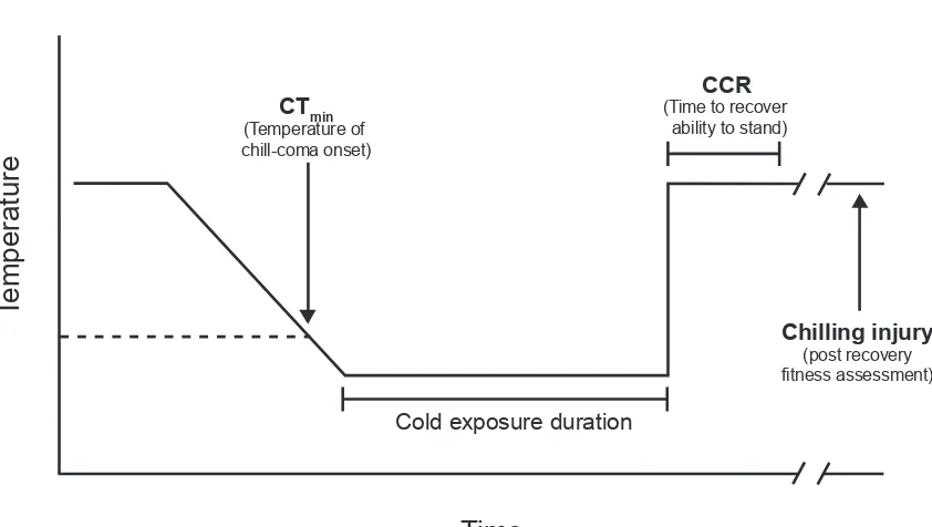
minThe insect CTmin is a temperature that marks the onset of chill-coma, a state of complete paralysis (Figure 1.1; Hazell and Bale, 2011; Mellanby, 1939). Provided the duration of cold exposure is short, chill-coma is reversible, as insects removed from the cold to warmer temperatures will recover the ability to move. Recovery from chill-coma has been quantified as the ambient temperature required for an insect to regain the ability to move (Fordyce and Shapiro, 2003), but is more commonly measured as the time required for the insect to recover the ability to stand following removal from the cold to room temperature, termed chill-coma recovery time (CCR; Figure 1.1; David et al., 1998). Both the CTmin and CCR are commonly-used measures of insect cold tolerance (e.g. Ayrinhac et al., 2004; Folk et al., 2007; Gaston and Chown, 1999; Gibert and Huey, 2001; Gibert et al., 2001; Ransberry et al., 2011). In this dissertation, recovery from chill-coma is always measured as a time to recovery at a static temperature, but some distinctions are made between the time required for the ability to stand (CCR), the recovery of muscle function, and recovery of ventilation (Chapter 3). A detailed introduction to the physiology thought to underlie chill-coma and chill-coma recovery is presented in Chapter 2.
1.3.2
Freeze tolerance and freeze avoidance
Figure 1.1. Terminology of cold tolerance measures typically quantified in a chill-susceptible insect in the laboratory. A typical experimental cold exposure is shown, where temperature is decreased at a constant rate and then held at a low temperature. The CTmin is measured as the temperature at which a loss of coordination occurs (typically an inability to stand or cling to a surface). At temperatures below the CTmin, insects enter chill-coma, which is a state of complete paralysis. Recovery is typically accomplished by removing the insect to room temperature. Chill-coma recovery time (CCR) is the time required to recover the ability to move or stand, and varies according to the duration and temperature of cold exposure. Incidence of chilling injury following a cold stress may be quantified by mortality or impairment of functions related to fitness, typically measured 24 h following removal from the cold stress.
CTmin (Temperature of chill-coma onset)
Temperature
Time
Cold exposure duration
CCR (Time to recover
ability to stand)
In freeze tolerant insects, ice formation is initiated in the extracellular space by ice nucleating agents or ice nucleating proteins, and initiation of freezing at relatively mild sub-zero temperatures may allow for slow and controlled freezing that minimizes physical damage (Layne et al., 1988; Zachariassen and Kristiansen, 2000; but see Sinclair et al., 2009). In freeze tolerant insects, the intracellular space is kept in a liquid state and cell membranes are protected by the accumulation of low molecular weight cryoprotectants such as glucose, glycerol, and trehalose (Storey, 1997). Ice formation in the extracellular space increases local osmotic pressure, which provides the driving force for water to cross cell membranes through aquaporins. The redistribution of water during extracellular freezing reduces cell volume and the likelihood of intracellular freezing (Lee and Denlinger, 2010). Frozen insects survive substantial redistribution of water from intracellular to extracellular spaces during freezing, but lose very little total body water to their environment because extracellular water is largely bound as ice (Irwin and Lee, 2002; Lundheim and Zachariassen, 1993).
Freeze avoiding insects depress the supercooling point (SCP; the temperature at which a fluid spontaneously freezes) of their body fluids and remain unfrozen at low sub-zero temperatures by eliminating potential ice nucleators from the body, producing antifreeze proteins that bind to ice crystals and inhibit their growth, and again by accumulating cryoprotectants (Lee and Denlinger, 2010). In freeze avoiding insects, high concentrations of cryoprotectants facilitate colligative supercooling point suppression. For example, overwintering prepupae of the emerald ash borer (Agrilus planipennis, Coleoptera: Buprestidae) accumulate nearly 4M glycerol in their hemolymph, produce antifreeze agents, and lose substantial body water, all facilitating depression of their SCP to -30 °C (Crosthwaite et al., 2011).
water. Animals that utilize cryoprotective dehydration over the winter remain in vapour pressure equilibrium with their environment (Sørensen and Holmstrup, 2011).
1.3.3
Chill susceptibility
The majority of insects globally are neither freeze tolerant nor freeze avoidant, but are chill-susceptible (Bale, 1996). At temperatures below the CTmin, chill-susceptible insects accumulate injuries and may die from processes that are unrelated to freezing of their body fluids (Bale, 1996; Baust and Rojas, 1985). Such insects are the focus of this dissertation.
Injuries sustained from exposure to low temperatures but unrelated to freezing are termed chilling injury. Chilling injury on a whole-organism level has been quantified in a several ways, the simplest of which is the incidence of mortality following a non-freezing cold exposure (Turnock, 1993; Turnock et al., 1983). Other common measures of chilling injury are functions related to fitness, such as the ability to move in a coordinated fashion. resume development, mate, or lay eggs following recovery from a cold exposure (typically measured 24 h after the stress; Figure 1.1; Koštál and Tollarová-Borovanská, 2009; Koštál et al., 2006; Marshall and Sinclair, 2010; Rinehart et al., 2000; Rojas and Leopold, 1996). The accumulation of chilling injury in chill-susceptible insects is both time- and temperature-dependent; sustained exposure to low temperatures causes more damage and greater mortality than brief exposure to relatively mild temperatures (Nedvĕd et al., 1998; Turnock et al., 1983).
Impairments of whole-organism fitness via cold shock and indirect chilling injury are associated with damage sustained in sensitive tissues, particularly the neuromuscular system and gut. Yocum et al. (1994) noted that flesh flies (Sarcophaga crassipalpis, Diptera: Sarcophagidae) that failed to emerge from their puparium following chronic cold exposure lacked the coordination to do so, and ascribed chilling injury to nervous system damage. Damage to the neuromuscular system can be further inferred from observation of insect gait following low temperature exposure; insects that have sustained chilling injury walk in an uncoordinated manner (Koštál et al., 2006). Cold shock treatments induce high levels of cellular apoptosis in the flight muscles of Drosophila melanogaster (Yi et al., 2007), as well as the muscle, fat body and gut tissues of flesh flies (Yi and Lee, 2004; Yi and Lee, 2011), so chilling injury may result from cold-induced cellular apoptosis.
1.3.4
Variation in cold tolerance
The cold tolerance of chill-susceptible insects can vary widely among species and populations, and many species have substantial capacity for cold tolerance plasticity. Cold tolerance variation has been particularly well characterized in the predominantly chill-susceptible genus Drosophila (Diptera: Drosophilidae). Temperate Drosophila species and populations are generally more cold tolerant (i.e. have lower CTmin and faster CCR) than those from the tropics, and much of this variation persists under common laboratory rearing conditions (Ayrinhac et al., 2004; David et al., 2003; Gibert and Huey, 2001; Kellermann et al., 2012). Cold tolerance of chill-susceptible insects is also highly phenotypically plastic. For example, the CTmin and CCR of Drosophila melanogaster can be altered in response to long-term exposure to variable temperatures in the field, or to laboratory rearing and acclimation temperatures (Nyamukondiwa et al., 2011; Overgaard and Sørensen, 2008; Ransberry et al., 2011). Tolerance of chill-susceptible insects to a severe cold exposure can also be altered by brief (c. 30 min) prior exposure to a relatively mild low temperature, a response termed rapid cold hardening (RCH; Czajka and Lee, 1990; Lee et al., 1987). Thus, chill-susceptible insects are not simply passive victims of their thermal environment, but can often successfully avoid chill-coma and damage from cold exposure through cold tolerance plasticity. The physiological mechanisms by which cold tolerance can be improved in chill-susceptible insects are largely unknown, in part because the mechanisms that drive the CTmin, CCR, RCH, and chilling injury are poorly understood.
1.3.5
Does a loss of ion homeostasis underlie insect chill-coma
and chilling injury?
days (Koštál et al., 2006). Similarly, adult firebugs (Pyrrhocoris apterus, Hemiptera: Pyrrhocoridae) progressively lose ion balance between their hemolymph and tissues and die following long-term exposure to -5 °C (Koštál et al., 2004).
Plasticity of cold tolerance appears to be associated with modulation of ion balance physiology. Cold-acclimated firebugs better maintain ion balance between their hemolymph and tissues, and show markedly better survival following cold exposure than warm-acclimated individuals (Koštál et al., 2004). Similarly, cold-acclimated cockroaches maintain muscle resting potentials at 5 °C and remain active (Koštál et al., 2006). Thus, cold exposure appears to disrupt ion homeostasis in critical tissues that drives both chill-coma and chilling injury. In turn, variation in insect cold tolerance may be mediated by variation in ionoregulatory physiology.
1.4 Insect ion and water balance
If exposure to low temperatures causes a loss of ion homeostasis, cold exposure is likely to impair functions of excitable tissues that are dependent on ion distribution, such as the nerves and muscles, as well as the renal epithelia that maintain whole-organism ionic and osmotic balance.
1.4.1
Ion homeostasis in excitable tissues
Figure 1.2. Overview of physiological mechanisms of ion and water homeostasis of the hemolymph (white) gut lumen (light grey) and cell cytoplasm (dark grey) of a typical phytophagous insect. Intracellular homeostasis of the nervous system (ns) and muscles (m) are primarily maintained by active pumping of the cations Na+, K+ and Ca2+. Nerve axons (a) are protected from changes in hemolymph composition by the blood-brain barrier, composed of glial cells (g) with tight septate junctions (black circles) that restrict paracellular ion and water leak. The outermost layer of the blood-brain barrier is the perineurial glia (pn), which secrete the neural lamella (nl), a thick extracellular matrix. Muscle cells are bathed directly in hemolymph, and ion balance is maintained across the muscle cell membrane. High levels of primary and secondary active ion transport drive transport of ions and water in the insect renal system, which is composed of the Malpighian tubules (Mt) and hindgut (hg). The midgut (mg) and foregut (fg) play minor roles in ion homeostasis that vary among insect taxa.
fg mg hg Mt Upper tubule Lower tubule Posterior hindgut t
Na+ Mg2+ K+ Cl
-Active transport Passive transport
H2O
H2O K+ Cl
-Na+ H2O Cl
-Direction of fluid flow Excitable tissues Renal system Nerve Muscle g pn Na+ K+ Na+ K+ nl a m ns Na+ K+ Ca2+ sr Ca2+
To precisely control the ionic environment of the neurons, the glial cells and the neurons themselves have high levels Na+/K+-ATPase, that promotes high [Na+] and low [K+] in the nerve extracellular fluid (Figure 1.2; Kocmarek and O’Donnell, 2011; Leiserson and Keshishian, 2011; Schofield and Treherne, 1975).
Ion balance in insect muscle differs from that of the nervous system; unlike neurons, insect muscle cells are directly bathed in hemolymph. Muscle resting potential is maintained by active transport of ions across the muscle cell, sarcoplasmic and endoplasmic reticulum membranes, and is predominantly determined by the K+ concentration gradient across the muscle cell membrane (Hoyle, 1953). In most insect groups, Na+/K+-ATPase is the primary regulator of muscle resting potential, and maintains high [K+] and low [Na+] inside muscle cells (Figure 1.2; Emery et al., 1998; Fitzgerald et al., 1996). Lepidoptera differ from this typical arrangement, as they possess little Na+/K+-ATPase in their muscle membranes, maintain extremely low extracellular [Na+] and instead appear to maintain K+ gradients by an H+-ATPase coupled to a K+/Cl -co-transporter (Djamgoz, 1987; Fitzgerald et al., 1996). Action potentials in insect muscle cells initiate contraction, and are generated by an inward depolarizing Ca2+ current (unlike vertebrates which primarily use Na+ for initial depolarization) and both fast and slow outwards (repolarizing) K+ currents (Ashcroft, 1981; Ashcroft and Stanfield, 1982; Collet and Belzunces, 2007; Washio, 1972). Cell membrane Ca2+-ATPase and the sarcoplasmic and endoplasmic Ca2+-ATPase (SERCA) stop muscle contraction by rapidly removing Ca2+ from the cytoplasm and returning cytoplasmic [Ca2+] to resting conditions (Sanyal et al., 2006; Vázquez-Martínez et al., 2003).
1.4.2
The insect renal system
and hindgut. The Malpighian tubules vary in number among insects, but are always composed of a single cell layer surrounding a tubule lumen. In some insects, such as the blood-feeding Rhodnius prolixus (Hemiptera: Triatominae) the tubules are composed of a single cell type, while in others (e.g. Drosophila melanogaster) principal and stellate cells play distinct transport roles. At the distal ends of the Malpighian tubule, transcellular transport of (primarily) Na+, K+ and Cl- drive net flux of water into the tubule lumen through transcellular (aquaporin) and/or paracellular routes, producing fluid nearly isosmotic to the hemolymph (Figure 1.2). Diffusion or coupled-transport of metabolic waste products and toxins facilitates their excretion (O’Donnell, 2008). Transport of water and ions by the Malpighian tubules is predominantly driven by V-ATPase H+ transport, which motivates Na+ and K+ transport through the cells via channels or secondary transporters such as the Na+/K+/2Cl- co-transporter and Na+/H+ exchanger (Beyenbach and Wieczorek, 2006; Beyenbach et al., 2010). In Drosophila, Na+/K+-ATPase in the basolateral (hemolymph-facing) membrane of the principal cells also plays an electrogenic role, and likely regulates the ratio of Na+ and K+ in the secreted fluid (Linton and O’Donnell, 1999; O’Donnell, 2008). The proximal region of the Malpighian tubules of some insects reabsorbs a small proportion of water, K+, and Cl-, but the majority of fluid and ions travel through the tubule lumen to the hindgut (O’Donnell and Maddrell, 1995).
1.5 Overview of dissertation
In this dissertation, I explore the physiological mechanisms underlying coma, chill-coma recovery, and indirect chilling injury, as well as the potential mechanisms underlying plasticity and evolution of cold tolerance, in chill-susceptible insects. My primary goals in undertaking this research are: (1) to determine whether oxygen limitation on thermal tolerance underlies the insect CTmin in normoxia, (2) to test the hypothesis that a loss of ion balance associated with chill-coma and chilling injury of chill-susceptible insects, (3) to characterize the tissue-level patterns by which ion homeostasis is both lost in the cold and recovered following a cold stress, and (4) to determine whether variation in the mechanisms of whole-organism ion homeostasis are likely to contribute to the well-documented variation in the cold tolerance of chill-susceptible insects.
In Chapter 2, I review the current state of knowledge on the mechanisms underlying insect chill-coma. Here, I present a conceptual model of neuromuscular failure, portions of which are directly tested in the chapters that follow. I suggest how chill-coma could manifest as a result of either OCLT (as in marine animals) or alternatively through the direct effects of low temperatures on ionoregulation. I also discuss three common measures of insect cold tolerance, the CTmin, chill-coma and CCR in the context of shared underlying mechanisms. It is my hope that the hypotheses raised in this chapter will continue to be tested beyond the work presented in this dissertation, both by others and myself.
contributes to a growing body of evidence that oxygen limitation theory does not hold in terrestrial animals, and makes it appear more likely that the thermal limits of insects are related to the direct effects of temperature on ion homeostasis.
In Chapter 4, I use G. pennsylvanicus to investigate the patterns of ion balance disruption that cause a depolarization of resting potential in muscle cells of insects during cooling and prolonged chilling. Here, I find that loss of resting potential and acquisition of cold-induced injury are not tied to ion balance failure of the muscle cells themselves. Instead, chilling causes a failure of ion homeostasis at the gut epithelia, leading to migration of Na+ and water from the hemolymph to the gut. This migration of water elevates K+ concentration in the remaining hemolymph, which in turn depolarizes the muscles. These findings allow for new hypotheses on the physiological mechanisms underlying chill-coma and chilling injury, and target the insect renal system as a potential driver of cold tolerance plasticity in insects.
variation in Drosophila is associated with modulation of hemolymph ion homeostasis. I find that cold-tolerant phenotypes are associated with low hemolymph Na+ and K+ concentrations. Measurement of whole-body Na+/K+-ATPase activity suggests that reductions in hemolymph ion balance are driven in part by reductions in the activity of this enzyme. Thus, broad patterns in hemolymph ion balance may explain variation in cold tolerance both within and among species of the Drosophila genus.
Collectively, the studies I present in this dissertation point to an important role of ion homeostasis in insect chill-susceptibility, and open exciting avenues of future study on the physiology setting insect thermal limits (Chapter 7). The broad applicability of the ideas presented here remains to be determined, but it is my hope that this work will either serve to inform better models of insect distribution in a changing climate, or inspire ideas that will one day do so.
1.6 Literature cited
Addo-Bediako, A., Chown, S.L. and Gaston, K.J. (2000). Thermal tolerance, climatic variability and latitude. Proceedings of the Royal Society B 267: 739–745.
Ashcroft, F.M. (1981). Calcium action potentials in the skeletal muscle fibres of the stick insect Carausius morosus. The Journal of Experimental Biology 93: 257–268. Ashcroft, F.M. and Stanfield, P. (1982). Calcium and potassium currents in muscle fibres
of an insect (Carausius morosus). The Journal of Physiology 323: 93–115.
Audsley, N., Jensen, D. and Schooley, D. A. (2013). Signal transduction for Schistocerca gregaria ion transport peptide is mediated via both cyclic AMP and cyclic GMP. Peptides 41: 74–80.
Ayrinhac, A., Debat, V., Gibert, P., Kister, A.-G., Legout, H., Moreteau, B., Vergilino, R. and David, J.R. (2004). Cold adaptation in geographical populations of
Drosophila melanogaster : Phenotypic plasticity is more important than genetic variability. Functional Ecology 18: 700–706.
Bale, J.S. (2002). Insects and low temperatures: from molecular biology to distributions and abundance. Philosophical transactions of the Royal Society of London. Series B, Biological sciences 357: 849–62.
Battisti, A., Stastny, M., Netherer, S., Robinet, C., Schopf, A., Roques, A. and Larsson, S. (2005). Expansion of geographic range in the pine processionary moth caused by increased winter temperatures. Ecological Applications 15: 2084–2096.
Baust, J.G. and Rojas, R.R. (1985). Insect cold hardiness: Facts and fancy. Journal of Insect Physiology 31: 755–759.
Beyenbach, K.W. and Wieczorek, H. (2006). The V-type H+ ATPase: molecular structure and function, physiological roles and regulation. The Journal of Experimental Biology 209: 577–589.
Beyenbach, K.W., Skaer, H. and Dow, J.A.T. (2010). The developmental, molecular, and transport biology of Malpighian tubules. Annual Review of Entomology 55: 351– 374.
Bozinovic, F., Calosi, P. and Spicer, J.I. (2011). Physiological correlates of geographic range in animals. Annual Review of Ecology, Evolution, and Systematics 42, 155– 179.
Cahill, A.E. (2013). How does climate change cause extinction? Proceedings of the Royal Society B: Biological Sciences 280: 20121890.
Chen, I.-C., Hill, J.K., Ohlemüller, R., Roy, D.B. and Thomas, C.D. (2011). Rapid range shifts of species associated with high levels of climate warming. Science 333: 1024– 1026.
Chown, S.L. and Gaston, K.J. (2008). Macrophysiology for a changing world. Proceedings of the Royal Society B: Biological Sciences 275: 1469–1478. Chown, S.L., Gaston, K.J. and Robinson, D. (2004). Macrophysiology: large-scale
patterns in physiological traits and their ecological implications. Functional Ecology 18: 159–167.
Collet, C. and Belzunces, L. (2007). Excitable properties of adult skeletal muscle fibres from the honeybee Apis mellifera. The Journal of Experimental Biology 210: 454– 464.
Crosthwaite, J.C., Sobek, S., Lyons, D.B., Bernards, M.A. and Sinclair, B.J. (2011). The overwintering physiology of the emerald ash borer, Agrilus planipennis Fairmaire (Coleoptera: Buprestidae). Journal of Insect Physiology 57: 166–173.
Crozier, L. (2003). Winter warming facilitates range expansion: cold tolerance of the butterfly Atalopedes campestris. Oecologia 135: 648–656.
Czajka, M.C. and Lee, R.E. (1990). A rapid cold-hardening response protecting against cold shock injury in Drosophila melanogaster. The Journal of Experimental Biology 148: 245–254.
David, J.R., Gibert, P., Pla, E., Petavy, G., Karan, D. and Moreteau, B. (1998). Cold stress tolerance in Drosophila: analysis of chill coma recovery in D. melanogaster. Journal of Thermal Biology 23: 291–299.
David, J.R., Gibert, P., Moreteau, B., Gilchrist, G.W. and Huey, R.B. (2003). The fly that came in from the cold: geographic variation of recovery time from low-temperature exposure in Drosophila subobscura. Functional Ecology 17: 425–430.
Deutsch, C.A., Tewksbury, J.J., Huey, R.B., Sheldon, K.S., Ghalambor, C. K., Haak, D.C. and Martin, P.R. (2008). Impacts of climate warming on terrestrial ectotherms across latitude. Proceedings of the National Academy of Sciences of the United States of America 105: 6668–6672.
Djamgoz, M. (1987). Insect muscle: intracellular ion concentrations and mechanisms of resting potential generation. Journal of Insect Physiology 33: 287–314.
Dollo, V.H., Yi, S.-X. and Lee, R.E. Jr. (2010). High temperature pulses decrease indirect chilling injury and elevate ATP levels in the flesh fly, Sarcophaga crassipalpis. Cryobiology 60: 351–353.
Doucet, D., Walker, V.K. and Qin, W. (2009). The bugs that came in from the cold: molecular adaptations to low temperatures in insects. Cellular and Molecular Life Sciences 66: 1404–1418.
Drobnis, E.Z., Crowe, L.M., Berger, T., Anchordoguy, T.J., Overstreet, J.W. and Crowe, J.H. (1993). Cold shock damage is due to lipid phase transitions in cell membranes: a demonstration using sperm as a model. Journal of Experimental Zoology 265: 432–437.
Eliason, E.J., Clark, T.D., Hague, M.J., Hanson, L.M., Gallagher, Z.S., Jeffries, K.M., Gale, M. K., Patterson, D.A., Hinch, S.G. and Farrell, A.P. (2011). Differences in thermal tolerance among sockeye salmon populations. Science 332: 109–112. Elnitsky, M.A., Hayward, S.A.L., Rinehart, J.P., Denlinger, D.L. and Lee, R.E. (2008).
Emery, A.M., Billingsley, P.F., Ready, P.D. and Djamgoz, M.B.A. (1998). Insect Na+/K+-ATPase. Journal of Insect Physiology 44: 197–209.
Esch, H. (1988). The effects of temperature on flight muscle potentials in honeybees and cuculiinid winter moths. The Journal of Experimental Biology 135: 109–117. Farrell, A.P., Hinch, S.G., Cooke, S.J., Patterson, D.A., Crossin, G.T., Lapointe, M. and
Mathes, M.T. (2008). Pacific salmon in hot water: applying aerobic scope models and biotelemetry to predict the success of spawning migrations. Physiological and Biochemical Zoology 81: 697–708.
Feder, M.E. (2007). Evolvability of physiological and biochemical traits: evolutionary mechanisms including and beyond single-nucleotide mutation. The Journal of Experimental Biology 210: 1653–1660.
Fischlin, A., Midgley, G.F., Price, J.T., Leemans, R., Gopal, B., Turley, C., Roundsevell, M.D.A., Dube, O.P., Tarazona, J. and Velichico, A.A. (2007). Ecosystems, their properties, goods and services. In Climate change 2007: Impacts, Adaptation and Vulnerability. Contribution of Working Group II to the Fourth Assessment Report of the Intergovernmental Panal on Climate Change (ed. Parry, M. L., Canziani, O. F., Palutikof, P. J., van der Linden, P., and Hanson, C.), pp. 211–272. Cambridge: Cambirdge University Press.
Fitzgerald, E.M., Djamgoz, M.B.A. and Dunbar, S.J. (1996). Maintenance of the K+ activity gradient in insect muscle compared in Diptera and Lepidoptera:
contributions of metabolic and exchanger mechanisms. The Journal of Experimental Biology 199: 1857–1872.
Folk, D.G., Hoekstra, L.A. and Gilchrist, G.W. (2007). Critical thermal maxima in knockdown-selected Drosophila: are thermal endpoints correlated? The Journal of Experimental Biology 210: 2649–2656.
Fordyce, J.A. and Shapiro, A.M. (2003). Another perspective on the slow-growth/high-mortality hypothesis: chilling effects on swallowtail larvae. Ecology 84: 263–268. Frederich, M. and Pörtner, H.-O. (2000). Oxygen limitation of thermal tolerance defined
by cardiac and ventilatory performance in spider crab, Maja squinado. American Journal of Physiology. Regulatory, Integrative and Comparative Physiology 279: R1531–R1538.
Gamperl, A.K. and Farrell, A.P. (2004). Cardiac plasticity in fishes: environmental influences and intraspecific differences. The Journal of Experimental Biology 207: 2539–2550.
Gaston, K.J. and Chown, S.L. (1999). Elevation and climatic tolerance: a test using dung beetles. Oikos 86: 584–590.
Gaston, K.J., Chown, S.L., Calosi, P., Bernardo, J., Bilton, D.T., Clarke, A., Clusella-Trullas, S., Ghalambor, C.K., Konarzewski, M., Peck, L.S., et al. (2009).
Macrophysiology: A conceptual reunification. The American Naturalist 174: 595– 612.
Gibert, P. and Huey, R.B. (2001). Chill-coma temperature in Drosophila: Effects of developmental temperature, latitude, and phylogeny. Physiological and Biochemical Zoology 74: 429–434.
Gibert, P., Moreteau, B., Pétavy, G., Karan, D. and David, J.R. (2001). Chill-coma tolerance, a major climatic adaptation among Drosophila species. Evolution 55: 1063–1068.
Goller, F. and Esch, H. (1990). Comparative study of chill-coma temperatures and
muscle potentials in insect flight muscles. The Journal of Experimental Biology 150: 221–231.
Han, E.-N. and Bauce, E. (1998). Timing of diapause initiation, metabolic changes and overwintering survival of the spruce budworm, Choristoneura fumiferana.
Ecological Entomology 23: 160–167.
Hanrahan, J. and Phillips, J.E. (1983). Mechanism and control of salt absorption in locust rectum. American Journal of Physiology. Regulatory, Integrative and Comparative Physiology 244: R131–R142.
Harrison, J.F., Camazine, S., Marden, J.H., Kirkton, S.D., Rozo, A. and Yang, X. (2001). Mite not make it home: tracheal mites reduce the safety margin for oxygen delivery of flying honeybees. The Journal of Experimental Biology 204: 805–814.
Hazell, S.P. and Bale, J.S. (2011). Low temperature thresholds: are chill coma and CTmin synonymous? Journal of Insect Physiology 57: 1085–1089.
Helmuth, B., Kingsolver, J.G. and Carrington, E. (2005). Biophysics, physiological ecology, and climate change: does mechanism matter? Annual Review of Physiology 67: 177–201.
Hosler, J.S., Burns, J.E. and Esch, H.E. (2000). Flight muscle resting potential and species-specific differences in chill-coma. Journal of Insect Physiology 46: 621– 627.
Hoyle, G. (1953). Potassium ions and insect nerve muscle. The Journal of Experimental Biology 30: 121–135.
IPCC (2007b). Climate Change 2007: Synthesis report: Summary for policymakers. Irwin, J.T. and Lee, R.E. (2002). Energy and water conservation in frozen vs.
supercooled larvae of the goldenrod gall fly, Eurosta solidaginis (Fitch) (Diptera: Tephritidae). Journal of Experimental Zoology 292: 345–350.
Jeong, S.-J., Ho, C.-H., Gim, H.-J. and Brown, M.E. (2011). Phenology shifts at start vs. end of growing season in temperate vegetation over the Northern Hemisphere for the period 1982–2008. Global Change Biology 17: 2385–2399.
Jepsen, J.U., Hagen, S.B., Ims, R.A. and Yoccoz, N.G. (2008). Climate change and outbreaks of the geometrids Operophtera brumata and Epirrita autumnata in subarctic birch forest: evidence of a recent outbreak range expansion. Journal of Animal Ecology 77: 257–264.
Johnston, I.A., Calvo, J., Guderley, H., Fernandez, D. and Palmer, L. (1998). Latitudinal variation in the abundance and oxidative capacities of muscle mitochondria in perciform fishes. The Journal of Experimental Biology 201: 1–12.
Kearney, M. and Porter, W. (2009). Mechanistic niche modelling: combining
physiological and spatial data to predict species’ ranges. Ecology Letters 12: 334– 350.
Kellermann, V., Loeschcke, V., Hoffmann, A.A., Kristensen, T.N., Fløjgaard, C., David, J.R., Svenning, J.-C. and Overgaard, J. (2012). Phylogenetic constraints in key functional traits behind species’ climate niches: patterns of desiccation and cold resistance across 95 Drosophila species. Evolution 66: 3377–3389.
Klok, C.J., Sinclair, B.J. and Chown, S.L. (2004). Upper thermal tolerance and oxygen limitation in terrestrial arthropods. The Journal of Experimental Biology 207: 2361– 2370.
Kocmarek, A.L. and O’Donnell, M.J. (2011). Potassium fluxes across the blood brain barrier of the cockroach, Periplaneta americana. Journal of Insect Physiology 57: 127–135.
Koštál, V. and Tollarová-Borovanská, M. (2009). The 70 kDa heat shock protein assists during the repair of chilling injury in the insect, Pyrrhocoris apterus. PLoS one 4: e4546.
Koštál, V., Vambera, J. and Bastl, J. (2004). On the nature of pre-freeze mortality in insects: water balance, ion homeostasis and energy charge in the adults of Pyrrhocoris apterus. The Journal of Experimental Biology 207: 1509–1521. Koštál, V., Yanagimoto, M. and Bastl, J. (2006). Chilling-injury and disturbance of ion
Lalouette, L., Koštál, V., Colinet, H., Gagneul, D. and Renault, D. (2007). Cold exposure and associated metabolic changes in adult tropical beetles exposed to fluctuating thermal regimes. The FEBS Journal 274: 1759–1767.
Layne, J.R.J., Lee, R.E. and Huang, J.L. (1988). Inoculation triggers freezing at high subzero temperatures in a freeze-tolerant frog (Rana sylvatica) and insect (Eurosta solidaginis). Canadian Journal of Zoology 68: 506–510.
Lee, R.E. (1991). Principles of insect low temperature tolerance. In Insects at Low Temperature (ed. Lee, R. E. and Denlinger, D. L.), pp. 17–36. New York: Chapman and Hall.
Lee, R.E. and Denlinger, D.L. (2010). Low Temperature Biology of Insects. (ed. Denlinger, D.L. and Lee, R.E.J.) New York: Cambirdge University Press. Lee, R.E., Chen, C. and Denlinger, D.L. (1987). A rapid cold-hardening process in
insects. Science 238: 1415–1417.
Lee, R.E., Damodaran, K., Yi, S.-X. and Lorigan, G.A. (2006). Rapid cold-hardening increases membrane fluidity and cold tolerance of insect cells. Cryobiology 52: 459– 463.
Leiserson, W.M. and Keshishian, H. (2011). Maintenance and regulation of extracellular volume and the ion environment in Drosophila larval nerves. Glia 59:1312–1321. Linton, S.M. and O’Donnell, M.J. (1999). Contributions of K+:Cl- cotransport and
Na+/K+-ATPase to basolateral ion transport in malpighian tubules of Drosophila melanogaster. The Journal of Experimental Biology 202: 1561–1570.
Lundheim, R. and Zachariassen, K.E. (1993). Water balance of over-wintering beetles in relation to strategies for cold tolerance. Journal of Comparative Physiology B: Biochemical, Systemic, and Environmental Physiology 163: 1–4.
Marcott, S.A., Shakun, J.D., Clark, P.U. and Mix, A.C. (2013). A Reconstruction of regional and global temperature for the past 11,300 years. Science 339: 1198–1201. Marshall, K.E. and Sinclair, B.J. (2010). Repeated stress exposure results in a
survival-reproduction trade-off in Drosophila melanogaster. Proceedings of the Royal Society B: Biological Sciences 277: 963–969.
Martins, E.G., Hinch, S.G., Patterson, D.A., Hague, M.J., Cooke, S.J., Miller, K.M., Lapointe, M.F., English, K.K. and Farrell, A.P. (2011). Effects of river temperature and climate warming on stock-specific survival of adult migrating Fraser River sockeye salmon (Oncorhynchus nerka). Global Change Biology 17: 99–114. Mellanby, K. (1939). Low temperature and insect activity. Proceedings of the Royal
Melzner, F., Mark, F.C. and Pörtner, H.-O. (2007). Role of blood-oxygen transport in thermal tolerance of the cuttlefish, Sepia officinalis. Integrative and Comparative Biology 47: 645–655.
Michaud, M.R. and Denlinger, D.L. (2004). Molecular modalities of insect cold survival: current understanding and future trends. International Congress Series 1275: 32–46. Nedvĕd, O., Lavy, D. and Verhoef, H.A. (1998). Modelling the time-temperature
relationship in cold injury and effect of high-temperature interruptions on survival in a chill-sensitive collembolan. Functional Ecology 12: 816–824.
Nyamukondiwa, C., Terblanche, J.S., Marshall, K.E. and Sinclair, B.J. (2011). Basal cold but not heat tolerance constrains plasticity among Drosophila species (Diptera: Drosophilidae). Journal of Evolutionary Biology 24: 1927–1938.
O’Donnell, M. (2008). Insect excretory mechanisms. Advances in Insect Physiology 35: 1–122.
O’Donnell, M.J. and Maddrell, S.H. (1995). Fluid reabsorption and ion trasnport by the lower Malpighian tubules of adult female Drosophila. The Journal of Experimental Biology 198: 1647–1653.
Overgaard, J. and Sørensen, J.G. (2008). Rapid thermal adaptation during field temperature variations in Drosophila melanogaster. Cryobiology 56: 159–162. Overgaard, J., Andersen, J.L., Findsen, A., Pedersen, P.B.M., Hansen, K., Ozolina, K.
and Wang, T. (2012). Aerobic scope and cardiovascular oxygen transport is not compromised at high temperatures in the toad Rhinella marina. The Journal of Experimental Biology 215: 3519–3526.
Peck, L.S., Pörtner, H.-O. and Hardewig, I. (2002). Metabolic demand, oxygen supply, and critical temperatures in the Antarctic bivalve Laternula elliptica. Physiological and Biochemical Zoology 75: 123–133.
Perry, A.L., Low, P.J., Ellis, J.R. and Reynolds, J.D. (2005). Climate change and distribution shifts in marine fishes. Science 308: 1912–1915.
Petersen, A.M. and Gleeson, T.T. (2009). Skeletal muscle substrate utilization is altered by acute and acclimatory temperature in the American bullfrog (Lithobates
catesbeiana). The Journal of Experimental Biology 212: 2378–2385.
Philip, B.N., Kiss, A.J. and Lee, R.E. (2011). The protective role of aquaporins in the freeze-tolerant insect Eurosta solidaginis: functional characterization and tissue abundance of EsAQP1. The Journal of Experimental Biology 214: 848–857. Phillips, J. (1964). Rectal absorption in the desert locust, Schistocerca gregaria Forskal
Phillips, J.E., Hanrahan, J., Chamberlin, M. and Thompson, B. (1986). Mechanisms and control of reabsorption in insect hindgut. Advances in Insect Physiology 19: 329– 434.
Phillips, J.E., Wiens, C., Audsley, N., Jeffs, L., Bilgen, T. and Meredith, J. (1996). Nature and control of chloride transport in insect absorptive epithelia. The Journal of
Experimental Zoology 275: 292–299.
Pörtner, H.-O. (2001). Climate change and temperature-dependent biogeography: oxygen limitation of thermal tolerance in animals. Naturwissenschaften 88: 137–146.
Pörtner, H.-O. (2002). Climate variations and the physiological basis of temperature dependent biogeography: systemic to molecular hierarchy of thermal tolerance in animals. Comparative Biochemistry and Physiology Part A: Molecular and Integrative Physiology 132: 739–761.
Pörtner, H.-O. (2010). Oxygen- and capacity-limitation of thermal tolerance: a matrix for integrating climate-related stressor effects in marine ecosystems. The Journal of Experimental Biology 213: 881–893.
Pörtner, H.-O. and Farrell, A. P. (2008). Physiology and climate change. Science 322: 690–692.
Pörtner, H.-O. and Knust, R. (2007). Climate change affects marine fishes through the oxygen limitation of thermal tolerance. Science 315: 95–97.
Pörtner, H.-O., Bennett, A.F., Bozinovic, F., Clarke, A., Lardies, M.A., Lucassen, M., Pelster, B., Schiemer, F. and Stillman, J.H. (2006). Trade-offs in thermal adaptation: the need for a molecular to ecological integration. Physiological and Biochemical Zoology 79: 295–313.
Ransberry, V.E., MacMillan, H.A. and Sinclair, B.J. (2011). The relationship between chill-coma onset and recovery at the extremes of the thermal window of Drosophila melanogaster. Physiological and Biochemical Zoology 84: 553–559.
Rinehart, J.P., Yocum, G.D. and Denlinger, D.L. (2000). Thermotolerance and rapid cold hardening ameliorate the negative effects of brief exposures to high or low
temperatures on fecundity in the flesh fly, Sarcophaga crassipalpis. Physiological Entomology 25: 330–336.
Rodgers, C.I., Armstrong, G.A.B. and Robertson, R.M. (2010). Coma in response to environmental stress in the locust: a model for cortical spreading depression. Journal of Insect Physiology 56: 980–990.