Electronic Thesis and Dissertation Repository
6-11-2013 12:00 AM
The biogeochemical cycling of gold under surface and
The biogeochemical cycling of gold under surface and
near-surface environmental conditions
surface environmental conditions
Jeremiah P. Shuster
The University of Western Ontario
Supervisor
Dr. Gordon Southam
The University of Western Ontario Graduate Program in Geology
A thesis submitted in partial fulfillment of the requirements for the degree in Doctor of Philosophy
© Jeremiah P. Shuster 2013
Follow this and additional works at: https://ir.lib.uwo.ca/etd
Part of the Biogeochemistry Commons, and the Geology Commons Recommended Citation
Recommended Citation
Shuster, Jeremiah P., "The biogeochemical cycling of gold under surface and near-surface environmental conditions" (2013). Electronic Thesis and Dissertation Repository. 1319.
https://ir.lib.uwo.ca/etd/1319
This Dissertation/Thesis is brought to you for free and open access by Scholarship@Western. It has been accepted for inclusion in Electronic Thesis and Dissertation Repository by an authorized administrator of
SURFACE ENVIRONMENTAL CONDITIONS
(Thesis format: Integrated Article)
by
Jeremiah Shuster
Graduate Program in Geology
A thesis submitted in partial fulfilment of the requirements for the degree of
Doctorate of Philosophy
The School of Graduate and Postdoctoral Studies The University of Western Ontario
London, Ontario, Canada
ii
The mobility of gold at near-surface environmental conditions, e.g., supergene weathering
environments, lateritic weathering systems, saline to hypersaline systems and placer gold
environments, takes place as oxidised, soluble gold complexes and as reduced elemental
gold. The transformation between aqueous and solid states of gold is attributed to the
varying geochemical conditions that occur in dynamic environments that are catalysed in part
by the biosphere. The primary focus of this research is the investigation of biogeochemical
processes that contribute to the cycling of gold using laboratory models to represent various
natural systems including chemolithotrophic bacteria, e.g., iron-oxidising bacteria, and
heterotrophic sulphate-reducing bacteria and nitrifying bacteria. Results from these studies
demonstrate that bacteria initiate the gold cycle by liberating gold through the chemical
weathering of gold-bearing minerals. Through oxidative-complexation, soluble gold
complexes, e.g., gold (I) thiosulphate and gold (III) chloride, could be produced; however,
destabilisation of these gold complexes coupled with bioprecipitation and biomineralisation
can immobilise gold thereby completing the cycle. Since the biosphere has an influence on
the geochemical conditions of natural environments, the duration of the mobility of gold as
soluble complexes is finite and represents a brief “snap shot” of gold’s occurrence.
Therefore under surface and near-surface environmental conditions gold will predominantly
occur as secondary gold. Secondary gold occurs as nanometre-size to micrometre-size
colloids, octahedral platelets, euhedral crystals and bacteriomorphic structures. Furthermore,
when bacteria develop as a structurally cohesive biofilms, reduction and enrichment of gold
can occur and produce macroscopic gold structures including foils, grains and nuggets.
Therefore, bacteria can have a profound effect on the occurrence of gold in natural
environments as long as nutrients necessary for microbial metabolism are sustained and gold
is in the system. The direct and indirect biogenic effects on gold biogeochemistry will persist
over geological time forming observed anomalous gold concentrations such as nugget
formations and supergene gold enrichment. Characterising the morphology of gold grains
and nuggets in association with understanding how biogeochemical conditions contribute to
gold immobilisation is important for gold exploration as it has practical application in
iii
Keywords:
gold (I) thiosulphate, gold (III) chloride, gold sulphide, colloidal gold,octahedral gold platelets, iron-oxidising bacteria, sulphate-reducing bacteria, Nitrobacter sp.
263, gold grain, gold nugget, flour gold, transparent gold, gold biogeochemistry,
iv
Chapter 3, entitled “The effect of iron-oxidising bacteria on the stability of gold (I)
thiosulphate complex” authored by J. Shuster, A. Smith, T. Bolin, L.C.W. MacLean and G.
Southam, has been prepared as a manuscript and intended for submission to Chemical
Geology. Shuster obtained microbial samples, performed all microscopy and biogeochemical
analysis, drafted the manuscript and prepared all figures. Smith assisted in microbial
culturing and experimental set up. Bolin provided assistance during x-ray absorption fine
structure data acquisition. MacLean performed absorption fine structure data analysis.
Southam advised on all aspects of this research.
Chapter 4 constitutes a manuscript entitled, “Bacteria contribute to gold nugget structure and
chemistry: evidence from in situ surface biofilms and casts”, authored by J. Shuster, C.W.
Johnston, N.A. Magarvey, R.A. Gordon, K. Barron, N. Banerjee, G. Southam. This
manuscript has been accepted with major revision by Geobiology (No. GBI-087-2012, April
21, 2012). Shuster performed all the microscopy and biogeochemical analysis, drafted the
manuscript and prepared all figures. Johnston and Magarvey assisted in bacterial
identification. Gordon provided assistance during synchrotron-based elemental mapping.
Barron obtained the gold grain samples. Banerjee and Southam provided advice on all
aspects of this research.
Chapter 5, entitled “The immobilisation of gold from gold (III) chloride by a halophilic
sulphate-reducing bacterial consortium”, has been accepted by the Geological Society (No.
ODEE-327R2, in press). The authors include J. Shuster, S. Marsden, L.C.W. MacLean, J.
Ball, T. Bolin and G. Southam. Shuster performed all bacterial experiments, microscopy,
biogeochemical analysis; drafted the manuscript and prepared all figures. Marsden assisted
with bacterial experiments. MacLean performed absorption fine structure data analysis. Ball
and Bolin provided assistance during x-ray absorption fine structure data acquisition.
v
vi
I first met Gordon Southam when I enrolled in his third year course. At the time I didn’t
exactly know what the study of geomicrobiology involved; however, after the first week of
classes I was hooked and decided that my undergraduate thesis project the following year had
to be in geomicrobiology. Little did I know, that decision was going to pave the path of my
journey into graduate school. Throughout my time working in the Southam lab, Gord was an
inspiring supervisor that taught me, as I perceive it, the art of science. His mentorship has
been so influential on my academic career that I am truly privileged and honoured to have
had the opportunity to work under his supervision. I value his friendship and I’m very
appreciative of his encouragement both in and out of the lab.
Going to the lab on daily basis and trying to make sense of results from a
sometimes-maverick experiment was only a part of the excitement. The other major source of fun was
working alongside the other “lablings” and “honourary lablings” throughout the years
including: Ian Power, Chris Omelon, Ian Foster, Laura Donkervoort, Dusa Vukosavljevic
(Dusa-moj), Andrea Fernandes (Pepper), Liane Loiselle, Jie Ren, Maija Raudsepp, Gord
Campell (Scampy), Nahed Mahrous, Jenine McCutcheon (J9), Alyssa Smith (No. 1), Alex
Pontefract, Louisa Preston, Kelly Summer, Matt Izawa and Jess Stromberg. Ian Power was
my senior graduate mentor when I first started working in the lab. His mentorship was so
influential that when Gord told me I was next in charge after Ian graduated I thought it would
be impossible to fill his shoes. In reverence, I felt as though the shoes were a tad loose at
times. I will always be grateful for Ian’s guidance in the lab and more importantly his
friendship. I admit that Ian’s “drink your medicine” philosophy at 4:00 pm on Friday
afternoons was the inspiration for my “lab meetings”. It was through him I realised that
camaraderie was part of the fabric of being a labling. I also want to thank Chris for his
enlightening perspective on various aspects of scientific research. I will never forget our
discussions related to The importance of stupidity in scientific research and Microbiological
laboratory hazards of bearded men. It wasn’t until a few months into my first year as a
graduate student that I really got to know Dusa. We were independently attempting to make
SRB media for the first time and were independently successful at doing it wrong. I’m
almost certain we were both secretly trying to see who could get it done first as though it
vii
lab bench and declaring in frustration that I needed a “coffee break”. To my surprise, Dusa
decided to join me. While sitting in the Rock Garden we contrived the idea to work together.
It worked. Since that time we were inseparable and Dusa-moj will always be my best friend.
To my lab princesses (Maija, Pepper, Liane, Jie, Nahed and Alex) and Scampy, I will always
cherish the memorable times we had in and out of the lab whether it was: deciphering G-slam
notes; trips to Argonne; cutting syringes with the sketchy drill; or figuring out a way to ignite
strawberries for a birthday cake. I’m proud to have seen you come into the lab and
accomplish your respective goals and I know that we will always be good friends. As the
number of lablings gradually began to dwindle, I sometimes wondered how J9
single-handedly endured my antics. I suppose she really had no choice in the matter since we also
shared the same office space. I enjoyed all the adventures we had together over the past two
years and look forward to many more in the future (fingers crossed). She is truly a great
friend. Collectively, I’d like to thank my labmates, honorary labmates and friends I’ve made
within the department. There are not enough words to express my gratitude for the love and
support I received from everybody outside the academic setting.
I would like to thank Liz Webb for being our acting supervisor and Neil Banerjee for his
unofficial mentorship. I also owe much thanks to Dr. Susan Koval and Dr. Norman Duke for
their support during my qualifying exam and more importantly their encouragement as my
advisory committee members. I would also like to thank Drs. Norman Duke, Robert Linnen,
Sheila Macfie and Chris Weisener for being my thesis examiners. Thanks to Todd Simpson,
Tim Goldhawk, Charlie Wu and Monique Durr for their reliable assistance at the
Nanofabrication Facility and the Biotron. For the collaborative work, I’d like to thank
Lachlan MacLean, Sian Marsden, Robert Gordon, Trudy Bolin, Nathan Magarvey, Chad
Johnston, Keith Barron and Neil Banerjee. Throughout my time as a graduate student I’ve
been fortunate and thankful to receive funding from the Queen Elizabeth II Graduate
Scholarship in Science and Technology, Western Graduate Research Scholarship, Robert &
Ruth Lumsden Award in Science and William S. Fyfe Graduate Scholarship in Natural
Resources. These generous contributions allowed me to further my research and develop as
viii
(Andre). Your love has been the support and motivation that has enabled me to accomplish
ix
Table of Contents
Abstract ... ii
Co-Authorship Statement... iv
Thesis dedication ... v
Acknowledgments... vi
Table of Contents ... ix
List of Tables ... xv
List of Figures ... xvi
List of Appendices ... xviii
List of Abbreviations and Symbols... xix
Chapter 1 ... 1
1 Introduction ... 1
1.1 Gold distribution ... 1
1.2 Gold geochemistry ... 2
1.3 Gold biogeochemistry ... 3
1.4 Study objectives ... 4
1.5 References ... 6
Chapter 2 ... 9
2 Microbial weathering of gold-bearing sulphide ore ... 9
2.1 Materials and Methods ... 11
2.1.1 Bacterial enrichment and enumeration ... 11
2.1.2 Gold-bearing metal sulphide sample preparation ... 11
2.1.3 Bacterial enrichment and gold-bearing metal sulphide characterisation .. 12
2.1.4 Bacterial weathering system assemblage ... 12
x
2.2 Results ... 14
2.1.1 Bacterial enumeration and characterisation ... 14
2.1.2 Gold-bearing metal sulphide characterisation ... 14
2.1.3 Geochemical analysis... 15
2.2 Discussion ... 15
2.3 Conclusion ... 19
2.4 References ... 24
Chapter 3 ... 28
3 The effect of iron-oxidising bacteria on the stability of gold (I) thiosulphate complex 28 3.1 Materials and Methods ... 30
3.1.1 Sample acquisition and bacterial enrichments ... 30
3.1.2 Gold stock solutions ... 31
3.1.3 Experimental systems ... 31
3.1.4 Scanning electron microscopy-energy dispersive spectroscopy ... 33
3.1.5 Transmission electron microscopy-energy dispersive spectroscopy ... 33
3.1.6 X-ray absorption near-edge spectroscopy ... 34
3.1.7 X-ray absorption near-edge spectroscopy analysis ... 35
3.2 Results ... 35
3.2.1 Bacterial enrichment ... 35
3.2.2 Chemical analysis ... 35
3.2.3 Scanning electron microscopy-energy dispersive spectroscopy ... 36
3.2.4 Transmission electron microscopy-energy dispersive spectroscopy ... 36
3.2.5 X-ray absorption near-edge structure (XANES) ... 37
xi
3.4 Conclusion ... 39
3.5 References ... 48
Appendices to Chapter 3 ... 53
A3 The effect of iron-oxidising bacteria on the stability of the gold (III) chloride complex ... 53
A3.1 Material and Methods ... 53
A3.1.1 Gold stock solutions ... 53
A3.1.2 Experimental-gold systems ... 53
A3.2 Results ... 54
A3.3 Discussion ... 55
A3.4 Conclusion ... 56
A3.5 References ... 59
Chapter 4 ... 60
4 Bacteria contribute to gold grain structure and chemistry: evidence from in situ surface biofilms and casts ... 60
4.1 Materials and Methods ... 61
4.1.1 Sample acquisition and processing ... 61
4.1.2 Gold grain surface characterisation ... 62
4.1.3 Gold grain cross-sections ... 62
4.1.4 Synchrotron-based spectroscopy and elemental mapping ... 62
4.1.5 Bacterial culturing ... 63
4.1.6 Bacterial phylogeny determination ... 63
4.1.7 Bacterial-gold experiments ... 63
4.2 Results ... 64
4.2.1 Gold grain surface characterisation ... 64
4.2.2 Gold grain cross-sections ... 65
xii
4.2.5 Nitrobacter sp. 263-gold experiments ... 66
4.3 Discussion ... 67
4.4 Conclusion ... 70
4.5 References ... 78
Chapter 5 ... 82
5 The immobilisation of gold from gold (III) chloride by halophilic sulphate-reducing bacterial consortium ... 82
5.1.Materials and Methods ... 84
5.1.1 Gold stock solutions ... 84
5.1.2 Bacterial enrichment and enumeration ... 85
5.1.3 Bacterial experiments... 85
5.1.4 Chemical control ... 86
5.1.5 Scanning electron microscopy-energy dispersive spectroscopy ... 87
5.1.6 Transmission electron microscopy ... 87
5.1.7 X-ray absorption near-edge spectroscopy ... 88
5.1.8 X-ray absorption fine structure data analysis... 88
5.2.Results ... 89
5.2.1 Bacterial enrichment and enumeration ... 89
5.2.2 Chemical analysis ... 89
5.2.3 Scanning electron microscopy-energy dispersive spectra analysis ... 90
5.2.4 Transmission electron microscopy analysis... 90
5.2.5 X-ray absorption near edge spectroscopy analysis ... 91
5.3.Discussion ... 91
5.4.Conclusion ... 95
xiii
Chapter 6 ... 108
6 The in vitro formation of gold nuggets and other fine- particles derived from the bacterial immobilisation of soluble gold complexes. ... 108
6.1 Materials and Methods ... 109
6.1.1 Host-sediment - biosphere ... 109
6.1.2 Gold... 111
6.1.3 Experimental system assemblage ... 111
6.1.4 Running and maintenance of the experimental fluvial system ... 112
6.1.5 Experimental system sampling ... 113
6.1.6 Scanning electron microscopy-energy dispersive spectroscopy ... 113
6.1.7 Transmission electron microscopy – Energy Dispersive X-ray Spectroscopy ... 114
6.2 Results ... 114
6.2.1 Aqueous geochemical analysis ... 114
6.2.2 Sand grains ... 114
6.2.3 Organics ... 115
6.2.4 Electron translucent gold ... 115
6.2.5 Gold grains ... 116
6.2.6 Gold leaf and gold nuggets ... 116
6.2.7 Flocculant ... 117
6.2.8 Experimental system gold balance ... 118
6.3 Discussion ... 118
6.4 Conclusion ... 123
6.5 References ... 133
Chapter 7 ... 138
7 General Summary... 138
xiv
7.3 Discussion ... 142
7.4 Conclusion ... 142
7.5 References ... 145
xv
List of Tables
Table 2.1. Chemical analysis of soluble metals from the bacterial weathering system and the
abiotic control system. ... 20
Table 3.1. Chemical analysis of constituents and experimental-gold systems. ... 41
Table 5.1. The immobilisation of gold from HAuCl4 solutions by halophilic, sulphate
reducing bacteria. ... 96
Table 6.1. List of constituents added to the experimental system. ... 124
Table 6.2. A record of measured and calculated amounts of gold in association with pH and
xvi
Figure 2.1. SEM-EDS characterisation of iron-oxidising bacteria and a gold-bearing,
polymetallic sulphide ore ... 21
Figure 2.2. SEM-EDS characterisation of the biogeochemically weathered gold-bearing, polymetallic sulphide ore ... 22
Figure 2.3. SEM-EDS characterisation of bacterial-catalyzed mineral dissolution and precipitation ... 23
Figure 3.1. Photograph of Rio Tinto, Spain ... 42
Figure 3.2. SEM-EDS characterisation of an entire enrichment-gold system ... 43
Figure 3.3. SEM-EDS characterisation of precipitate from a spent media-gold system ... 44
Figure 3.4. TEM-EDS of acicular iron oxide minerals and colloidal gold sulphide. ... 45
Figure 3.5. TEM-EDS characterisation of a bacterial-gold system ... 46
Figure 3.6. XANES analysis of entire enrichment-, spent media-, iron oxide-gold system ... 47
Figure 4.1. SEM characterisation of Rio Saldana gold grains ... 71
Figure 4.2. SEM-EDS characterisation of grain surfaces ... 72
Figure 4.3. SEM-EDS characterisation of an iron-coated gold grain ... 73
Figure 4.4. BSE-SEM characterisation of a bacterial cast in gold ... 74
Figure 4.5. SEM-EDS characterisation of a FIB milled gold grain surface ... 75
Figure 4.6. BSE-SEM characterisation of a FIB-etched gold grain and synchrotron-generated x-ray emission spectra elemental maps... 76
xvii
Figure 5.1. Photograph of Basque Lake #1... 97
Figure 5.2. A light micrograph and SEM characterisation of sulphur-reducing bacteria ... 98
Figure 5.3. SEM-EDS characterisation of a bacterial-gold system ... 99
Figure 5.4. SEM-EDS characterisation of carbonate minerals associated with sulphur-reducing bacteria ... 100
Figure 5.5. TEM characterisation of a biofilm exposed to gold ... 101
Figure 5.6. TEM characterisation of regions within a biofilm exposed to gold ... 102
Figure 5.7. XANES analysis of bacterial-gold systems ... 103
Figure 6.1. SEM characterisation of bacterial enrichments ... 125
Figure 6.2. A light micrograph and SEM-EDS characterisation of a host sediment ... 126
Figure 6.3. A light micrograph and TEM-EDS characterisation of a gold-encrusted biofilm ... 127
Figure 6.4. TEM-EDS characterisation of translucent gold ... 128
Figure 6.5. SEM-EDS characterisation of gold grains ... 129
Figure 6.6. SEM-EDS characterisation of gold leaf and nuggets ... 130
Figure 6.7. SEM characterisation of creviced regions on a gold grain ... 131
Figure 6.8. SEM-EDS characterisation of a flocculate material and gold ... 132
xviii
Table A1. ICP-AES analysis of soluble gold from experimental-gold systems. ... 57
Figure A1. BSE-SEM characterisation of colloidal gold precipitate from a fresh media-gold
xix
List of Abbreviations and Symbols
A. Acidithiobacillus
approx. Approximately
ARD Acid Rock Drainage
atm Atmosphere
AunSm- Gold sulphide chemical formula with variable number of atoms
(aq) Aqueous
BLAST Basic Local Alignment Search Tool
BSE Back Scatter Electron
cm Centimetres
DNA Deoxyribonucleic Acid
EDS Energy Dispersive Spectrometer
e.g. for example
Eh Reduction potential
et al. and others
eV Electron volts
f foreword (universal bacterial primer)
FEG-SEM Field Emission Gun-Scanning Electron Microscope
FIB Focused Ion Beam
g Grams
g Gravitational-force
Gt Gigatonnes
hr. Hours
ICP-AES Inductively Coupled Plasma-Atomic Emission Spectroscopy
i.e. in other words
kb Kilo base pairs (nucleotides)
kJ·mol-1 Kilojoules per mole
km Kilometres
kV Kilovolts
m Metres
xx
mM Millimolar
min. Minutes
mL Millilitres
mm Millimetres
MPN Most Probable Number
N North
nm Nanometres
PCR Polymerase Chain Reaction
pH -log[H+]
pL Picolitres
ppm Parts per million
r reverse (universal bacterial primer)
rpm Revolution per minute
RT Room temperature
S South
S Svedberg unit
SEM Scanning Electron Microscope
SEM-EDS Scanning Electron Microscopy-Energy Dispersive Spectroscopy
sp. Species (singular)
spp. Species (plural)
SRB Sulphur-reducing bactera
TEM Transmission Electron Microscope
TEM-EDS Transmission Electron Microscopy-Energy Dispersive Spectroscopy
t Tonnes
W West
(wt/vol) Weight/ Volume
XAFS X-ray absorption fine structure
XANES X-ray Absorption Near-Edge Spectroscopy
x by
× times
xxi
µL Microlitres
µm Micrometres
0
Elemental
° Degrees
˚C Degrees Celsius
' Minutes
" Seconds
Chapter 1
1
Introduction
Gold has been one of the most renown and sought-after natural materials since antiquity.
Although generally considered as a hedge investment, it can be argued that modern
applications of gold’s physical properties, e.g., ductility, high conductivity and resistance to
corrosion, have contributed to the persistent socioeconomic and cultural demand for this
commodity. Like other precious metals, the rarity of gold in the Earth’s crust creates
challenges for finding economically viable source materials. A better understanding of the
biogeochemistry of gold under near-surface to surface environmental conditions will possibly
provide new strategies for exploration, i.e., vectoring anomalous values for undiscovered
deposits, and possibly greater recovery from mining processes, e.g, biomining.
1.1
Gold distribution
Relative to other metals, gold is unevenly distributed throughout the Earth’s crust and has an
average concentration of approximately 1.5 µg.kg-1 (Frimmel, 2008). Gold deposits,
however, are globally distributed and contain approximately 104 times more concentrated
gold relative to the low crustal concentration (Boyle, 1979; Frimmel, 2008). Assuming that
the mass of continental crust is approximately 2.97 × 10¹⁹ t (Frimmel, 2008), an estimated 45
Gt of gold is thought to be present in the modern continental lithosphere. To date, 183 000 t
of gold has been extracted and therefore represents only a fraction of the theoretical amount
of gold present (Frimmel, 2008). Although factors controlling gold distribution and
deposit-level concentrations are variable and still debated, gold deposits can be associated with
magmatic, hydrothermal or sedimentary origins (Boyle, 1979, 1986; Hannington, 1994;
Sillitoe, 1994; Sillitoe et al., 1996; Groves et al., 2005; Bierlein et al., 2006; Pitcairn et al.,
2006a,b). The geological settings in which gold deposits occur are indicative of different
modes that gold were sourced, transported and concentrated. Gold deposits are categorised
as either primary or secondary gold sources. Primary gold is generally associated with quartz
veins and a host of metal sulphide minerals while secondary gold is associated with
environments where mechanical sorting occurs, i.e., eluvial, colluvial or fluvial erosion
Marsden and House, 1992; Hannington, 1994; Stillitoe et al., 1996).
1.2 Gold geochemistry
Gold can be dissolved as aqueous gold (I) and (III) complexes when an inorganic complexing
ligand and an oxidising agent are both present in aqueous solution. Gold (I) complexes have
linear stereochemistry with the gold atom having a coordination number of two. In contrast
to the first oxidation state of gold, gold (III) complexes commonly have a square-planar
stereochemistry with the gold atom having a coordination number of four (Puddephatt,
1978). Depending on the reaction conditions and the concentration of ligands in solution,
gold (I) generally forms AuCl2- and Au(S2O3)23- complexes while gold (III) forms AuCl4
-(Puddephatt, 1978; Williams-Jones et al., 2009). It is important to note that at ambient
temperatures, i.e., surface to near-surface conditions, gold (I) chloride complexes are
metastable and will eventually disproportionate to elemental gold and gold (III) chloride
because the third oxidation state is preferred (Puddephatt, 1978; Reaction 1.1).
3 AuCl → 2 Au0 + AuCl3 (1.1)
It should be noted that organic ligands, such as cyanide, are by-products of active microbial
metabolism (Vlassopoulos and Wood, 1990) and are known to contribute to gold dissolution
(Fairbrother et al., 2009). However, from a geological context, the formation of soluble gold
complexes is primarily dependent on the geochemical conditions of the surrounding
environment. Furthermore, gold mobility as soluble gold complexes within surface and
near-surface hydrological regimes will depend on the type and the availability of ligands present
(Puddephatt, 1978; Mann, 1984; Webster, 1985; Benedetti and Boulegue, 1991). Therefore,
under oxidising conditions where an abundance of metal sulphides exist in association with
gold, gold (I) thiosulphate complexes could conceivably be formed during biooxidation of
these materials. In this environment, oxidised sulphur compounds would likely be more
prominent as a complexing ligand than chloride (Mann, 1984; Webster, 1986; Benedetti and
Boulegue, 1991; Reaction 1.2). Under saline to hypersaline environmental conditions the
occurrence of gold (I) thiosulphate would be unlikely since chloride would be the dominant
ion present (Mann, 1984). Gold (I and III) chloride complexes are generally stable under
acidic conditions when a solution contains high chloride content and a strong oxidizing
conditions (Mann, 1984; Reaction 1.3). Furthermore, it should be noted that the oxidation of
elemental gold to form soluble gold (I) and gold (III) complexes depends on the other
chemical factors, i.e., pH, Eh and the concentration and type of complexing ligands
(Puddephatt, 1978). Surface and near-surface hydrologic regimes are dynamic systems that
are influenced by the physical and chemical conditions of the environment in which they
occur. For example, the formation and transport of soluble gold complexes in ground waters
located proximal to mesothermal gold deposits are affected by different ground water salinity
gradients. In these environments, the convective mixing of varying brines can affect the
distributions and stability of gold complexes (Carey et al., 2003; Williams-Jones et al., 2009).
Therefore, the ionic state of gold in any given surficial environment must be considered to be
dynamic as the formation of various soluble gold complexes can occur such as gold (I)
thiosulphate and gold (III) chloride.
2 Au0 + 2 S4O62- + 2 H2O + Fe2+→ 2 Au(S2O3)23- + 4 H+ + FeO2 (1.2)
Au0 + 4 Cl- + 0.5 O2→ AuCl4- + 3 OH- (1.3)
1.3 Gold biogeochemistry
Bacteria are ubiquitous in nature and are considered a “simple” example of life that is
capable of carrying out metabolic processes. However, as a group, they have existed and
evolved for billions of years, which has given them the opportunity to adapt to a wide range
of environmental conditions (Rothschild and Mancinelli, 2001). Metabolically active
bacteria are capable of altering the geochemical conditions of their surrounding environment
ranging from micrometre to global scales (see Beveridge, 1981; MacLean et al., 2007;
Wanger et al., 2006; Southam and Saunders, 2005).
Goldschmidt (1937) was the first to suggest the significance of biogeochemical processes
that enabled the selective concentration of metals from the surrounding environment. Further
contributions to this idea by Lowenstam (1981) identified the role of bacteria as nucleation
sites for mineral formations. Similarly Beveridge (1981) highlighted the importance of
bacterial cell envelopes, i.e., gram-positive and gram-negative cell walls, in determining the
cell morphology and in providing an interface between the internal cellular environment and
the surrounding external milieu (Beveridge and Fyfe, 1985; Schultze-Lam et al., 1996;
lipopolysaccharides that contain carboxylate and phosphoryl groups. Gram-positive bacteria
contain metal-reactive ionic groups at their cell water interface in association with the
peptidoglycan layer that also incorporates teichoic and teichuronic acids. The net negative
charge of bacterial cell surfaces is responsible for metal binding (Daughney et al., 1998;
Beveridge and Fyfe, 1985; Southam and Saunders, 2005). This passive mineral precipitation
acts as a mode by which mineral precipitates are nucleated and mediated by biological
systems. Collectively, the functional groups found on both gram-negative and gram-positive
bacteria are also capable of ionisation and metal binding (Beveridge and Fyfe, 1985;
Daughney et al., 1998; Karthikeyan and Beveridge, 2002; Southam and Saunders, 2005;
Lengke et al., 2006; Fairbrother et al., 2012 Kenny et al., 2012; Song et al., 2012).
Therefore, bacterial envelopes can function as kinetic factors by donating electrons that
contribute to the reduction of soluble gold complexes forming elemental gold. Furthermore,
Karthikeyan and Beveridge (2002) also suggested that cells from biofilms lyse due to toxicity
effects of increased metal concentrations thereby releasing intracellular material. This
increased amount of “organic debris” can then act as additional substrates on which a greater
quantity of metal ions can be reduced and eventually precipitate relative to non-lysed
bacterial cells (Karthikeyan and Beveridge, 2002).
A microbe-driven biogeochemical cycle for gold has been proposed (Reith et al., 2006). In
this biogeochemical cycle, the dissolution and precipitation of gold in surface to near-surface
environments are often directly linked to the transformation of iron and sulphur compounds
(Southam and Saunders, 2005). Bacteria can actively oxidise metals or use metals as
terminal electron acceptors. In both modes, bacteria interact with a metal stressed
environment in which the metals are used in dissimilatory metabolic reactions (Bonneville et
al., 2003; Chatellier et al., 2001; Roden and Urratia, 2002; Southam, 2001).
Biomineralisation has the potential to continue to occur in any environment as long as
metabolic growth is maintained, reproducing biomass that is lost to biomineralisation
(Southam and Saunders, 2005).
1.4
Study objectives
Since microorganisms contribute to the biogeochemical conditions of surrounding
and indirect role in the mobility of gold. The main objective of these studies was to
understand the biogeochemical cycling of gold under surface and near-surface environmental
conditions with particular emphasis on the immobilisation of gold through the reduction of
soluble gold (I) thiosulphate or gold (III) chloride complexes leading to secondary gold
enrichment. In this context, five laboratory studies representing different natural systems
were conducted.
Chapter 2 focuses on the biological-induced weathering of gold-bearing metal sulphide
minerals to demonstrate that acidophilic iron-oxidising bacteria “liberate” primary gold.
Through this process the biogeochemical cycling of gold is initiated as gold can be mobilised
as particles or as dissolved soluble complexes. The interaction between soluble gold (I)
thiosulphate with acidophilic iron-oxidising bacteria and associated biogeochemical
conditions, attributed to their active metabolism, is examined in Chapter 3. In addition, the
same experiments are performed using soluble gold (III) chloride (Appendix A). A suite of
gold grains is extensively characterised in Chapter 4 in association with a series of laboratory
experiments involving the reaction of a pure bacterial culture, obtained from a gold grain
surface, with gold (III) chloride. These combined studies address how bacteria contribute to
the structure and chemistry of gold grains. Chapter 5 focuses on the interaction of gold (III)
chloride with halophilic sulphur-reducing bacteria and how varying salinity, i.e., excess
chloride ions, could potentially inhibit secondary gold biomineralisation. Investigations in
Chapter 6 demonstrate the completion of gold biogeochemical cycling under surface and
near-surface environmental conditions and the in vitro bioaccumulation of secondary gold
into gold nuggets. Finally, in Chapter 7 I discuss the combined contributions of these
experiments to the study of gold biogeochemistry. Understanding gold biogeochemical
cycling under surface and near-surface environmental conditions is important because it
provides the basis for developing innovative and sustainable modes of mineral exploration
and recovery.
1.5 References
Benedetti, M., Boulegue, J., 1991. Mechanism of gold transfer and deposition in a supergene environment. Geochimica et Cosmochimica Acta, 55, 1539-1547.
Beveridge, T.J., 1981. The interaction of metals in aqueous solution with bacterial cell walls from Bacillus subtilis. Ann Arbor Scientific Publishing. Michigan, USA.
Beveridge, T.J., Fyfe, W.S., 1985. Metal fixation by bacterial cell walls. Canadian Journal of Earth Science, 22, 1893-1898.
Bierlein, F.P., Groves, D.I., Goldfarb, R.J., Dube, B., 2006. Lithospheric controls on the formation of provinces hosting giant orogenic gold deposits. Mineralium Deposita, 40, 874-886.
Bonneville, S. Behredns, T., Van Cappellen, P., Hyacinthe, C., Roling, W.F.M., 2003. Reduction of Fe (III) colloids by Shewanella putrefaciens: A kinetic model. Geochemica et Cosmochimica Acta, 70, 5842-5854.
Boyle, R.W., 1979. The geochemistry of gold and its deposits. Geological Survey of Canada Bulletin, 280, 584.
Boyle, R.W., 1986. Gold deposits in turbidite sequences: Their geology, geochemistry and history of theories of their origin, in Keppie, J.D., Boyle, R.W., Haynes, S.J., eds., Turbidite-hosted Gold Deposits Special Paper Geological Association of Canada, 32, 1-13.
Carey, M.L., McPhail, D.C., Taufen, P.M., 2003. Groundwater flow in playa lake
environments: Impact on gold and pathfinder element distributions in groudwaters surrounding mesothermal gold deposits, St. Ives area, Eastern Goldfields, Western Australia. Geochemistry-Exploration, Environment, Analysis, 3, 57-71.
Chatellier, X., Fortin, D., West, M., Leppard, G., Ferris, F., 2001. Effect of the presence of bacterial surfaces during the synthesis of Fe oxides by oxidation of ferrous ions. European Journal of Mineralogy, 13, 705-714.
Cuneen, R., Sillitoe, R.H., 1989. Paleozoic hot spring sinter in the Drummond Bassin, Queensland, Australia. Economic Geology, 84, 135-142.
Daughney, C.J., Fein, J.B., Yee, N., 1998. A comparison of the thermodynamics of metal adsorption onto two common bacteria. Chemical Geology, 144, 161-176.
Fairbrother, L., Brugger, J., Shapter, J., Laird, J.S. Southam, G., Reith, F., 2012. Supergene gold transformation: Biogenic secondary and nano-particulate gold from arid Australia. Chemical Geology, 321, 17-31.
Frimmel, H.E., 2008. Earth’s continental gold endowment. Earth and Planetary Science Letters, 286, 45-55.
Groves, D.I., Condie, K.C., Goldfarb, R.J., Hronsky, J.M.A., Vielreicher, R.M., 2005. Secular changes in global tectonic processes and their influence on the temporal distribution of gold-bearing mineral deposits. Economic Geology, 100, 203-224.
Hannington, M.D., 1994. Shallow marine hydrothermal systems in modern arc settings. Geological Association of Canada Newletters-The Gangue, 43, 6-8.
Karthikeyan, S. and Beveridge, T.J., 2002. Pseudomonas aeruginosa biofilms react with and precipitate toxic soluble gold. Environmental Microbiology, 4, 667-675.
Kenny, J.P.L., Song, Z., Bunker, B.A., Fein, J.B., 2012. An experimental study of Au removal from solution by non-metabolising bacterial cells and their exudates. Geochimica et Cosmochimica Acta, 87, 51-60.
Lengke, M.F., Fleet, M.E., Wanger, G., Ravel, B., Southam, G., 2006. Precipitation of gold by reactions of aqueous gold (III) chloride with cyanobacteria at 25-80 degrees Celsius: Studied by X-ray absorption spectroscopy. Canadian Journal of Chemistry, 85, 651-659.
Lowenstam, H., 1981. Minerals formed by organisms. Science, 211, 1126-1131.
MacLean, L.C.W., Pray, T.J., Onstott, T.C., Brodie, E.L., Hazen, T.C., Southam, G., 2007. Mineralogical, chemical and biological characterisation of an anaerobic biofilm collected from a borehole in a deep gold mine in South Africa. Geomicrobiology Journal, 24, 491-504.
Mann, A.W., 1984. Mobility of gold and silver in lateritic weathering profiles; some observations from Western Australia. Economic Geology and the Bulletin of the Society of Economic Geologist, 79, 38-49.
Marsden, J., House, I.H., 1992. The chemistry of gold extraction. Ellis Horwood Publishing. New York. 13-58.
Mossman, D.J., Harron, G.A., 1983. Origin and distribution of gold in Huronian Supergroup, Canada-the Case for Witwatersrand-type paleoplacers. Journal of Precambrian
Geology, 20, 543-583.
Pitcairn, I.K., Teagle, D.A.H., Craw, D., Olivo, G.R., Kerrich, R., Brewer, T.S., 2006a. Sources of metals in orogenic gold deposits: insights from the Otago and Alpine Schists, New Zealand. Economic Geology, 101, 1525-1546.
Pitcairn, I.K., Warwick, P.E., Milton, J.A., Teagle, D.A.H., 2006b. A method for ultra-low level analysis of gold in rocks. Analytical Chemistry, 78, 1280-1285.
Puddephatt, R., 1978. The Chemistry of Gold. Elsevier Publishing Company. New York. 31-87.
Reith, F., Rogers, S.L., Falconer, D., Craw, D. and Southam, G., 2006. Biomineralisation of gold: Biofilms on bacterioform gold. Science, 313, 233-236.
Roden, E.E., Urratia, M.M., 2002. Influence of biogenic Fe (II) on bacterial crystalline Fe (III) oxide reduction. Geomicrobiology Journal, 19, 209-251.
Schultze-Lam, S., Fortin, D., Davis, B.S., Beveridge, T.J., 1996. Mineralisation of bacterial surfaces. Chemical Geology, 132, 171-181.
Sillitoe, R.H., 1994. Erosion and collapse of volcanoes: Causes of telescoping in intrusion-centered ore deposits. Geology, 22, 945-948.
Sillitoe, R.H., Hannington, M.D., Thompson, J.F.H., 1996. High sulphidation deposits in the volcanogenic massive sulphide environment. Economic Geology, 91, 204-212.
Song, Z., Kenney, J.P.L., Fein, J.B., Bunker, B.A., 2012. An x-ray fine structure study of Au adsorbed onto the non-metabolising cells of two soil bacterial species. Geochimica et Cosmochimica Acta, 86, 103-117.
Southam, G., 2001, Quantification of sulphur and phosphorus within secondary gold rims on Yukon placer gold. Geology, 26, 339-342.
Southam, G. and Saunders, J.A., 2005. The geomicrobiology of ore deposits. Economic Geology, 100, 1067-1984.
Vlassopoulos, D. and Wood, S.A., 1990. Gold speciation in natural waters: 1. Solubility and hydrolysis reactions of gold in aqueous solution. Geochimica et Cosmochimica Acta, 54, 3-12.
Wanger, G., Southam, G., Onstott, T.C., 2006. Structural and chemical characterisation of a natural fracture surface from 2.8 kilometres below land surface: Biofilms in the deep subsurface. Geomicrobiology Journal, 23, 443-452.
Webster, J.G, 1985. Thiosulphate in surficial geothermal waters, North Island, New Zealand. Applied Geochemistry, 2, 5-6.
Chapter 2
2
Microbial weathering of gold-bearing sulphide ore
Many studies have demonstrated that the oxidation of sulphide minerals, e.g., pyrite,
chalcopyrite, and arsenopyrite, in aqueous conditions generally involves either chemical or
electrochemically catalysed reactions (see Steger and Desjardin, 1978; Lowson, 1982;
Nicholson et al., 1988; Buckley and Walker, 1988; Nesbitt et al., 1995; Nordstrom and
Southam, 1997; Nesbitt and Muir, 1998; Rimstidt and Vaughan, 2003; Chandra and Gerson,
2010). The oxidation of metal sulphides has geochemical significance since it can lead to the
formation and release of sulphuric acid into the environment and, in the case of arsenopyrite,
arsenic and arsenous acids (Nesbitt et al., 1995). In near-surface weathering environments,
significant dissolution of (iron) sulphide minerals has been attributed to the metabolic
activity of lithoautotrophic, iron-oxidising bacteria (Singer and Stumm, 1970). The
importance of the biosphere in catalysing metal sulphide oxidation and dissolution has been
shown in a wide range of environments (see Ohmura et al., 1993; Bhatti et al., 1993;
Nordstrom and Southam, 1997; Fowler and Crundwell, 1999; Edwards et al., 2000a,b;
Rodriquez et al., 2003a,b,c; Jones et al., 2003; Mielke et al., 2003; Chan et al., 2009;
Thurston et al., 2010; Africa et al., 2012). In every one of these systems, the oxidation of
ferrous iron is the rate-determining step promoting the abiotic oxidation of metal sulphide
minerals (Singer and Stumm, 1970). Under laboratory conditions, iron-oxidising bacteria
have been shown to oxidise soluble ferrous iron up to six orders of magnitude faster relative
to abiotic iron oxidation (Lacey and Lawson, 1970; Tyagi et al., 1993). However, in natural
environments ferrous iron oxidation rates are likely to occur somewhere between abiotic
rates and this maximal rate since metabolic growth of iron-oxidising bacteria are influenced
by other environmental factors, i.e., temperature, essential nutrients for growth, hydrological
activity and pH (Nordstrom and Southam, 1997). Arredondo et al. (1994) demonstrated that
leaching of chalcopyrite occurred by direct attachment of bacteria to mineral surfaces.
However, Jones et al. (2003) demonstrated that direct bacterial attachment to metal sulphide
Gold from primary sources is generally associated with a wide range of metal sulphide
minerals (Boyle, 1979). The ore associated with the Capillitas mine, located in the Province
of Catamarco, Argentina is characterised as a sequence of highly-sulphidised epithermal
veins, i.e. the San Salvador vein. These veins contain metal sulphide minerals including
chalcopyrite, pyrite, pyrrhotite, galena, enargite and arsenopyrite. Gold generally occurs as
tens of micrometre-size, irregular grains associated with these sulphide minerals within a
quartz matrix (Marquez-Zavalia and Craig, 2004). Millimetre- and centimetre-size cavities
are also common features within the veins. The surficial oxidation and supergene enrichment
horizons of the Capillitas mine are 50 m and 150 m thick, respectively (Marquez-Zavalia and
Craig, 2004). Understanding the oxidation of gold-bearing, highly-sulphidised, epithermal
ore deposits, such as the Capillitas mine, is important from a geochemical exploration
perspective because weathering can lead to the redistribution of gold within near-surface
environments (Cooke and Simmons, 2000). Southam and Saunders (2005) highlighted how
bacteria influence the geochemistry of ore deposits at near-surface conditions. Studies have
demonstrated that the transport of metals in solution was a consequence of bacterial
contributions to the dissolution of sulphide minerals. However, bacteria also mediate the
formation of secondary mineral precipitates through the oxidation or reduction of soluble
metals (Nordstrom and Southam, 1997; Rawlings, 2002; Johnson and Hallberg, 2003).
Through the oxidation of metal sulphide minerals, rock permeability and porosity is
increased which contributes to low temperature transport of metals (Enders et al., 2005).
Studies by Rainbow et al. (2006) demonstrated that microbes do in fact contribute to the
supergene oxidation of highly-sulphidised, epithermal gold-silver deposits.
The effect of iron-oxidising bacteria on a highly-sulphidised, gold-bearing, polymetallic
sulphide ore is an important question because these organisms contribute to metal sulphide
mineral dissolution that could lead to supergene weathering in near-surface environments.
Therefore, in this study, biotic and abiotic systems representing aqueous weathering
environments were constructed to characterise bacterial colonisation and the physical and
chemical weathering effects attributed to iron-oxidising bacteria on a gold-bearing
2.1 Materials and Methods
2.1.1 Bacterial enrichment and enumeration
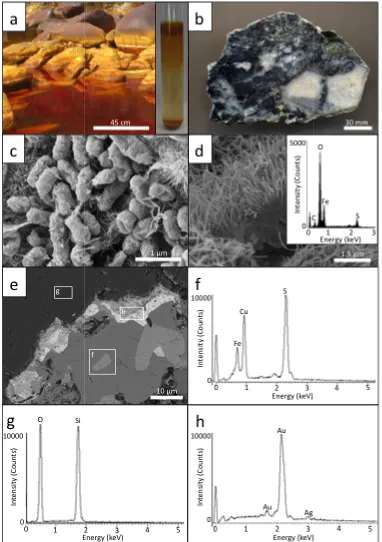
Submersed cobbles in Rio Tinto, Spain (Fig. 2.1a) (37° 35' 33.27" N, 6° 33 1.84" W)
contained mineralised biofilms approximately 3 cm thick (Fig. 2,1a). See
Fernandez-Remolar et al. (2005) for elemental composition and mineralogy of cobbles and coatings.
This biofilm was sampled along with river water. The pH of the river water was 2.9
(measured using Electron Microscopy Sciences colourpHast Indicator Strips 0.0-4.0). The
unconsolidated (yellow-orange, fine-grained, iron oxide) biofilm was dispersed within the
sampled river water using a vortex before culturing. Primary bacterial enrichments were
prepared by inoculating 0.5 mL of the dispersed consortium into Fisherbrand® 13 × 100 mm
borosilicate glass test tubes containing 4 mL of modified media defined by Silverman and
Lundgren (1959) with 0.5 mL of 33.3 g/100 mL ferrous sulphate heptahydrate. The pH of the
growth medium and the iron supplement were adjusted to pH 2.3 using 2 M sulphuric acid
(Denver Instrument Basic pH/Eh Meter calibrated to pH 2 and 4 reference standards using
potassium biphthalate buffer at room temperature). Precision of pH measurements was ±0.03
pH units. The test tubes were covered with sterile, plastic push caps to prevent
contamination and were incubated for three weeks under aerobic conditions at room
temperature (RT, approx. 22˚C). Replicate bacterial enrichments were used for electron
microscopy (see section 2.1.3), aqueous geochemical analysis and to count viable cells using
the Most Probable Number (MPN) statistical method described by Cochran (1950).
2.1.2 Gold-bearing metal sulphide sample preparation
A gold-bearing metal sulphide ore was obtained from the San Salvador vein from the
Capillitas mine, Argentina (Marquez-Zavalia and Craig, 2004; 27° 27' S, 66° 30' W). The
sample was cut into two pieces each approximately 3 mm in thickness. One side of each
piece was polished to expose a “fresh” mineral surface using a 500 nm aluminium silicate
2.1.3 Bacterial enrichment and gold-bearing metal sulphide
characterisation
Bacterial cells from the fluid phase of a three week bacterial enrichment was obtained (see
section 2.1.4) and fixed with 2%(aq) glutaraldehyde for 24 hours, dehydrated in sequential
25%(aq), 50%(aq), 75%(aq) and 3 × 100% ethanol series and dried using a Tousimis Research
Corporation Samdri-PVT-3B critical point drier. The sample was then placed on an
aluminium stub with a 12 mm carbon adhesive tab. All samples were coated with a 5 nm
osmium deposition using a Denton Vacuum Desk II sputter coater to reduce charging effects
during sample characterisation. Bacteria from the enrichment and gold-bearing sulphide
samples were characterised using a LEO Ziess 1540XB Field Emission Gun-Scanning
Electron Microscope (FEG-SEM) equipped with an Oxford Instruments’ INCAx-sight
Energy Dispersive Spectrometer (EDS). The FEG-SEM, operating at an accelerating voltage
of 3 or 10 kV, was used for surface imaging and qualitative elemental composition,
respectively.
2.1.4 Bacterial weathering system assemblage
Three 5 mL bacterial enrichments, incubated for 3 weeks, were homogenised using a vortex
to help “liberate” loosely bound “individual” cells from the iron oxyhydroxide minerals that
precipitate on the interior surface of the borosilicate glass test tube (Fig. 2.1a, inset). The
majority of the iron oxyhydroxide precipitates, dislodged from the walls of the test tubes,
were allowed to settle to the bottom of the tubes for 1 min. to ensure that a 10 mL bacterial
suspension possessed a limited amount of iron oxyhydroxide (confirmed using light
microscopy). The bacterial weathering system was constructed in a sterile, 125 mL volume
Erlenmeyer flask by adding the 10 mL fluid phase “inoculum” to 90 mL of basal medium
defined by Silvermann and Lungdren (1959) with no addition of ferrous sulphate
heptahydrate.
The polished gold-bearing metal sulphide ore samples were sonicated three times for 5 min.
each to remove the osmium coating from imaging (see section 2.1.3), sterilised using 100%
ethanol and rinsed with filter-sterilised, deionised water and attached to sterile polyethylene
(see section 2.1.2). The purpose of suspending the sample was to ensure that bacteria in
solution were attaching and not settling onto mineral surfaces. The other gold-bearing metal
sulphide sample was suspended in a separate, sterile, 125 mL Erlenmeyer flask containing
the same basal medium without the addition of bacteria, representing an abiotic control.
Both systems were covered with sterile aluminium foil to prevent contamination and reduce
evaporation. The first laboratory model represents the biogeochemical conditions
encountered in near-surface environments in which sulphide-bearing rocks are weathered via
biogeochemical processes. The abiotic control system was used to evaluate the catalytic
effect of having bacteria in the reaction systems and to determine whether or not the acidic
conditions could be responsible for the dissolution of sulphide minerals. Both reaction
systems were incubated for 60 days at RT. The pH was re-measured after the incubation
period using a Denver Instrument Basic pH/ Eh Meter as described in section 2.1.1.
2.1.5 Gold-bearing metal sulphide re-characterisation
After 60 days incubation, the surfaces of bacterial and abiotic control gold-bearing metal
sulphide ores were re-characterised along with an iron oxyhydroxide precipitate that
developed at the bottom of the flask in the bacterial weathering system. Scanning Electron
Microscopy (SEM) conditions and sample preparation were the same as the methods
described for the characterisation of the bacterial cells and gold-bearing metal sulphide prior
to bacterial exposure (see section 2.1.3).
2.1.6 Aqueous geochemical analysis
Duplicate 5 mL aliquots were sampled from both systems and were filtered using a 0.1 µm
pore-size filter to remove any solid material. These solutions represented the initial aqueous
geochemical conditions of the bacterial weathering system and the abiotic control at the start
of the experiment. After 60 days, additional duplicate 5 mL aliquots were sampled and
represented the geochemical conditions at the end of the experiment. All samples were
acidified to pH 1 using concentrated (71%) nitric acid and analysed for soluble silver,
arsenic, gold, copper, iron, mercury, molybdenum, nickel, lead and zinc using Perkin-Elmer
Optima 3300-DV Inductively Coupled Plasma-Atomic Emission Spectroscopy (ICP-AES).
2.2 Results
2.1.1 Bacterial enumeration and characterisation
The MPN count of the iron-oxidising bacterial enrichments was 2.4 × 10⁴ bacteria/mL.
Evidence of active iron-oxidising bacterial metabolism from the enrichment was indicated by
the increased acidity, i.e., pH = 2.1, and the formation of a red-orange, iron oxyhydroxide
precipitate that coated the inside surface of the borosilicate test tubes (Fig. 2.1a, inset). SEM
characterisation indicated that the bacterial enrichment contained predominantly rod-shaped
bacteria and a lesser amount of spirilla occurring on the surface of nanometre-size, acicular
precipitates composed of iron and oxygen that formed micrometre-scale sheets (Fig. 2.1c, d).
2.1.2 Gold-bearing metal sulphide characterisation
Prior to exposure in the bacterial weathering system, SEM analysis demonstrated that the
gold-bearing metal sulphide contained natural crevices that occurred within and along metal
sulphide mineral boundaries within a quartz matrix. Backscatter SEM micrographs and EDS
spectra indicated that sulphide minerals contained a range of elements including arsenic,
copper, iron, manganese, lead and tin and were consistent with previous studies identifying
metal sulphide minerals (see section 2; Marquez-Zavalia et al., 1999; Marquez-Zavalia and
Craig, 2004; de Brodtkorb, 2009). Gold and silver occurred along sulphide mineral
boundaries as irregular, tens of micrometre-size “grains” (Fig. 1e-h).
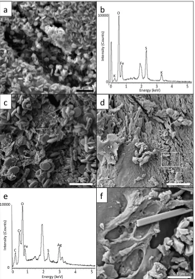
After 60 days of exposure in the bacterial weathering system, an abundance of bacteria were
observed on articulated surfaces of metal sulphide minerals and extensive biofilms were also
observed within crevices (Fig. 2.2a). A coating composed of iron, phosphorus and oxygen
occurred as a laminate structure that coated the polished surface of the ore. Bacteria were
directly attached to this iron phosphate coating (Fig. 2.2b-d). Although rod-shaped bacteria
were the dominant morphotype, a greater proportion of spirilla relative to the inoculum were
observed. More importantly, both types of cells had extracellular, secondary mineral
precipitation. These nanometre-size precipitates were composed of iron and sulphur (Fig.
2.2e, f). SEM analysis indicated that a micrometre-size, discoidal mineral precipitate
phosphate coating (Fig. 2.3a, b). The once polished metal sulphide surfaces now appeared
weathered. The discoidal precipitate occurred on the surface of weathered minerals along
with bacteria (Fig. 2.3c). A fragment of metal sulphide mineral was also found at the bottom
of the flask along with the discoidal precipitates. This demonstrates that weathering may
have occurred along grain boundaries, releasing mineral fragments from the polish rock
surface. The metal sulphide at the bottom of the flask had a rough surface texture similar to
the weathered mineral surfaces observed on the suspended sample. However, bacteria
attachment was not observed but bacterial-size impressions were present (Fig. 2.3d-f).
2.1.3 Geochemical analysis
Detailed results from ICP-AES analysis are found in Table 2.1. Prior to the addition of the
gold-bearing metal sulphide sample, the fluid phase of the bacterial weathering system
contained iron and sulphur from the bacterial inoculum. After 60 days, the fluid phase of the
bacterial weathering system contained trace metal concentrations including silver, arsenic,
copper, iron, lead, sulphur and zinc. Interestingly, the iron concentration had decreased
while sulphur slightly increased relative to the initial respective concentrations. In the abiotic
control system copper, iron, lead, and zinc were present but concentrations were less than
those of the bacterial weathering system. The pH of the biological weathering system
dropped from pH 2.3 to pH 2.1 while the abiotic system remained the same.
2.2 Discussion
Iron-oxidising bacteria are capable of oxidising both iron and sulphur (Fowler and
Crundwell, 1999) and are commonly found in near-surface weathering environments. The
presence of iron-oxidising bacteria has a profound effect by catalysing the dissolution of
metal sulphides (see Erhlich, 1964; Singer and Stumm, 1970; Hedrich et al., 2011). The
contribution of the biosphere in acidic, oxidised environments is important for the physical
and chemical weathering of metal sulphides because it potentially leads to the liberation and
mobility of soluble gold complexes or solid gold particles. The bacterial weathering system,
used in this study, represented an amplified natural system containing a high water:rock ratio
such as an oxidised zone of a gold-bearing, sulphide ore body exposed to near-surface
Sasaki et al. (1995) and Sasaki and Konno (2002) demonstrated that jarosite-group minerals
form as a by-product of actively metabolising iron-oxidising bacteria, i.e., Acidithiobacillus
ferrooxidans. Since sodium and potassium were limited constituents in the media defined by
Silvermann and Lundgren (1959), it is likely that the nanometre-size, acicular iron
oxyhyroxide precipitate formed in the bacterial enrichments were likely hydroniumjarosite
(Fig. 2.1d, inset).
Since the gold-bearing metal sulphide ore was suspended in solution, the iron-oxidising
bacteria must have attached onto the sample from the fluid-phase rather than settling onto the
surface due to gravitational forces. More importantly, the preferential attachment of enriched
iron-oxidising bacterial consortia onto metal sulphide mineral grains instead of quartz
surfaces was consistent with many previous studies (Ohmura et al., 1993; Sand et al., 1995;
Escobar et al., 1996; Edwards et al., 2000a,b; Sampson et al., 2000; Kinzler et al., 2003;
Mielke et al., 2003; Rodriquez et al., 2003a,b,c; Sand and Gehrke, 2006; Africa et al., 2012).
Evidence of extensive biofilm development including extracellular polymeric substances
(Fig. 2.2a) within creviced regions suggests that active bacterial growth occurred on the
metal sulphide components of the sample. Evidence of bacterial metabolism was also
demonstrated by the formation of secondary minerals occurring on extracellular surfaces.
These nanometre-size, secondary minerals containing iron and sulphur were consistent with
previous studies demonstrating extracellular iron mineralisation (Fortin et al., 1996).
Evangelou (1994) proposed that phosphate coatings could act as a “protective barrier” to
reduce polymetallic sulphide oxidation. Conversely, Jones et al. (2003) demonstrated that
bacterial-catalysed oxidation of arsenopyrite can occur in a phosphate-rich culture medium
even when an abiotic, iron phosphate precipitate coated arsenopyrite mineral surfaces
(Reaction 2.1). The laminated structure of the iron phosphate precipitate observed in this
study was consistent with observations by Jones et al. (2003). Both studies demonstrated the
continuous biogeochemical weathering of metal sulphides under acidic and oxidised
conditions despite the presence of this coating. Although it is intuitive that some soluble iron
would have come from the inoculum, the formation of the secondary iron phosphate and iron
oxyhydroxide mineral coatings presumably resulted from microbial activity. The discoidal
EDS spectra and its morphology (see Grishin et al., 1988; Herbert, 1997; Sasaki and Konno,
2002; Henao and Godoy, 2010). Sasaki and Konno (2002) highlighted that the mode and
rate of iron oxidation were critical for the formation of different crystal morphologies of
jarosite-group minerals; therefore, bacteria and the presence of associated organics, i.e.
extracellular polymeric substances, could play an important role in jarosite formation (Chan
et al., 2009; Sasaki et al., 1995; Sasaki and Konno, 2002). The jarosite overlaying the iron
phosphate patina indicated that the iron phosphate formed first and was less soluble than
jarosite. It is interesting to note the two morphological differences between jarosite that
formed in the enriched iron-oxidising bacterial consortia relative to the bacterial weathering
system. Both systems contained the same basal medium but different sources of energy, i.e.,
soluble ferrous sulphate heptahydrate and solid iron sulphide minerals. Based on the abiotic
control, the active metabolism of iron-oxidising bacteria caused the widespread dissolution
and release of the metal sulphide minerals (and their associated gold grains) from the
polished rock surface.
Fe3+ + PO43-→ FePO4 (2.1)
The dissolution of iron sulphide minerals would lead to an increased concentration of soluble
iron and sulphate in solution. However, the decreased iron concentration and slightly
increased sulphur concentrations in the bacterial weathering system are underestimates of the
amount of iron sulphides that “dissolved” due to the growth of iron-oxidising bacteria. The
decreased iron and sulphur concentrations are attributed to the precipitation of iron phosphate
and jarosite (which also contains sulphur; Reaction 2.2). While these sulphide mineral
coatings appeared to occur as a physical barrier between bacteria and sulphide mineral
surfaces, these precipitates possess high surface area and appear to have enabled or promoted
increased attachment of bacteria (Fig. 2.3c). The weathered texture of the suspended metal
sulphide sample and the fragment found at the bottom of the flask suggests that abiotic
processes, e.g., ferric iron leaching, were also contributing to sulphide mineral dissolution.
Furthermore, results from ICP-AES analysis confirmed that Ag, As, Cu, Pb and Zn occurred
in the fluid phase of the bacterial weathering system (see Table 2.1). The initial iron
concentration was proportional to the amount of iron from the inoculum. Copper, iron, lead
and zinc were found in solution in the abiotic control and could be attributed to the acidic
bacterial weathering system. This indicates that active microbial metabolism was responsible
for the increased dissolution of polymetallic sulphide minerals. Although soluble gold
concentrations were not detected in the fluid phase of the bacterial weathering system, low
levels of soluble silver were present. As bacteria contributed to the oxidation and dissolution
of polymetallic sulphide minerals, thiosulphate leaching of silver from gold grains
“embedded” within sulphide minerals intuitively occurred (Reaction 2.3). It is possible that
leaching and dissolution of gold could have also occurred in a similar manner (Aylmore and
Muir, 2001). However, gold concentrations could have been below the 0.01 ppm detection
limit of ICP-AES analysis. Furthermore, gold was not observed in association with the
weathering pits on the ore surface suggesting that it was released along with the metal
sulphides. Unfortunately, the fine grained, i.e., tens of micrometre-sized, gold grains were
not found at the bottom of the flask along with the weathered sulphide material and iron
oxides.
3 Fe3+ + 2 SO42- + K+ + 6 H2O→ KFe3(OH)6(SO4)2 + 6 H+ (2.2)
Ag2S + 4 S2O32- + 2 SO32- + 6H+→ 2 Ag(S2O3)23- + 3 S + 3 H2O (2.3)
The results presented here have important implications for understanding gold mobility under
surface and near-surface biogeochemical conditions and for the development of industrialised
biomining practices. Bioleaching is the biotechnological application of living processes to
recover precious and base metals from primary sources including ore and mineral
concentrates. As an efficient alternative method for gold extraction, it reduces the harmful
effect associated with the use of fossil fuels in conventional extraction methods (Mannion,
1992; Rawlings and Johnson, 2007; Johnston et al., 2013). The effects of microbes on
copper enrichment at ore deposits and from mine tailings have been studied (Southam and
Saunders, 2005; Merson, 1992) and gold enrichment by Pedomicrobium in placer deposits
has also been highlighted (Mann, 1992). Therefore, in the case of gold-bearing metal
sulphides, the same principle of gold “liberation” using iron-oxidising bacteria can be
applied. Using arsenopyrite as a model of a gold-bearing metal sulphide, bacterial-catalysed
oxidation of iron sulphide minerals can promote gold mobility as suspended particles in
solution or as soluble complexes if gold is completely dissolved (Reaction 2.4 and 2.5). The