www.euacademic.org
Rapid detection of Multi Drug Resistant-
Tuberculosis using Line Probe Assay (LPA) in
Sudan
MUATAZ M. ELDIRDERY Tropical Medicine Research Institute National Center for Research, Sudan
INTISAR E. ELRAYAH Tropical Medicine Research Institute National Center for Research, Sudan Faculty of Applied Medical Science, Shaqra University, KSA
MONA OA. AWAD ELKARIM Blood Transfusion Service, National Blood Transfusion Center, Sudan FATIMA A. KHALID Tuberculosis Research Center, University of Kassala, Sudan
ASRAR M A/SALAM ELEGAIL NUHA Y. IBRAHIM EMAN O M. NOUR RAHMA H. ALI National Tuberculosis Reference Laboratory National Laboratory of Public Health, Sudan
HAMDAN M. HAMDAN Consultant of Tuberculosis control program, Sudan NIHAD M A. ELHAJ ATIF A. ELAGIB Tropical Medicine Research Institute National Center for Research, Sudan
Abstract:
approved for use in low income settings and can be used to screen smear-positive sputum specimens for rapid detection of Rifampicin and Isoniazid resistance in 1-2 days. A total of 300 smear positive sputum specimen were collected from Pulmonary Tuberculosis patients ,182 specimens from new cases and 118 from previously treated TB patients in a national reference TB laboratory in Khartoum, Sudan used in this study.The performance of a commercial line probe assay (Genotype MTBDRplus) was assessed for rapid detection of rifampicin and isoniazid resistance directly on smear-positive sputum. Sudan. Results were compared with LJ culture and drug susceptibility testing (DST).The overall prevalence of MDR in all study groups was 38% by DST and 37.3% by LPA. Results of LPA were compared with DST, out of 300 smear-positive sputum specimens only 112 samples were found MDR by (DST) and (LPA), 186 sample sensitive by both and 2 sample were MDR by DST but sensitive by LPA. No significant differences were found between the drug sensitivity test (DST) and Line Probe Assay (LPA) technique, (P = 0.50). The sensitivity of Line Probe Assay was (98.3%) and the specificity was 100% while the positive predictive value was (100%) and negative predictive value was (98.9%). Out of 112 specimen screened by LPA, 94(83.9%) of RIF Resistant samples were found linked with Rpob gene, codon 530-533. On the other hand, all of INH Resistant samples were linked with KatG gene, codon 315 (S 315T1). Line Probe Assay rapid technology and provide results in 2 days, comparing by DST it provide results in 14 weeks; therefore it can be useful diagnostic method in high TB burden countries.
Key words: Line Probe Assay, Mycobacterium tuberculosis, Sudan.
INTRODUCTION:
TB (range, 8.6 million–9.4 million) globally, equivalent to 126 cases per 100 000 population [2]. Furthermore the emergence of multidrug-resistant tuberculosis (MDR-TB) and more recently, extensively drug-resistant TB (XDR-TB) is a major threat to global TB control [3]. Sudan is a country has a high burden of tuberculosis (TB) with an estimated 50,000 incident cases during 2009, when the estimated prevalence was 209 cases per 100,000 of the population. Few studies have been undertaken on TB in Sudan and the prevalence of drug resistant disease is not known yet [4]. The rapid identification of MDR-TB is essential for proper patient management [5, 6]. The World Health Organization (WHO) recommended the use of molecular line probe assays (LPAs) for rapid screening of MDR-TB in low and middle income settings [7]. LPA is use multiplex polymerase chain reaction (PCR) amplification and reverse
hybridization to identify M. tuberculosis complex and mutations
to genes associated with Rifampicin and Isoniazid resistance. LPA can be performed directly from acid fast bacilli (AFB) smear-positive sputum or from culture isolates, and provide results in 1-2 days, it is highly sensitive and specific for detection of Rifampicin resistance (≥97% and ≥99%) and Isoniazid resistance (≥90% and ≥99%) on culture isolates and smear-positive sputum. Overall agreement with conventional DST for detection of MDR-TB was 99% [8].
MATERIALS AND METHODS:
Study setting:
National Reference Tuberculosis Laboratory Khartoum, Sudan. Samples were collected from the patients after obtained their written informed consent. The study was approved by Tropical Medicine Research Institute (TMRI) Ethics committees.
Specimen collection and processing:
Smear-positive sputum was collected from patients at risk of MDR-TB at the National Reference Tuberculosis Laboratory were consecutively screened for acid fast bacilli (AFB) using Ziehl-Neelsen (ZN) smear microscopy, All consenting smear positive patients were enrolled. Two or three sputum samples (spot or morning) were collected per patient in 50 ml sterile conical centrifuge tubes. All manipulations with potentially infectious clinical specimens were performed in a Class II safety cabinet in a BSL 2 or 3 Laboratory. Sputum was processed with sodium hydroxide (NaOH) method 4%). Any processed specimen remaining was stored at 2-8°C for the duration of the study to allow for re-testing of specimens giving discrepant results.
Conventional laboratory testing:
Conventional laboratory testing sputum specimens submitted for smear, culture and DST was treated using 4% sodium hydroxide (NaOH) method, for decontamination because the specimens susceptible to contamination by more rapidly growing normal flora. Two or three drops of the decontaminated sample were inoculated in the Lowenstein-Jensen (LJ) solid medium. and were incubated at 37°C for 8 weeks [9]. Drug susceptibility test was done for 300 isolates, LJ media contain drugs (Rifampicin, Isoniazid, Streptomycin and Ethambutol),
for drug susceptibility test of Mycobacterium tuberculosis
prepared without drugs as a control (10). The suspension was prepared by transferring of a certain amount of bacterial
growth from the culture by 10 µL loop into the suspension
bottle (Peugeot bottles contains glass beads). Homogenization was made by vortexing the bottle, the turbidity of suspension was adjusted to McFarland standard No.1. Then four serial dilutions were made from the original suspension of each
sample (10-1, 10-2 10-3 and 10-4). Two to three drops was
inoculated from dilution 10-2 in media which contain drugs
and10-4 in plain media, then was incubated at 37°C, the bottles
was tightening loosely for 24 hours to allow evaporation and then well tighten. Contamination was examined for 72 hours and the contaminated bottles were discharged. The first
examination for growth of Mycobacterium tuberculosis was done
after two weeks from inoculation, and the second examination after month from the first examination. The resistance was calculated as the ratio of the number of colonies on the drug containing medium and those of control media, the strain was considered as resistant if the ratio is greater or equal to 1% [11].
Line probe assay (LPA)
refrigerated at 2-8°C overnight after DNA extraction to allow repeat testing if required. All steps were done in National Reference TB laboratory - Sudan.
STATISTICAL ANALYSIS:
Statistical Package for Social Science (SPSS) version 16.0 ( Inc. Chicago, IL.USA) was used to analyze the results. Mc-Nemar test was used to compare between two techniques (DST and LPA), P. value less than 0.05 was considered statistically significant.
Sensitivity, specificity, positive predictive value and negative predictive value were calculated according to the follow statistical formula
Sensitivity = True positive / True positive + False
negative X100
Specificity = True negative / True negative + False
positive X100
Positive Predictive Value: = True positive / True
positive + False positive X100
Negative Predictive Value: = True Negative / True
Negative + False Negative X100
RESULTS:
Drug susceptibility Test (DST):
Line Probe Assay (LPA):
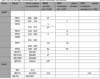
Line probe assay was done for three hundred positive sputum specimen, 112 cases were found MDR and 188 were sensitive. Out of 112 specimen screened by LPA, 94(83.9%) of RIF
Resistant samples was found linked with Rpob gene, codon
530-533 and the remaining linked with other codons; 510-517, 513-519, 518-525 and 526-529, 1(.89%), 2(1.8%), 3(2.7%), 12(10.7%) respectively. On the other hand, all of INH Resistant samples were linked with KatG gene, codon 315 (S 315T1).
INTERPRETATION OF LPA RESULTS:
Results of LPA were compared with drug susceptibility testing (DST). Out of 300 smear-positive sputum specimens only 112 was found MDR by conventional method (DST) results and Molecular (LPA), 186 sample sensitive by both and 2 sample were MDR by DST but sensitive by LPA. No significant differences were found between the drug sensitivity test (DST)
and Line Probe Assay (LPA) results, P = 0.50. The sensitivity of
Line Probe Assay was (98.3%) and the specificity was 100% while the positive predictive value was (100%) and negative predictive value was (98.9%), table (1).
Table 1 Performance of line probe assay (LPA) in smear positive sputum specimens compared with conventional drug susceptibility testing (LJ DST).
LPA, line probe assay
DST, Drug Susceptibility Test in LJ Media
Techniques LPA
DST
INHR
RIFR
INHR
RIFS
INHS
RIFR
INHS
RIFS
INHR RIFR 112 0 1 1
INHR RIFS 0 0 0 7
INHS RIFR 0 1 0 0
RIFS, rifampicin susceptible; RIFR, rifampicin resistant
INHS, isoniazid susceptible; INHR, isoniazid resistant.
Table 2 Performance of line probe assay (LPA) in detecting rifampicin, isoniazid and multidrug-resistance from smear positive sputum specimens
Rifampicin Isoniazid Multi-drug
resistance
No. resistant/No. susceptible strains
115/185 113/187 112/188
Sensitivity, % (95% CI)
98.3% (93.86% 99.79%)
92.6% (86.35% to 96.54%)
98.3% (93.81% 99.79%) Specificity, % (95% CI) 98.9% (96.15% 99.87%) 99.4% (96.93% to 99.99%)
100% (98.04% to 100.00%) Overall accuracy, %
(95% CI)
98.7% 96.7% 99.3%
PPV, % (95% CI)
98.3% (93.86% 99.79%)
99.1% (95.17% to 99.98%)
100% (96.76% to 100.00%) NPV, %
(95% CI)
98.9% (96.15% 99.87%)
95.2% (91.06% 97.78%)
98.9% (96.21% to 99.87%)
Table 3 Pattern of gene mutations detected by Line Probe Assay (LPA) in drug resistant M. tuberculosis strains.
Gene Band Gene region
or mutation MDR strains (n = 112)
RIF mono
resistant strains
(n = 115)
INH mono
resistant strains (n = 121) rpoB
WT1 506 509 2*
WT2 510 513
1
1 WT3
513 517 5*
WT4 516 519 2
WT5 518 522 3 3
WT6
521 525 WT7
526 529
12 12
WT8 530 533 94 97
MUT1 D516V 1
MUT2A H526Y 4*
MUT2B H526D
MUT3 S531L 10*
katG
WT 315
MUT1 S315T1 112 113
InhA
WT1 15/ 16
WT2 8
MUT1 C15T
MUT2 A16G
MUT3A T8C
MUT3B T8A
1 MDR strain had both rpoB WT2 and WT3 mutations, 2 MDR strain had both rpoB WT3 and WT4 mutations, 3 MDR strain had both rpoB WT5 and WT6 mutations and 12 MDR had both rpoB WT6 and WT7 mutations. * More than one mutation in rpoB Gene.
DISCUSSION:
In this study 182(60.7%) of the Pulmonary TB patients were diagnosed for the first time while118 (39.3%) had history of P-TB reflecting the active transmission of P-TB. The findings of the present study indicated that the molecular methods were highly consistent with the conventional culture and DST method. In this study (41) patients were new cases for MDR-TB and (73) patients were retreatment cases for MDR-TB. There was no XDR-TB case recorded in this study. This is the first study in Sudan to ascertain levels of drug resistant tuberculosis by LPA.
Result of Drug susceptibility test (DST) showed 114 cases were MDR and 186 were sensitive. LPA result showed that 112 cases were MDR TB and 188 cases were sensitive. The sensitivity and specificity of LPA for the detection of MDR TB for clinical isolates was 98.3% and 100% respectively.
MTBDRplus) for rapid detection of Rifampicin and Isoniazid resistance, and the results were compared with (DST). The Sensitivity and specificity of LPA were 92.3%, 96.2% for detection of MDR compared with DST.
In this study the RIF resistance was associated with
mutation in the region of rpoB 530-533, mostly S531L
mutation, this agrees with the study of Telenti et al.,[19] and
Barnard et al.,[20], they found that this mutation was more
frequently in MDR strains in South Africa. The present findings provide the basis for rapid detection of Rifampicin resistance, and it is considered a marker of multidrug-resistant tuberculosis. On another hand all of INH resistant samples in this study were linked with KatG gene, codon 315 (S 315T1) as was shown in many high TB burden countries presumably related to ongoing transmission of these strains [21].
In DST it was noticed that 73% of re-treatment cases and 15% of new patients were found to have strains resistant to streptomycin thus suggests that the efficacy of this drug is limited in this setting. It is an injectable drug that necessitates regular attendance at a health clinic and the high levels of resistance observed suggest its role in the treatment of TB in Sudan should be reappraised. This study agrees with study done by Zaki and Ibrahim [22], as out of the 45 patients testing revealed that 48.9% (n=22) were resistant to streptomycin, also agrees with study by Magzoub [23] who used Lowenstein Jensen proportion method to test the antimicrobial sensitivity
of 200 isolates of Mycobacterium tuberculosis complex against
streptomycin, 43 (21.5%) were resistant to STM, it recorded the highest proportion of resistant comparing with all drugs.
CONCLUSION:
of resistance to Rifampicin and Isoniazid with highly sensitivity and specificity. It achieves a substantial reduction in diagnostic delay and it has the potential to revolutionize MDR TB diagnosis, therefore it can be useful diagnostic method in high TB burden countries.
ACKNOWLEDGEMENTS:
This study was funded by International Atomic Energy Agency (IAEA) RAF 60/40, National Center for Research and National TB Control Program. Also Extends thanks to Tropical Medicine Research Institute (TMRI) staff and National Reference Laboratory Staff.
Competing Interests
The authors have no competing interests.
REFERENCES:
1- Martin G and Lazarus A (2000). Epidemiology and
Diagnosis of Tuberculosis. Recognition of At-risk Patients is Key to Detection, Postgrad. Med. 108:42-44, 47-50, 53-54.
2- WHO Global Tuberculosis Report 2014.
3- Shah NS, Wright A, Bai GH. (2007). Worldwide emergence
of extensively drug-resistant tuberculosis. Emerg Infect Dis. 13:380–7.
4- Sharaf Eldin GS, Fadl-Elmula I, Ali MS, Ali BA, Abdel Latif
GA, Mallard K, Bottomley C and McNerney R (2011) Tuberculosis in Sudan: a study of Mycobacterium tuberculosisstrain genotype and susceptibility to
anti-tuberculosis drugs BMC Infectious Diseases,
5- American Thoracic Society (1994). Treatment of tuberculosis and tuberculosis infection in adults and children. Am J Respir Crit Care Med. 1359-1374.
6- British Thoracic Society-Tuberculosis Committee (1998).
Chemotherapy and management of tuberculosis in the United Kingdom. Thorax. 53:536-548.
7- WHO (2008) Policy Statement. Molecular Line Probe Assays
for Rapid Screening of patients at risk of multidrug
resistant tuberculosis (MDR-TB),
.http://www.who.int/tb/dots/laboratory/lpa_policy.pdf.
8- Ling D, Zwerling A and Pai M (2008). GenoType MTBDR
assays for the diagnosis of multidrug-resistant tuberculosis: a meta-analysis. Eur Respir J, 32:1165-1174.
9- WHO (1998). Laboratory Services in Tuberculosis Control
Culture Part III PP (57-75).
10-National TB Control Programme (2009). Manual of
Standard Operating Procedures (SOPs). Culture of
Mycobacterium tuberculosis and Drug Susceptibility Testing
on solid Medium Intermediate Reference Laboratory for Tuberculosis - Central TB Division, (Ministry of Health & Family Welfare) New Delhi-Version No. 01.01 Date: 01/04.
11-Sethi S, Sharma S, Sharma SK, Meharwal SK, Jindal SK
and Meera S (2004). Drug susceptibility of Mycobacterium
tuberculosis to primary anti- tubercular drugs by nitrate reductase assay. Indian J Med Res. 120, PP 468-471.
12-World Health Organisation: Policy Statement. Molecular
Line Probe Assaysfor Rapid Screening of patients at risk of
multidrug resistant tuberculosis(MDR-TB),
2008.http://www.who.int/tb/dots/laboratory/lpa_policy.pdf.
13-Hain Lifescience. Genotype® MTBDRplus product insert.
Version 1 http://
14-Sharma M, Sethi S, Mishra B, Sengupta C, Sharma SK
(2003). Rapid detection of mutations in rpoB gene of
rifampicin resistant Mycobacterium tuberculosis strains by
line probe assay. Indian J Med Res.117:76-80.
15-Barnard M, Albert H, Coetzee G, O’Brien R, Bosman ME
(2008). Rapid molecular screening for multidrug-resistant tuberculosis in a high-volume public health laboratory in South Africa. Am J Respir Crit Care Med, 177(7):787-792.
16-Aurin TH, Munshi SK, Kamal SM, Rahman MM, Hossain
MS, Marma T, Rahman F, Noor R (2014). Molecular approaches for detection of the multi-drug resistant tuberculosis (MDR-TB) in Bangladesh. PLoS One. 9(6):e99810.
17-Yadav RN, Singh BK, Sharma SK, Sharma R, Soneja M,
Sreenivas V, Myneedu VP, Hanif M, Kumar A, Sachdeva KS, Paramasivan CN, Vollepore B, Thakur R, Raizada N, Arora SK and Sinha S. (2013). Comparative evaluation of GenoType MTBDRplus line probe assay with solid culture method in early diagnosis of multidrug resistant tuberculosis (MDR-TB) at a tertiary care centre in India. PLoS One. ;8(9):e72036. doi: 10.1371/journal.pone.0072036.
18-Albert H, Bwang F, Mukkada S, Nyesiga B, Ademun JB,
Lukyamuzi G, Haile M, Hoffner S, Joloba M and O’Brien N
(2010). Rapid screening of MDR-TB using molecular Line
Probe Assay is feasible in Uganda. AlbeBMC Infectious Diseases. 10:41.
19-Telenti A, Imboden P, Marchesi F, Lowrie D, Cole S, Colston
MJ, Matter L, Schopfer K and Bodmer T. (1993). Detection
of rifampicin-resistance mutations in Mycobacterium
tuberculosis. Lancet. 341, 647–650.
20-Barnard M, Albert H, Coetzee G, O’Brien R, Bosman ME.
21-Van Rie A, Warren R, Mshanga I, Jordaan A, Spuy van der
GD, Richardson M,Simpson J, Gie RP, Enarson DA, Beyers
N, van Helden PD, Victor TC (2001).Analysis for a limited
number of gene codons can predict drug resistance of
Mycobacterium tuberculosis in a high-incidence community.
J Clin Microbiol, 39:636-641.
22-Zaki AM and Ibrahim N Y (2004). A study on Prevalence of
Drug Resistance in Drug Default Pulmonary Tuberculosis. Trop Med Int Health. 9(12):1305-11.
23-Magzoub EMN (2008). Isolation, Identification and
Antimicrobial Sensitivity of Mycobacterium tuberculosis