Clinical Optometry
C A S E S T U DY
open access to scientific and medical research
Open Access Full Text Article
Dovepress
Sudden unilateral visual loss as an initial
presentation of chronic myelogenous leukemia
Len V Hua Salisa K Williams
Pacific University Eye Clinic, Forest Grove, OR, USA
Correspondence: Len V Hua
Pacific University College of Optometry,
2043 College Way, Forest Grove, OR 97116, USA
Tel +1 503 352 3059 Fax +1 503 352 2929
Email [email protected]
Abstract: Leukemia is a cancer of the white blood cells. Many patients with leukemia are unaware of their disease until routine blood work up for other conditions reveals incidental findings leading to a diagnosis of leukemia. Up to 50% of patients with leukemia have ocular manifestations. In fact, floaters or decreased vision may be the initial symptom of leukemia.
Case study: A 51-year-old Caucasian female patient with sudden unilateral visual loss in the left eye was found to have bilateral retinal neovascularization and Roth spot hemorrhages. Blood work up and cytological analyses confirmed the Philadelphia chromosome, which has been implicated in the development of chronic myelogenous leukemia (CML). Further testing confirmed a diagnosis of chronic phase CML. After a two-month course of imatinib and close monitoring by a hematological oncologist, her vision has improved and the retinal vasculariza-tion has significantly resolved.
Conclusions: Eye care professionals are in a unique position to identify this devastating dis-ease early on. A delay in diagnosis of the disdis-ease may lead to conversion into the acute phase, which has a poor prognosis for survival. A prompt referral to internal medicine and oncology for co-management is crucial.
Keywords: chronic myelogenous leukemia, retinal hemorrhages, Roth spot, imatinib (Gleevec®)
Philadelphia chromosome, vision loss
Background
Leukemia is a group of blood disorders defined by neoplastic production of white blood cells. It is classified into myelogenous and lymphocytic origins, which are further sub-divided into four types based on acute or chronic manifestations: acute myelogenous leukemia (AML), acute lymphocytic leukemia (ALL), chronic myelogenous leukemia (CML), or chronic lymphocytic leukemia (CLL).1 Chronic myelogenous leukemia (CML, chronic myelocytic or chronic myeloid leukemia) accounts for approximately 15% to 20% of leukemias in adults and is associated with a reciprocal chromosomal translocation t(9; 22) (q34; q11) resulting in a BCR-ABL fusion gene (Philadelphia chromosome).2 This translocation results in a new hybrid protein (BCR-ABL) with overactive tyrosine kinase activity and is implicated in the development of CML.3 Approximately 95% of patients with CML have this Philadelphia chromosome, which has been the target for drug design and treatment of this disorder.1 CML is estimated to have an annual incidence of 1 to 2 cases per 100,000, with a slight male predominance. The age at presentation is approximately 50 to 60 years, with exposure to ionizing radiation being the only known risk factor.4
Clinical Optometry downloaded from https://www.dovepress.com/ by 118.70.13.36 on 21-Aug-2020
For personal use only.
Number of times this article has been viewed
This article was published in the following Dove Press journal: Clinical Optometry
Hua & Williams Dovepress
Ocular complications
Leukemia can cause ocular complications in three ways: direct tissue infiltration by excess white blood cells, infec-tions by immunosuppression, and abnormal homeostasis of blood cells such as anemia, thrombocytopenia and hypervis-cosity.5 Approximately 50% or more of all leukemias have some form of ocular manifestations.6 Leukemic retinopathy is a common manifestation of leukemia and is found in both the acute and chronic forms.6,7 The authors present a rare case of retinopathy secondary to CML in which unilateral visual symptoms were the initial presentation that led to its diagnosis and timely management.
Case report
A 51-year-old Caucasian female presented to the Pacific University Optometry Clinic in October 2009 with a chief complaint of severe blurry and cloudy vision OS for the last two months. She did not seek care because of lack of health insurance. Medical history was unremarkable, and without any ocular injury or surgery. Family medical history was negative for hypertension, glaucoma, or blindness, with the exception that her brother had diabetes with major compli-cations. Her last eye examination was in August 2009 at a franchised optometric clinic. At that time, her examination was unremarkable, with distance visual acuity of 20/20 OD, OS and OU without correction.
At the current visit, her distance visual acuity was 20/20 OD and 20/400 OS with no improvement with pinhole. Both eyes showed smooth, accurate, full, and equal extra-ocular muscle movements in all fields of gaze. No apparent pupillary defect was observed. Finger counting confronta-tion visual fields were full OD and OS. Anterior segment findings, as observed with biomicroscopy, demonstrated no abnormalities. Her conjunctiva was pale but no jaundice was noted. Goldmann applanation tonometry was 16 mm Hg OD and 17 mm Hg OS at 2:15 p.m.
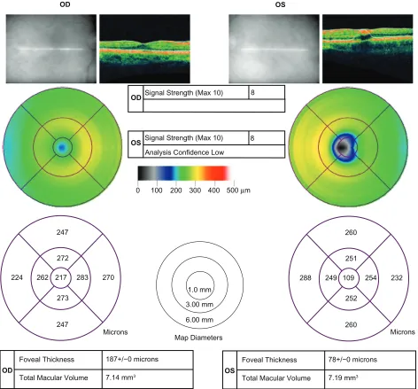
Dilated fundus examination showed multiple vascular abnormalities including retinal hemorrhages and Roth spots OU (Figures 1A, 1B). Optical coherence tomography of the macula revealed foveal vacuoles or holes and thinning, OS significantly greater than OD (Figure 2). The cup-to-disc ratios were normal O.30V/0.30H both eyes.
A diagnosis of bilateral retinopathy with potential hema-tologic abnormalities was made. The patient was educated about the seriousness of the retinal findings, with the poten-tial for a similar effect to the fellow eye. She was referred to a retinal specialist for consultation regarding the possible etiology. In the interim, she went to a local health clinic
where her blood pressure and blood sugar were determined to be normal. A week later, she was examined by a retinal specialist, who confirmed the possibility of hematologic abnormalities, and was referred to internal medicine for blood work up the next day.
Laboratory data showed that she had a critical high white blood cell (WBC) count of 150,000/µL [normal reference
Figure 1a Fundus photography of the right eye shows multiple areas of vascular neovascularizations and engorged, tortuous veins at initial examination.
Figure 1b Fundus photography of the left eye shows clusters of Roths spots including one at the fovea, and engorged, tortuous veins at initial examination.
Clinical Optometry downloaded from https://www.dovepress.com/ by 118.70.13.36 on 21-Aug-2020
Unilateral visual loss in chronic myelogenous leukemia Dovepress
OD
OS
Signal Strength (Max 10)
Signal Strength (Max 10)
Analysis Confidence Low
8
8
OD
OD
247
272
262 217 224
247 273
270
1.0 mm
3.00 mm
6.00 mm Microns
Map Diameters Microns
283
260
251
249 109 288
260 252
232 254
Foveal Thickness
Total Macular Volume
187+/−0 microns
7.14 mm3
0 100 200 300 400 500 µm
OS
OS
Foveal Thickness
Total Macular Volume
78+/−0 microns
7.19 mm3
Figure 2 Optical computed topography (OCT) scans of both eyes indicate slight macular thinning OD and significant macular thinning (hole) OS at initial examination.
(ref): 3,900–10,600/µL], high platelet count of 448,000/µL [ref: 140,000–440,000/µL], low red blood cell count of 3,240,000/µL [ref: 3,800,000–5,200,000/µL] and low hemo-globin 9.0 g/dL [ref: 12.0–16.0 g/dL]. Manual blood cell differential indicated a remarkable high metamyelocyte of 8% [ref: 1%].
A bone marrow aspiration and biopsy were performed. Fluorescence in situ hydridization (FISH) was done on the peripheral blood cells and a t(9; 22) translocation was found. The t(9; 22) translocation results in the juxtaposition of the BCR (breakpoint cluster region) gene on chromosome 22 and the ABL (Abelson) oncogene on chromosome 9. At diagnosis, this so-called Philadelphia chromosome is
observed by routine cytogenetic karyotyping in greater than 95% of patients with CML. Her FISH result of t(9; 22) translocation combined with morphologic findings were diagnostic of CML. The patient needed treatment with imatinib (Gleevec) daily, but she did not have medical insurance. Fortunately, the local cancer center provided her with immediate treatment of hydroxyurea (50 mg/kg/day) and allopurinol (300 mg/day) while she awaited approval by a drug assistance program for imatinib. During this time, the patient’s white blood cell count decreased further. The patient was ultimately granted approval by an assistance program for a one-month supply of imatinib. She was started on imatinib 800 mg per oral (PO) daily, and discontinued
Clinical Optometry downloaded from https://www.dovepress.com/ by 118.70.13.36 on 21-Aug-2020
Hua & Williams Dovepress
Figure 3a Fundus photography of the right eye shows resolutions of multiple areas of neovascularizations two months after beginning treatment.
Figure 3b Fundus photography of the left eye shows resolutions of Roths spots including one at the fovea two months after beginning treatment.
hydroxyurea and allopurinol. She was then monitored weekly at the cancer center.
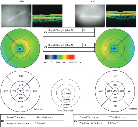
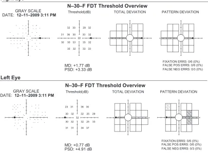
Two months after her initial diagnosis, her visual acuity in the left eye had improved from 20/400 to 20/40 without correction. She had been taking a lower dose of imatinib (400 mg/d) for the past two months, because she could not tolerate the higher dose of 800 mg/d after two weeks. Fun-dus photography revealed significant resolution of retinal hemorrhages in both eyes (Figures 3A, 3B). Optical coher-ence tomography (OCT) macular analyses also indicated nor-malization of the macular thickness in both eyes (Figure 4). Frequency doubling technology (FDT) visual field testing revealed no defects OD and few area of loss OS (Figure 5). The patient still complained of halos and distortions in her left eye and some tolerable side effects, such as nausea and musculoskeletal pain, from the medication. She is being monitored closely by her oncologist.
Discussion
The differential diagnosis for common bilateral retinal hemorrhages includes diabetic retinopathy, central or branch retinal vein occlusion, hypertensive retinopathy, sickle cell retinopathy, and leukemic retinopathy. The patient was first suspected to have diabetic retinopathy because her brother has a medical history of severe diabetic complications. A complete blood count with differentials and cytological analyses confirmed CML and leukemic retinopathy.
CML is a relatively asymptomatic disease in up to 50% of patients. Many patients are diagnosed as a result of symptoms from splenomegaly, anemia or routine blood testing inciden-tal to their disease.8 Among symptomatic patients, systemic symptoms of fatigue, weight loss, excessive sweating, easy bleeding and abdominal fullness are common.9 The patient in this case later recalled that she had lost some weight and constantly felt fatigued, but the symptoms were not severe enough to seek medical attention until she experienced decreased vision in her left eye that did not get better after two months. Fortunately, she presented for an eye examination and was subsequently diagnosed and treated. Most patients with untreated CML progress to acute leukemia within 3 years. Once a patient reaches the acute state, additional chemotherapy has little efficacy.8
Our patient presented with some of the features of leu-kemic retinopathy which include retinal neovascularization, white-centered hemorrhages (Roth spots), and retinal venous tortuosity. Roth spot hemorrhages refer to retinal hemor-rhages with leukemic infiltration or platelet fibrin deposits
that are often seen in patients with bacterial endocarditis, pernicious anaemia, and leukemia. Neovascularizations may arise secondary to retinal nonperfusion and ischemia from the hyperviscosity.10 As seen in the fundus photography of the left eye (Figure 1B), a cluster of hemes with white pre-cipitates (Roth spots) was located in the fovea causing visual symptoms of floaters and decreased vision. OCT finding
Clinical Optometry downloaded from https://www.dovepress.com/ by 118.70.13.36 on 21-Aug-2020
Unilateral visual loss in chronic myelogenous leukemia Dovepress
OD
OS
Signal Strength (Max 10)
Signal Strength (Max 10)
9
10
OD
OD
250
276
262 211 228
235 267
258
1.0 mm
3.00 mm
6.00 mm
Microns Map Diameters Microns
281
247
256
266 205 273
247 255
221 245
Foveal Thickness
Total Macular Volume
181+/−0 microns
7.03 mm3
0 100 200 300 400 500 µm
OS
OS
Foveal Thickness
Total Macular Volume
179+/−0 microns
7.02 mm3
Figure 4 Optical computed topography (OCT) scans of both eyes indicate resolutions of macular thinning OU two months after beginning treatment.
revealed significant thinning of the macula, probably due to a reversible macular cyst or hole, as has been reported in other leukemic cases (Figure 2).11
Treatment
The treatment of CML is based upon specifics of the three disease phases: chronic stable phase, accelerated phase, and blast crisis. Chronic phase CML is managed by BCR-ABL tyrosine kinase inhibitors (TKIs) such as imatinib (Gleevec), interferon alpha, cytotoxic agents such as hydroxyurea (Hydrea®), and allogeneic hematopoietic cell transplanta-tion (HCT). Although allogeneic HCT is the only curative therapy, Gleevec is the initial treatment of choice for all patients with chronic phase CML.12 The majority of patients
with initial chronic phase CML treated with Gleevec showed no disease progression and remained on treatment for years.8 Our patient was treated initially with hydroxyurea with allopurinol, which was then followed by imatinib treatment. During imatinib treatment for two months, her vision in the left eye had improved as did the retinal findings as evidenced in recent fundus photography (Figures 3A, 3B) and OCT (Figure 4).
Imatinib treatment is generally well tolerated, the most common side effects in patients with chronic CML are superficial edema, muscle cramps, diarrhea, nausea and musculoskeletal pain. Neutropenia, thrombocytopenia and anemia are rare, but more severe, adverse events in patient with advanced disease.13 Ocular side effects of cystoids
Clinical Optometry downloaded from https://www.dovepress.com/ by 118.70.13.36 on 21-Aug-2020
Hua & Williams Dovepress
macular edema and macular ischemia had been reported in patients treated with Imatinib that resembled the signs of active disease and could complicate the differential diagnosis of visual disturbances.14,15
A number of studies have focused on the prognosis of CML and found the strongest single predictor of outcome is the stage of disease at the time of diagnosis. Patients with chronic phase at the time of diagnosis have the best long-term prognosis with treatment.16 The overall prognosis for patients with retinopathy is no different from those without retinopathy. The retinopathy typically resolves with treat-ment of the underlying disease, as seen in this case. An update from the International Randomized Interferon study versus the STI571 (IRIS) study revealed an overall survival rate of 86% with imatinib versus less than 50% at 5 years for those who discontinued imatinib (due to intolerance or lack of efficacy).17 This confirmed the effectiveness of imatinib in controlling the disease. However, the treatment must be maintained chronically or the disease will relapse.8 Furthermore, some patients do not respond to or tolerate imatinib, thus second generation TKIs (nilotinib, dasatinib, bosutinib) have been developed and shown efficacy in some of these patients.18
A more severe ophthalmic manifestation worthy of noting is optic nerve leukemic infiltration, which causes decreased vision. This is an ophthalmic emergency and requires prompt radiation therapy to preserve vision. Magnetic resonance imaging, B-scan ultrasonography, and lumbar puncture with cerebrospinal fluid analysis are helpful diagnostic tools in determining the presence and extent of infiltration.19
Conclusion
In conclusion, CML is a relatively asymptomatic disease in up to 50% of patients. However, approximately 50% or more of all leukemias have some form of ocular manifestations. Eye care professionals are amongst the first health care providers to detect the disease, work with internists and refer to oncolo-gists for timely management. A thorough case history is crucial in identifying some of the systemic symptoms such as fatigue, weight loss and excessive sweating. A thorough dilated eye examination is required for detection of retinal abnormalities. Every patient with retinal infiltrates requires a systemic and central nervous system workup.18 The patient in this case was fortunate to discover her CML early in the chronic phase. Her prognosis is good as she continues with medical management.
N–30–F FDT Threshold Overview
N–30–F FDT Threshold Overview Right Eye
Left Eye
GRAY SCALE DATE: 12–11–2009 3:11 PM
GRAY SCALE DATE: 12–11–2009 3:11 PM
Threshold(dB)
Threshold(dB)
TOTAL DEVIATION
TOTAL DEVIATION
PATTERN DEVIATION
PATTERN DEVIATION MD: +1.77 dB
PSD: +3.33 dB
MD: +0.77 dB PSD: +4.91 dB
FIXATION ERRS: 0/6 (0%)
FALSE NEG ERRS: 0/3 (0%) FALSE POS ERRS: 0/6 (0%)
FIXATION ERRS: 0/6 (0%)
FALSE NEG ERRS: 0/3 (0%) FALSE POS ERRS: 0/6 (0%)
32 32
32
32
32 30
30 30 30 30
33 36
35 31
33
33 33 33
29
32 25 29
32 32
30
31
31 34 37
25 33
30 32 32
23 31 36 30
Figure 5 Frequency Doubling Technology (FDT) Visual Field Tests showed normal VF OD and a few areas of loss OS two months after beginning treatment.
Clinical Optometry downloaded from https://www.dovepress.com/ by 118.70.13.36 on 21-Aug-2020
Clinical Optometry
Publish your work in this journal
Submit your manuscript here: http://www.dovepress.com/clinical-optometry-journal
Clinical Optometry is an international, peer-reviewed, open access journal publishing original research, basic science, clinical and epidemiological studies, reviews and evaluations on clinical optometry. All aspects of patient care are addressed within the journal as well as the practice of optometry including economic and business analyses. Basic and clinical
research papers are published that cover all aspects of optics, refraction and its application to the theory and practice of optometry. The manuscript management system is completely online and includes a very quick and fair peer-review system, which is all easy to use. Visit http://www.dovepress. com/testimonials.php to read real quotes from published authors.
Unilateral visual loss in chronic myelogenous leukemia Dovepress
Dovepress
Acknowledgements
The authors would like to thank Anthony V Ho, MD and his staffs at the Rose Quarter cancer center for co-management of our patient and providing updated reports. The authors also thank Jenny Smythe, OD for critical inputs and reviewing the manuscript. The authors report no conflicts of interest in this work.
References
1. Pejovic T, Schwartz PE. Leukemias. Clin Obstet Gynecol. 2002;45(3): 866–878.
2. Rowley JD. Letter: A new consistent chromosomal abnormality in chronic myelogenous leukemia identified by quinacrine fluorescence and Giemsa staining. Nature. 1973;243(5405):290–293.
3. Konopka JB, Watanabe SM, Witte ON. An alteration of the human c-abl protein in K562 leukemia cells unmasks associated tyrosine kinase activity. Cell. 1984;37(3):1035–1042.
4. Faderl S, Talpaz M, Estrov Z, et al. The biology of chronic myeloid leukemia. N Engl J Med. 1999;341(3):164–172.
5. Schachat AP, Markowitz JA, et al. Ophthalmic manifestations of leu-kemia. Arch Ophthalmol. 1989;107(5):697–700.
6. Reddy SC, Jackson N, Menon BS. Ocular involvement in leukemia–a study of 288 cases. Ophthalmologica. 2003;217(6):441–445. 7. Kincaid MC, Green WR. Ocular and orbital involvement in leukemia.
Surv Ophthalmol. 1983;27(4):211–232.
8. Thompson CB. Attacking Cancer at Its Root. Cell. 2009;138(6): 1051–1054.
9. Savage DG, Szydlo RM, Goldman JM. Clinical features at diagnosis in 430 patients with chronic myeloid leukaemia seen at a referral centre over a 16-year period. Br J Haematol. 1997;96(1):111–116.
10. Rosenthal AR. Ocular manifestations of leukemia. A review. Ophthal-mology. 1983;90(8):899–905.
11. Inkeles DM, Friedman AH. Retinal pigment epithelial degeneration, partial retinal atrophy and macular hole in acute lymphocytic leu-kemia. Albrecht Von Graefes Arch Klin Exp Ophthalmol. 1975; 194(4 Suppl):253–261.
12. Baccarani M, Saglio G, Goldman J, et al. Evolving concepts in the management of chronic myeloid leukemia: recommendations from an expert panel on behalf of the European LeukemiaNet. Blood. 2006;108(6):1809–1820.
13. Moen MD, McKeage K, Plosker GL, et al. Imatinib: a review of its use in chronic myeloid leukaemia. Drugs. 2007;67(2):299–320. 14. Georgalas I, Pavesio C, Ezra E. Bilateral cystoid macular edema in a
patient with chronic myeloid leukaemia under treatment with imanitib mesylate: report of an unusual side effect. Graefes Arch Clin Exp Ophthalmol. 2007;245(10):1585–1586.
15. Roth DB, Akbari S, Rothstein A. Macular ischemia associated with imatinib mesylate therapy for chronic myeloid leukemia. Retin Cases Brief Rep. 2009;3(2):161–164.
16. Sokal JE, Cox EB, Baccarani M, et al. Prognostic discrimination in “good-risk” chronic granulocytic leukemia. Blood. 1984;63(4): 789–799.
17. O’Brien SG, Guilhot F, Goldman JM. International randomized study of interferon versus STI571 (IRIS) 7-year follow-up: sustained sur-vival, low rate of transformation and increased rate of major molecular response (MMR) in patients (pts) with newly diagnosed chronic myeloid leukemia in chronic phase (CMLCP) treated with Imatinib (IM). Blood (ASH Annual Meeting Abstracts). 2008;112:186.
18. Quintas-Cardama A, Kantarjian H, Cortes J. Imatinib and beyond– exploring the full potential of targeted therapy for CML. Nat Rev Clin Oncol. 2009;6(9):535–543.
19. Gordon KB, Rugo HS, Duncan JL, et al. Ocular manifestations of leukemia: leukemic infiltration versus infectious process.
Ophthalmology. 2001;108(12):2293–2300.
Clinical Optometry downloaded from https://www.dovepress.com/ by 118.70.13.36 on 21-Aug-2020