Analysis
of
the
5’
Jwnctions
of
R2
Insertions
With
the
28s
Gene:
Implications for Non-LTR Retrotransposition
Janet
A. George, William D. Burke
and
ThomasH. Eickbush
Defmrtment of Biology, University of Rochester, Rochester, New York 14627
Manuscript received September 14, 1995 Accepted for publication December 6, 1995
ABSTRACT
R2 elements are non-long terminal repeat retrotransposable elements that insert into 28s rRNA genes of most insect species. The single open reading frame of R2 encodes a protein with both endonuclease activity, which cleaves the target site, and reverse transcriptase activity, which uses this cleavage to prime reverse transcription. This target-primed reverse transcription mechanism is also used by group I1 introns. Little is known of the mechanism by which the 5’ end of R2 is integrated after reverse transcription. We have determined the 5’junction sequence of 94 R2 elements from 14 different species of Drosophila. Only 37% of the full-length elements contained precise 5’junctions; the remainder contained deletions of the 28s gene and/or insertions of additional sequences. Because the 5’junctions of truncated copies were similar to full-length elements, no sequences at the 5’ end of R2 appear to be required for element integration. A model in which the R2 reverse transcriptase is capable of switching templates from the
R2 RNA transcript t o the upstream 28s gene can best explain the observed 5’junction sequences. This template jumping is analogous to the template switching of retroviral reverse transcriptases during formation of the double-stranded integration products.
N
ON-LONG terminal repeat (non-LTR) retrotrans- posable elements (also referred to as LINE-like elements) are widely distributed, highly abundant mo- bile elements of eukaryotes. They differ from the LTR- containing retrotransposable elements by the absence of terminal repeats, and their encoded open reading frames (ORFs) lack any identifiable integrase domain (EICKBUSH 1994). Molecular phylogenetic studies using the sequence of their reverse transcriptase domain indi- cate that non-LTR retrotransposable elements form a monophyletic group, distinct from the LTR-retrotrans- posable elements (XION(: and EICKBUSH 1988a, 1990; MCCLURE 1993). In fact, the non-LTR elements appear to be more similar to the group I1 introns of fungi mitochondria (LAMBOWITZ and BEI.FORT 1993) and the retrons of bacteria (INOIJYE and I N O W E 1993), than to the LTR-retrotransposable elements.The mechanism used by the non-LTR retrotranspos- able elements to insert within the genome is still poorly understood. Integration of these elements has been shown to result from the reverse transcription of an RNA intermediate by the demonstration that intron sequences placed within an element are precisely re- moved during the generation of-new copies in vivo (Ev-
ANS and PALMITER 1991; JENSEN and HEIDMANN 1991; PELISSON ~1 ccl. 1991). This retrotransposition mecha- nism must differ in several important steps from that
Coorre$ponding nulhvr; Thomas H . Eickbush, Department of Biology, University of Rochester, Rochester, NY 14627.
E-mail: [email protected],Fn..I.ochester,edl~
Genetic\ 1 4 2 X5.3-Xfi9 (March, IlJ!)6)
of retroviruses and LTR retrotransposons, in that the non-LTR elements do not have the structural compo- nents (terminal repeats, tRNA primer binding sites) or encode the proteins (RNase H and integrase) required by the retroviral mechanism (MARTIN 1991; EICKBUSH 1994). Because the 3’ ends of non-LTR elements are usually intact while the 5’ ends are often truncated, it has been suggested that the reverse transcriptase en- coded by each non-LTR element might be able to di- rectly polymerize its reverse transcript (cDNA) onto a chromosome at a nick or break on the chromosome (SCHWARESOMMER et al. 1987; FINNECAN 1989; HUTCHI-
SON et al. 1989; BUCHETON 1990). This model has been greatly supported by studies of the sequence specific R2 element of Bombyx mom’ (BURKE et al. 1987; XIONC and EICKEKJSH 198%). The 120-kD polypeptide en- coded by the single ORF of R2 has been shown to be able to nick the target DNA and use the 3’ OH group exposed by this nick to prime reverse transcription of an R2 transcript (LUAN et al. 1993). The R2 enzyme is also capable of adding nontemplated nucleotides be- fore engaging the RNA in the reverse transcription reac- tion, thus sometimes generating short repeat sequences at the 3’ end of the element, a characteristic feature of non-LTR retrotransposable elements (LUAN and EIGK- BUSH 1995). Following reverse transcription of the RNA template, the second strand of the target DNA is cleaved.
854 J. A. George, W. D. Burke and T. H. Eickbush
encode their own endonuclease or utilize preexisting nicks o r breaks on the chromosome for their insertion is not known. Evidence for the former has been sug- gested by the presence in some non-LTR retrotranspos- able elements of a protein domain with similarity to the the major human apurinic endonuclease (MARTIN et al. 1995). Direct support for the generality of the R2 mechanism is the recent finding that the mobility of the group I1 intron, a12, also depends upon the priming of reverse transcription by the target DNA after specific endonuclease cleavage (ZIMMERLY et al. 1995).
While the in vitro experiments have revealed the ini- tial steps in the integration reaction, they have not helped to resolve the mechanism by which the cDNA of the non-LTR element is attached to the upstream target DNA, nor have they indicated how the RNA tem- plate is removed from the DNA and the second DNA strand synthesized. In this report we have taken a differ- ent approach to study how the R2 element attaches to the upstream target sequences. R2 elements specifically insert at a unique site in the 28s rRNA genes of their host (Figure 1A). Just as the sequence specificity of R2 for this site has greatly aided the biochemical analysis, this specificity has made it possible to score a large number of naturally occurring integrated products all resulting from insertions into identical target se- quences. A striking amount of sequence variation was seen at the 5’ e n d of the R2 elements providing insights into the mechanism used by R2 to complete its integra- tion reaction.
MATERIALS AND METHODS
Strains and DNA isolation: The geographical strains of D. melanogasterused have been previously described ( JAKUBCZAK et al. 1992). Stocks of species from the melanogusterand obscura species groups were obtained from the National Drosophila Species Resource Center (Bowling Green State University). Species from the testacea and quinarz‘a groups were obtained from J O H N J A E N I K E (University of Rochester). Genomic DNA was isolated from 10 to 50 adults of each stock as previously described (EICKBUSH and EICKRUSH 1995).
PCR amplification and sequence determination: The 5’ ends of R2 elements were obtained by PCR amplification of genomic DNA using primers complementary to the R2 and 28s gene positions shown in Figure 1A. Primer 1,5’-TCNCKC- CARTANGGNACCAT-3’ (N, any nucleotide; Y, T or C; R, A or G; W, A or T; K, G or T ) , encoded the reverse complement of the amino acid sequence, MVPYWRE, starting at position 1108 of the D. melanogaster K 2 element ( JAKUBCZAK et al.
1990). Primer 2, 5’-CATRTGNACNCCNARNCC3’, encoded the reverse complement of the amino acid sequence GLGVHM starting at position 403 of the K 2 element. Primers 1 and 2 were used in combination with primer 4,5’-CTAAGT- CGACTGCCCAGT-3‘, complementary to the 28s gene se- quence starting 62 bp upstream of the R2 insertion site. Taq
DNA Polymerase (Bethesda Res. Lab.) was used under condi- tions specified by the supplier. Approximately 0.2 p g of geno- mic DNA was amplified in 30 cycles of 94” for 1 min, 55” for 1 min and 72” for 3 min. Primer 1 worked well for species within the melanogaster species group and was used for the
subsequent sequencing studies. Primer 2 gave the most repro- ducible results for species from the obscuru, testeceu and quin- aria groups and was therefore used in the sequencing stud- ies. For determination of the nucleotide sequence of the junc- tions, the PCR-amplified products were purified from primer sequences by extraction of the DNA bands from agarose gels and cloned into a modified M13mp18 vector, which after digestion with XcmI, is suitable for direct cloning of PCR prod- ucts (BURKE et al. 1995). Multiple clones of both orientations from each species were sequenced by the single-stranded di- deoxy chain termination method (SANGER et al. 1977).
For direct determination of the length variation associated with the R2 5’ junctions primer 3, 5”CCTCTGCTCTCAAA- TAC-3‘, which was complementary to a region within the 5’ untranslated region of the D. rnelanoguster R2 element starting 12 bp from its 5’ junction with the 28s gene, was used in combination with primer 5, 5’-TCAGAACTGGCACGGAC3’, which was complementary to the 28s gene sequence starting 168 bp upstream of the R2 insertion site. To visualize the products on sequencing gels, the PCR amplifications were conducted as above with the addition of 60 nM PV2-dATP. After amplification, the products were denatured by adding an equal volume of 95% formamide, 20 mM EDTA, 0.05% bromophenol blue and 0.05% xylene cyanole, incubated at 72” for 5 min, and subjected to electrophoresis next to a sequence ladder on a standard 6% urea-acrylamide sequenc- ing gel.
RESULTS
Early characterization of cloned rDNA repeats from
D.
melanogaster indicated that allR2
insertions (origi- nally termed type I1 insertions) contained similar 3’junctions with the 28s gene, while their 5’ ends were subject to large truncations (LONG et al. 1980; DAWID a n d REBBERT 1981; ROIHA et al. 1981). A more extensive PCR analysis of the 3‘ end of R2 elements from six species of the melanogaster species subgroup confirmed that the 3‘ junctions of
R2
elements in each species were identical except for the length of a poly (A) tract defining the junction with the28s
gene (EICKBUSH and EICKBUSH 1995). Similar results have been obtained in a study of the 3‘ ends of =elements from 19 additional species of the Drosophila genus (W. C. LATHE and T. H. EICKBUSH, unpublished observations). As shown in Fig- ure 1B these 3’junctions are consistent with the initial cleavage generated in the bottom (coding) strand of the 28s gene by the R2 endonuclease in vitro (LUAN et al. 1993).Unlike the extensive analysis of the 3’junctions of
R2
elements, relatively few 5’junctions have been character- ized (BURKE et al. 1987;JAKUsczAK et al. 1990). To clone multiple 5’ R2/28S gene junctions, PCR amplification of genomic DNA was conducted using the primer locations shown in Figure 1A. O n e oligonucleotide, primer 4, was complementary to the28s
gene upstream of the inser- tion site, and a second oligonucleotide was complemen- tary to one of two regions of theR2
element encoding conserved amino acid sequences of the OW, primers 1 o r 2 (see MATERIALS AND METHODS).5' Junction of R2 Elements
A
rDNA repeat
18s 5.8s 28s R2 element
8.53
I-"
5 4 3
I b b 4 4 2 41 ' 1
5'UTR
I
rn
ORFI CCHH
0.2 kb
28s gene
S . . . CTCTCrrAAGCTAGCCAAAT
...
33'... CACACAATTCCATCGCllTA
...
+
.f
First strand cleavage Reverse transcription +
+
v
?R2 element
It2 element in the rDNA unit. (A) Diagram o f an R2 inserted rDNA unit of l?. mdmnpuskr. The location of thc 18.5, 5.8s and 28s genes are indicated by M, the spacer regions by -, and the R2 element by R. The .5' ewl of the 1<2 clrmcnt has hecw cxpantlrd to show the locatioll o f the PCR primers used in this study. Various regions of the R2
element h a \ ~ r heen indicated a s follows: 0 , 5' untranslatetl region (5' UTR); stippled box, location of the region encoding the ORF: tlarkrr stippled box, location of the sequences encoding a putative cysteine, histidine nucleic acid-binding motif. PCR
primers and their oricmtxtion arc indicated by the arrowheads. ( R ) Cleavage site of the R2 element and the known steps of the intcgration 1-cartion. 1,ocation o f the H Z entlonuclcasc cleavage sites on the two stlands of t h e 28s gene arc indicated by the arrows. The first step of the integration reaction is the clravagc of the bottom strand and the use o f the released 3' OH group
t o prime rwerse transcription. I n Ilrosophila cDNA synthesis ;~lwavs begins with a run o f 18-25 T nucleotides, thus the final 3'
junction of the R2 rlemrnt is the same i n a l l Drosophila species. The second step of the integration reaction is a cleavage of the r~ppcr target DNA strmds 2 hp rqxtrcam o f the lint cleavage. The mechanism for the attachment o f the R2 sequences to
the upstream 28s gcnc sequences is n o t known, and the junction itself is extremely variable. The only common feature is that most elements hcgin w i t h two G nucleotides (see Figures 2 and 4).
. . < .
14 species of Drosophila. This included an initial
27
junctions from I). mrl~~nogm/rr, to obtain a thorough sampling of'.junctions from one species, followed by the recovery of two to eight .5' junctions from 13 other species of Drosophila t o determine if the variation seen in I). m ~ I m o g m / r r was characteristic of R2 elements in other species. To sample representative species near I). mrlonogm/rr eight of these other species were chosen from the m~l~~nogcrs/rrspecies group: I). simulnns, I). srrh- p l l i n ,
I).
mtwri/innrr andI).
pnltubn of the mpIannsps/pr subgroup; I). /dtnhnshii of thr /nltnlwrshii subgroup; and I). onrrnnssnp, I). hijwr/inn/o and I). vnrinns of the nnnntrs- s n p subgroup. To sample species throughout the genus, the remaining five species were selected from three other species groups: I). prreimi1i.s andI).
j~.sSpudoo&nlrclfrom the ol)srlrrtr species group;
I).
/ p . s / f r r p o from the ks/n-r w specics group; and
I).
rrrpns andI).
jdlrni from thequinnrin species group. Comparison of the conserved sequences within the R2 elements themselves will he discussed in a subsequent report.
Most if not all of the 5' junctions obtained by this PCR amplification were derived from
K2
elements in- serted in typical units of the rDNA locus. In silu hyhrid- izations in I). mdnnogastm have indicated that all copies of K2 are located in the nucleolar organizer (PEA(:O(:I(856 J. A. George, W. D. Burke and T. H. Eickbush
28s gene
D.
melanogaster
TCAACGGCGGGAGTAACTATGACTCTCTTAA TCAACGGCGGGAGTAACTATGACTCTCTTAA TCAACGGCGGGAGTAACTATGACTCTCTTAA
TCAACGGCGGGAGTAACTATGACTCTCT--- TCAACGGCGGGAGTAACTAAGACTCTCTT--
GGGAGTAT TCAACGGCGGGAGTAACTATGACTCTCTTAA
TT TCAACGGCGGGAGTAACTATGACTCTCTTAA
G
CCC-AACACCP
T C A A C m A G T A A C T A - - -
TCAACGGCGGGAGTAACTAAGACTCTCT---
CTTT
TCAAC---
TCAACGGCGGGAGTAACTATGACTCTCTTAA TCAACGGCGGGAGTAACTATGACTCTCTTAA
GGG
G TCAACGGCGGGAGTAACTATGACTCTCTTAA
GTGATAACTAGTC TCAACGGCGGGAGTAACTATGACTCTCT---
TCAACG4CGGGAG--- GCACAGA TCAACGGCGGGAGTAACTATGACTCTCTT--
TCAACGGCGGGAGTAACTATGACTCTCTTAA
GAGA TCAACGGCGGGAGTAACTATGACTCTCTTAA
G
TCAACGGCGGGAGTAACTATGACTCTCTTAA TCAACGGCGGGAGTAACTATGACTCTCTTAA
R2 element
No. clones
=A- - -TCATGGGGTAT 4
ZGGGA--GTCATGGGGTAT 1
ZGGGATCATCATGGGGTAT 1
GGGGA- - -TCATGGGGTAT 1
GGGGA- - -TCATGGGGTAT 1
GGGGA- - -TCATGGGGTAT 1
XXX2- - -TCATGGGGTAT 1
ZGGGA- - -TCATGGGGTAT 1
-=A-”TCATGGGGTAT 1
GGGGA-- -TCATGGGGTAT 3
(28 bp) GAGGGGGAG 1
(511 bp) GAAAGGAGG 1
(2070 bp) GTGTTAGAG 1
(2258 bp) CGCCCTTCG 1
(2271 bp) GTCCTTATC 1
(2522 bp) GGTAGCCAG 1
( 132 bp) ACCTCCTCG 1
(1931 bp) ACTATTTGC 3
(2508 bp) CTCACGCAA 1
(2557 bp) GGGCCCGAG 1
FIGC‘KE %-Variation in the nucleotide sequences at the 5’junction of 112 elements i n 11. nwlanogaster. The number of PCR clones that were identical to the sequence shown is indicated at the far right of the figure. Nucleotides to the left of the first vertical line are 28s gene sequences. Nucleotides to the right of the second vertical line are R2 sequences. Nucleotides between the two vertical lines are additional inserted sequences. Segments of these insertions that are at least five nucleotides in length and that are identical to 28s gene sequences are underlined. For the 5’ truncated elements, the number of nucleotides deleted is given in the parenthesis [based on the numbering in JAKUBCZAK et ccl. (1990)l.
1.6 kb upstream of the insertion site (data not shown). Finally, sequence variation between R2 copies from the same species was
<
1% (data not shown), indicating none of the copies are degenerate in sequence. There- fore, based on their uniform sequence and the conser- vation of flanking restriction sites within the28s
gene, the cloned 5’junctions reported here are derived from the uniform family of R2 elements present in the rDNA locus of each species.D. melunoguster 5‘ junctions: The sequences of the 27 5’ junctions obtained from the standard Oregon R laboratory strain of 19. melanogaster are shown in Figure 2. Fifteen of the 5’ junctions were derived from full- length R2 elements, while 12 represented the junctions of 5”truncated elements. Because of the PCR approach used, the 12 R2 truncations shown in Figure 2 should not be considered representative of either the abun- dance or the size of
R2
truncations in D. melanogaster.Such estimates have been previously made based on Southern blots (see JAKCJBCZAK et al. 1992, Figure 6). Most of the truncated copies recovered by our PCR amplifications contained 1.9- to 2.5-kb deletions. The recovery of such large deletions was not expected, be- cause they would not contain the regions bound by PCR primers 1 and 2 (see Figure 1 ) . The recovery of such clones resulted from the cross-hybridization of the de- generate PCR primers to sequences located 2.8 (primer 1) and 3.0 kb (primer 2) from the
5’
end of the R2element.
The 5’ junctions of the D. melanogaster K2 elements were highly variable in sequence. While variation associ- ated with the 5’ truncations was expected because such
truncations presumably represent aberrant integration events, it was surprising to find that the 15 full-length elements contained 10 different 5’ ends. Indeed, defin- ing the precise 5’ boundary of the full-length R2 ele- ments with the
28s
gene was somewhat arbitrary. The sequence feature most associated with the end of the full-length R2 elements was GGGGA. Based on the se- quence of only a few R2junctions, we and others have previously suggested that the first two G residues in the GGGGA sequence are derived from the28s
gene (DAWID and REBBERT 1981; ROIHA et al. 1981; JAKUB- CZAK rt ul. 1990). In this interpretation, the 2 bp that are part of the staggered cut in the target DNA (Figurele)
are not lost during R2 integration. The expanded data set of-5’junctions shown in Figure 2 clearly argues against this suggestion. Two-thirds of the full-length junctions contained either deleted28s
sequences and/ or the presence of additional nucleotides upstream of these two G residues, strongly suggesting that these G residues are part of the inconling K2 sequences. There- fore in Figure 2, all sequences to the right of the second vertical line are defined as R2 sequences, while all se- quences to the left of the first vertical line are defined as being derived from the 28s target DNA. Those se- quences between the two vertical lines represent addi- tional sequences (insertions). While the origin of these additional sequences is not known, in several cases, seg- ments of these insertions (underlined nucleotides) were identical to segments of the 28s gene 13-27 bp upstream of the cleavage site.strain, as well as to compare this variation to that in other strains of I). mplcrnognstrr, a more comprehensive method of assaying the 5' variation in a strain was used. In this approach, direct estimates o f the length variation present at the 5' end of full-length It2 elements were made using a primer (primer 3, Figure 1) complemen- tary to the It2 element only 12 bp from the consensus
5' end of the element, GGGGA. The 28s gene primer
was complementary to sequences starting 168 bp
up
stream of the target site (primer 5 in Figure 1 ), resulting in amplified DNA of a convenient length to be scored on a 6% polyacrylamide sequencing gel. Full-length X2 elements with no insertions at their 5' junctions and with only the two bases of the staggered cut deleted from the target DNA would give rise to a PCR product 197 bp in length. [Because Taq polymerase has the tendency to add an additional nucleotide at the end of the template (CLARK 1988), the actual products gener- ated would be a mixture of 197 and 198 bp.]The PCR amplification products obtained from labo- ratory strains Oregon R, Canton S and seven geographi- cal isolates of 11. mrlclnognskrare shown in Figure 3. The I12 elements in the Oregon R strain (lane 2 ) gave rise to products that were predominantly 196-201 bp in length (referred to as -1 to +4). A large number o f other length hands were also detected ranging from -50 to +65 in length. The different length PCR prod- ucts obtained from the Oregon R strain were consistent with the 15 cloned full-length 5' junctions shown in Figure 2. Of the sequenced full-length products, 60% (9/15) would give rise to PCR lengths from - 1 to +4. The six remaining filll-lengtl~junctio~~s shown in Figure 2 would give rise to relative lengths of +8, -2, - 3 and three at -26.
The length variation found in the other eight strains of D. melan~ognstm tested in Figure 3 was similar to the variations found in Oregon R. The predominant length products were -1 to +4 in all strains, and most strains contained variants in both the -25 and the +35 regions of the gel. The pattern of other length variants was unique for each strain with some strains containing more insertions (lane 8) and other strains containing more deletions (lane 9). It is interesting to note that a Japanese strain (QD18) had the simplest profile of length variants (lane 5). We have previously shown that strain QD18 had the lowest total number of R2elements in a survey of 37 D. mdanogastprstrains (JAKURCLAK et nl. 1992). The length variation seen in Figure 3 should he regarded as an underestimate of the actual variety of 5' .junctions present in each strain because many of the sequencedjunctions contain combinations of both insertions and deletions of the 28s gene (Figure 2). For example, the nine sequenced clones from Oregon R containing lengths from -1 t o +4 represented six dif- ferent types of-junctions. We conclude that most strains of 11. m.rlnnognstpr have a broad range of 28s gene dele-
-
+35- 0
97 bP)
-
-25-
-50"-
I c
rrrr ?!
"r
b"
"
rr""
G A T C 1 2 3 4 5 6 7 8 9
FIGL'KE 3.-PCR assay to detect sequence heterogeneity at the 5' end of I t 2 elements. PCR amplifications using primers
3 and .5 (Figure 1) were conducted in the presence of "P- dATP. The products were denatured and run on an 6% poly- acrylamide sequencing gel. Most hands appear as doublets presumably because of the tendency for Tuq DNA polymerase to add an additional A nucleotide at the end of the polymer- ization ( C I A R K 1988). Approximate lengths were determined by comparison with the sequcnce of m l 3 shown on the left side of the figure. Lane 1, Canton S; 2, Oregon R 3, Australia
(Ma-17); 4, Oalifornia (Lemon Cove 84); 5 , Japan (QD18); 6,
Nethcrlands (163); 7 , Raleigh NC (CAM 105); 8, Oklahoma (RL); 9, Kenya (.5/17/88b#2). Further description of these strains can be found i n JAKIW:%AK et a/. (1992).
tions and insertions of additional nucleotides at the 5' end of their full-length R2 elements, and that the 15 cloned junctions of full-length elements shown in Fig- ure 2 are typical of those present in the species.
858 J. A. George, W. D. Burke and T. H. Eickbush
285 Gene R2 Element
0. mauritiana No. clones
TCAACGGCGGGAGTAACTATGACTCTCTT GGGGATCTCTTT TCAACGGCGGGAGTAACTATGACTCTCTTAA
TCAACGGCGGGAGTAACTATGACTCTCTTAA
1
m-W G G G T W
TCAA---
0. simulans
TCAACGGCGGGAGTAACTATGACTCTCT---
TCAACGGCGGGAGTAACTATGACTCTCTTAA
I
D. sechellia
TCAACGGCGGGAGTAACTATGACTCTCTTAA T"""""""""""""""
TCAACGGCGGGAGTAACTATGACTCT--- TCAACGGCGGGAGTAACTATGACTCTCT---
TA
A
D. yakuba
TCAACGGCGGGAGTAACTATGACTCTCTTAA TCAACGGCGGGAGTAACTATGACTCTCTTAA TCAACGGCGGGAGTAACTATGACTCTCTTAA TCAACGGCGGGAGTAACTATGACTCTCTTAA
TCAACGGCGGGAGTAACTATGACTCTCT--- """"""-(42 bp)"""--"-
ATGA
TTAA-GGCGGGAGTAACTATGACTCTCT--- CTGG
GGGGATCTGGGGTAATTGC GGGGAX-TAATTGC
(663 bp) AATCAGCTCG
(696 bp) GCTCGCGTCC
I
GAGGGATCTGGGGTAATTG(571 bp) AGCTAAGACA
GGGGATCTGGGGTAATTGC GGGGATC-AGGGTAATTGC
(352 bp) ATCATGGGTA
(357 bp) GGGTACCCTG
GGGGAAACATGGGGTAAAG GGGGGAACATGGGGTAAAG
(382 bp) GGTCCTTTA
(542 bp) GTAAACACA
(684 bp) CTCTAACCA
(-2.3 k b ) ATCACAAGT -GGAGAACATGGGGTAAAG
0. takahashii
TCAACGGCGGGAGTAACTATGACTCTCTTAA
TCAACGGCGGGAGTAACTATGACTCTCTTAA
I
G
I
GGTGAACTGGTGTTTAGATGGTGAACTGGTGTTTAGAT
GGTGAACTGGTGTTTAGAT GGTGAACTGGTGTTTAGAT --TGAACTGGTGT?TAGAT
0. ananassae
T C A A C G G C G G G A G T A A C T A T ~ A A ~ GGAGACTCTCT~ GGAGAATATGGATTTGATT
TCAACGGCGGGAGTAACTATGACTCTCTTAA GGAGAATATGGATTIGATT
0. varians
TCAACGGCGGGAGTAACTATGACTCTCTTAA GGAGAATATGGATTTGATT
""---"-(32 bp)"""---
1
GGAGAATATTTI
GGAGAATATGGATTTGAW0. persirnilis
TCAACGGCGGGAGTAACTATGACTCTCTCTTAA TCAACGGCGGGAGTAACTATGACTCTCTTAA
GGAAGATATGGGTCTGAAT
(382 bp) GGTCCTTTAA
TCAACGGCGGGAGTAACTATGACTCTCTTAA (382 bp) GGTCCTTTAA
0. pseudoobscura
TCAACGGCGGGAGTAACTATGACTCTCTTAA c2aAT
TCAACGGCGGGAGTAACT---
TCAACGGCGGGAGT--- TTWTCTCTTAA
0. testacea
TCAACGGCGGGAGTAACTATGACTCTCTTAA TCAACGGCGGGAGTAACTATGACTCTCTTAA TCAACGGCGGGAGTAACTATGACTCTCT--- TCAACGGCGGGAGTAACTAT--- C A TCAACGGC--- GGAGA TCAACGGCGGGAGTAACTATGACTCTCTTAA GGTCTTCCGTTGGGCTTAC 0. falleni
0. recens
TCAACGGCGGGAGTAACTATGACTCTCTTAA~
TCAACGGCGGGAGTAACTATGACTCTCTTAA G
TCAACGGCGGGAGTAACTATGACTCTCTTAA GGAAGGAGGAATTAAAGGG TCAACGGCGGGAGTAACTATGACTC---
TCAACGGCGGGAGTAACTATGACTCTCTTA- TCAACGGCGGGAGTAACTATGACTCTCTTAA
""---"-(42 bp)---""---- TTC
GGAAGATATGGGTCTGAAT
(457 bp) GAGGAAGAGT
GGAAGATATGGATCTGAAT GGAGGAATTAAGTGATCTA GGAGGAATTAAGTGATCTA GGAGGAATTAAGTGATCTA GGAGGAAWAAGTGATCTA GGAGGAATTAAGTGATCTA
(-2.0 k b ) CTTAAATGC
GGAGGATCAAAGGACTGAG
GGAGGACCAATGGGGCGAG GGAGGACCAAAGGGCTGAG
(96 bp) AGGAATTAAA
(140 bp) GGCGCAGGGT
(247 bp) CGGCCCCTAG
(323 bp) GGGGAACGTA
1 1 2 1 2 5 1 2 1 3 2 1 1 1 1 1 1 1 1 1 1 4 1 1 5 1 1 1 1 1 1 1 2 1 1 2 1 1 2 1 1 1 1 2 1
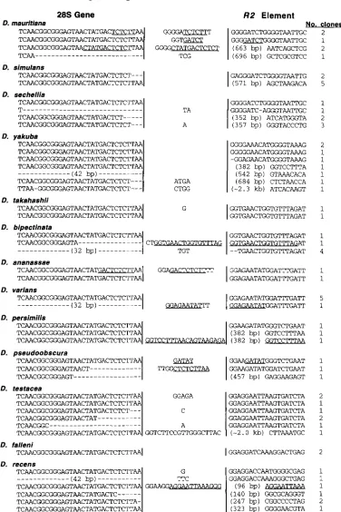
FIGURE 4.-Variation in the nucleotide sequences at the 5'junction of R2 elements in 13 species of Drosophila. The number of PCR clones that were identical to the sequence shown is indicated at the far right of the figure. Nucleotides to the left of the first vertical line are 28s gene sequences. Nucleotides to the right of the second vertical line are R2 sequences. Nucleotides between the two vertical lines are additional inserted sequences. Segments of these insertions, at least five nucleotides in length, that are identical to 28s gene sequences or R2 sequences are underlined. For the 5' truncated elements, the number of nucleotides deleted, based on the sequence of full-length elements, is given in the parenthesis. In the case of D. yukubu and D.
5‘ Junction of R2 Elements 859
2.
3.
ments were recovered that the 5’ ends of the
R2
ele- ments could be defined by sequence comparisons within each species. In the remaining species the 5’ ends were defined by comparingR2
sequences between closely related species. Only in the case of D. simulanswas the exact 5’ end somewhat unresolved in that a GA dinucleotide could represent an insertion or the 5’ end of the R2 element. In Figure 2 this GA dinucleotide was defined as part of the R2 sequence, because in all other species R2 elements contained at least five pu- rines in the first six nucleotide positions.
The sequence variants found in all 13 species were of the same general type as found in D. melanogaster.
Therefore, the following summary of the variation seen at R2 junctions was based on the combined data from all 14 Drosophila species.
1. Deletion of 28s sequences: The
R2
insertions in all species appear to result in the deletion of2
bp, con- sistent with the elimination of the 2 bp within the staggered cut generated by the R2 endonuclease from B. mori (Figure 1B). The elimination of these 2 bp is readily supported by the junction sequences within D. melanogaster (Figure 2), D. pseudoobscura, D.testacea and D. recens (Figure 4). In the case of the other species, this conclusion is inferred by compari- sons between species from the same group or sub- group. In addition to this 2-bp deletion, 41% of the junctions (39/94) contained deletions of the 28s
gene that extended beyond the staggered cut. Nearly half of these deletions were relatively short, 1-6 bp in length, with 3-bp deletions being most abundant. The other half of the 28s deletions ranged in length from 11 to 42 bp with most of these longer deletions in the range of 11-18 or 26-32 bp.
Deletion of R2 sequences: Unlike the deletions of the target site, only a limited number of junctions contained short deletions of the R2 sequences (sin- gle examples of a 1- and 28-bp deletion in D. melane gaster, a I-bp deletion in D. yakuba, and four exam- ples of a 2-bp deletion in D. bipectinata). The remaining examples of the
R2
deletions (38 cases) represented deletions of at least 96 bp. N o particular sequence appeared to be associated with the loca- tions of these major truncations, although a number did occur at one or two G nucleotides. While these G’s could be interpreted as being derived from the 28s gene sequence, most of these junctions had in- sertions or deletions of the28s
gene beyond the terminal G nucleotides, thus these bases do not ap- pear to be derived from the 28s target site.Insertion of extra nucleotides: 45% (42/94) of the junctions contained from 1 to 20 nucleotides be-
tween the 28s gene and
R2
element sequences. In all but one of the longer insertions, a substantial segment of these additional sequences have identitywith
28s
orR2
gene sequences near the junction. Instances where this identity was at least five nucleo- tides in length have been indicated by underlining in Figures2
and 4. The only long insertion that does not appear to have a counterpart in the R2 or 28s gene is the 5’ truncated copy in D. testacea. However, the exact location of this junction is somewhat am- biguous because an entire D. testaceaR2
element has not been sequenced. The 5’junction of this D. testa- cea clone was therefore defined by its homology to a region 2.0 kb from the 5’ end of the R2 elements from D. melanogaster.A limited number of R2 5’ junction sequences have also been obtained from B. moriand several other widely divergent insects (BURKE et al. 1987; W. D. BURKE and T. H. EICKBUSH, unpublished observations). R2 trunca- tions, 28s gene deletions and duplications similar to those described in Drosophila were seen to a variable degree in all species. Insertions could not be scored in all species because of an insufficient number of junc- tions to precisely define their 5’ ends.
DISCUSSION
We have previously shown that over 85% of the
R2
elements that have been partially sequenced from spe- cies of Drosophila contained neither termination co- dons nor frameshifts disrupting the ORF (EICKBUSH and EICKBUSH 1995). The rate of nucleotide substitu- tions at synonymous sites in this O W averaged 9.6 times the rate at replacement sites, suggesting continuous se- lection on the encoded proteins of these elements (EICKBUSH et al. 1995). These data suggest retrotranspo- sition plays an essential role in maintaining R2elements in the28s
genes of its host. Indeed, our evidence to date suggests that R2 elements turnover rapidly in the rDNA loci. Using a genomic blot assay, the pattern of 5’ truncatedR2
elements was shown to differ for each strain of D. melanogaster tested ( JAKUBCZAK et al. 1992). In this report the pattern of full-length elements was also shown to vary for each strain (Figure 3). Thus indi- vidual copies of R2 do not appear to remain within the rDNA locus for extended periods.Based on these findings, it is likely that the variation detected at the 5’ end of
R2
elements presented in this report resulted from the process of integration rather than from subsequent recombination or mutation events. Indeed, it was only the 5’ junction of theR2
element that exhibited such variation in sequence. The 3’junction of R2 elements from these same species vary only in the length of the A-rich homopolymer at the precise junction (EICKBUSH and EICKBUSH 1995; W. C.
LATHE and T. H. EICKBUSH, unpublished observations).
860
1.
A. George, MT. D. Burkc and T. H. Eickbushthe initiation o f reverse transcription (LUAN and EKK- I<L'sI< 199.5). Truncations of IC2 sequences o r deletions o f 28s gene sequences have never been detected at the
3' end o f the R2 elements. This dramatic difference between the 3' and 5' junctions suggests that it is the rctrotransposition mechanism itself that is generating the many different .5'junctions. Indeed, the amount of variation we have scored in this report is likely to be an underestimate of the variation generated by the integra- tion mechanism. PCR bias during the amplifications of the junction sequences, and the concerted evolution processes of gene conversion and unequal crossovers acting on the rDNA units would all act to reduce the apparent variation that is scored.
The extensive sampling of the
R2
5' junctions and scoring of sequence variants within and between species cnables us t o evaluate various models for the attach- ment o f the R2 sequences to the upstream 28s gene. Indeed, over two-thirds of the 5'junctions obtained in this report were unique, suggesting that there is variabil- ity inherent in the mechanism by which R2 sequences are attached t o the upstream 28s gene sequence.Equally important, our results indicate that there are probably no sequences at the 5' end of the
R2
element that arc needed for integration. "hile i t was known that many 5' truncated R2 copies could be found in the 28s genes ofD.
w l t n q p ~ t n - , it was generally as- sumed that these insertions represented aberrant inte- grations. This report has shown for the first time that the 28SR2junctions of these 5' truncated copies have identical properties to the junctions of full-length R2elements. For example, 37% of the full-length elements could he defined as precise, ie., contain neither dele- tions o f the 28s gene beyond the 2-hp stagger nor inser- tions of additional nucleotides, compared to 29% of the 5' truncated elements. Another 39% of the full- length element.. contained deletions of the 28s gene compared to 45% o f the 5' truncated elements. Finally, 46% of the full-length element.. and 45% of the trun- cated element. contained additional nucleotides in- serted between the R2 and 28s gene sequences. What-
ever mechanism is used to attach the R2 sequences to the upstream 28s gene in Drosophila, it either does not utilize specific sequences at the 5' end of the element, o r multiple internal sequences can readily substitute for these sequences.
Several models for the attachment of
R2
to the u pstream 28s gene sequences are shown in Figure 5. When combined with the known cleavage sites of the R2 endonuclease (Figure 1 B) , the 5'junctions reported here provide evidence for o r against each of these mod- els. In model A the 3' OH exposed by second-strand cleavage serves as the primer for second-strand synthesis of the 112 element. The signal for this second step could be the reverse transcriptase running off the end of the RNA template, if R2 transcription begins at the
5'
endA
Cleavage primed synthesisD
DNA repairFIGURE 5.-Possible models for the attachment of R2 se- quences to the upstream 28s gene sequences. In all models the first step of the reaction is target DNA-primed reverse transcription of the R2 template. Thick lines, 28s gene target sequences; dotted line, cDNA and second strand synthesis of
R2 element sequences; stippled line, R2 RNA template; oval,
x2 protein. (A) Cleavage of the second DNA strand exposes a 3' OH group to prime synthesis of the second strand of the R2 element using the cDNA strand as template. (E) Ligation of the 3' e n d of the cDNA to the upstream 28s sequences. (C) cDNA synthesis extends to 28s gene sequences upstream of the R2 sequences. Recombination between this cDNA and the 28s gene attaches the cDNA to the upstream 28s gene. (D) After cDNA synthesis and second strand cleavage n o fur- ther step are catalyzed by the R2 protein. Unknown DNA repair processes link the two ends together. (E) At the end of reverse transcription the R2 polymerase switches from the RNA to the upper strand of the upstream DNA sequence, displacing the lower DNA strand. In all models except model A, second strand synthesis and final polishing of the ends are conducted by DNA repair enzymes.
5’Junction of R2 Elements 861
This model readily explains the deletion of the 2-bp stagger, however it does not explain why in over 40% of the observed junctions additional
28s
gene se- quences are also eliminated.A second model for the attachment of R2 sequences is ligation of the cDNA strand itself to the bottom strand of the upstream 28s gene (model B). This model is similar to that previously proposed for the cin4 elements
of maize (SCHWARZ-SOMMER et al. 1987) and the I ele- ments of Drosophila (FINNEGAN 1989). This mechanism is unlikely since it would not result in the deletion of either the 2-bp staggered cut or the upstream 28s gene sequences.
A third possible mechanism for 5’ attachment differs from models A and B in that it requires 28s gene se- quences be present at the 5‘ end of the R2 template (model C). Reverse transcription of this template be- yond the 5’ end of the R2 elements into the 28s gene would allow homologous recombination of the cDNA strand with the upstream 28s gene sequences. This model is also not supported by the junction sequence data because it would not generate 5’ truncated copies unless the donor RNAs are derived from preexisting 5‘ truncated R2 elements. The very different patterns of 5’ truncated elements seen in different strains of
D.
melanogaster ( JAKUBCZAK et al. 1992), and the variety of
nucleotide additions and deletions seen at the 5’ end of R2 elements strongly argues against model C.
A fourth model for R2 integration (model D) is that after reverse transcription and second strand cleavage, the free DNA ends are joined by cellular DNA repair mechanisms. This model predicts that the attachment of the two ends would occur well after cleavage, allowing sufficient time for exonuclease activity to de- lete part of the 28s gene. Such a repair mechanism also makes the important prediction that full-length and 5’ truncated elements would have similar junctions. Dou- ble-stranded break repair studies conducted fungi or mammalian systems support a homologous gap-repair mechanism (SZOSTAK et al. 1988; BOLLAG et al. 1989). If such a gap repair model were to be applied to R2 integration, a preexisting R2/28S junction already in the rDNA locus would serve as a template for the attach- ment of the 5’ end of the R2 element to the upstream
28s
gene sequence. This seems unlikely since such a process would give rise to greater uniformity in se- quence than what we have observed at the 5’ end of full-length elements and would suggest that truncated copies must rely on alternative mechanisms. Repairs of double-stranded breaks in higher eukaryotes can alsooccur by nonhomologous means (ROTH and WILSON
1988; THACKER et al. 1992; LUKACSOVICH et al. 1994). However, such repairs are based on short sequence re- peats and usually involve the deletion of nucleotides. The attachment of
R2
sequences at the 5’ end do not involve short repeats and frequently result in nucleotideadditions. However, given the many uncertainties in the cellular repair of higher eukaryotes, particularly those involving an RNADNA heteroduplex (Figure 5), more general DNA repair models to complete R2 retrotrans- position remain possible.
The model for the attachment of the
R2
sequence to the upstream28s
gene sequences we suggest as most supported by the 5’junctions is model E. This model requires that the R2 reverse transcriptase jump from theR2
RNA template to the top strand of the upstream28s
gene sequence. A similar model has been proposed for the retrotransposition of the Zelement (BUCHETON 1990). One attraction of this model is that it is consis- tent with the known template switching properties of reverse transcriptases (WHITCOMB and HUGHES 1992). Thus the switch from RNA to a DNA template could be viewed as similar to the template switching that occurs during first- and second-strand DNA synthesis in the retroviral retrotransposition mechanism. Model E is also consistent with the in vitro properties of the R2 reverse transcriptase. We have found that the R2 reverse transcriptase can readily switch to another RNA mole- cule when it reaches the end of the R2 template (D. D. LUAN and T. H. EICKBUSH, unpublished data). Unfortu- nately we have not been able to demonstrate that this switch can occur efficiently to the upstream28s
target sequence.862 J. A. George, W. D. Burke and T. H. Eickbush
have a short sequence of
28s
gene repeated. Duplica- tions can be explained if the reverse transcriptase at- tempts to switch to the target DNA template, falls off, and then must reattach to the target DNA. Model E cannot explain (nor can the other models) the tandem duplication of R2 sequences at the 5'junction.Can the template switching model for the attachment of element sequences to the upstream target site also be applied to other non-LTR elements? Unlike R 2 , most
non-LTR element insertions result in target site duplica- tions. These duplications are typically variable in length in the case of elements that do not have sequence spe- cific integration sites and are usually precise in length for those elements that do have sequence-specific inte- gration sites (EICKBUSH 1992). This difference from
R2
can be explained if cleavage of the second target DNA strand by these elements (ie., the strand not used to prime reverse transcription) takes place downstream of the first strand cleavage, rather than upstream as it does with the
R2
element. Second-strand cleavage down- stream of the first strand releases a single-stranded 3' overhang onto which template switching can occur. The R2 endonuclease may be unusual in that it cuts 2bp upstream from the first cleavage, resulting in a re- cessed 3' end that may be more difficult to support a template switch. Uniform template switching to the end of an exposed 3' overhang would give rise to a target site duplication of precise length. Template switching to variable locations within the 3' overhang would give rise to variable length target site duplications. While continued analysis of the 5' and 3' junctions of non- LTR elements can provide indirect support for or against the template switching model, direct character- ization of the template switching properties of the re- verse transcriptase from
R2
and other non-LTR ele- ments are ultimately needed.This work was supported by a National Institutes of Health grant (GM42790) to T.H.E. We thank DANNA EICKBUSH for comments on the manuscript and ROBERT BAMBARA and CHRIS LAWRENCE for help- ful discussions.
LITERATURE CITED
BOI.IAC;, R. J., A. S. WALDMAN and R. M. LISKAY, 1989 Homologous recombination in mammalian cells. Annu. Rev. Genet. 23: 199- 225.
BUCHETON, A. 1990 I transposable elements and I-R hybrid dysgene- sis in Drosophila. Trends Genet. 6: 16-21.
BURKE, W. D., C. C. CALAIANG and T. H. EICKBUSH, 1987 The site- specific ribosomal insertion element type 11 of Bomb)% mori
(R2Bm) contains the coding sequence for a reverse transcriptase- like enzyme. Mol. Cell. Biol. 7: 2221-2230.
BURKE, W. D., F. MULLER and T. H. EICKBUSEI, 1995 R4, a non-
of nematodes. Nucleic Acids Res. 2 3 4628-4634.
LTR retrotransposon specific to the large subunit rRNA genes
C I ~J. ,M, 1988 Novel non-templated nucleotide addition reac- tions catalyzed by prokaryotic and eukaryotic DNA polymerases. Nucleic Acids Res. 16: 9677-9686.
DAWID, I. B., and M. L. REBBERT, 1981 Nucleotide sequence at the boundaries between gene and insertion regions in the rDNA of
D. melanogastm. Nucleic Acids Res. 9: 501 1-5020.
DESTEFANO, J. J., R. A. BAMBARA and P. J. FAY, 1994 The mechanism of human immunodeficiency virus reverse transcriptase-catalyzed strand transfer from internal regions of heteropolymeric RNA
templates. J. Biol. Chem. 269: 161-168.
EICKBUSH, D. G., and T. H. EICKBUSH, 1995 Vertical transmission of the retrotransposable elements RI and R2 during the evoln- 671-684.
tion of the I)ro.rophila melanogastprspecies subgroup. Genetics 139:
EIC:KBUSH, D. G., W. C. LA'I'HE 111, M. P. FRANCINO and T. H. EICKRLISH, 19% KI and R2retrOtrdnSpoSabk elements o f Drosophila evolve at rates similar to those of nuclear genes. Genetics 139: 685-
695.
EICKBUSH, T. H., 1992 The non-LTR class of retrotransposable ele- ments. New Biol. 4 430-440.
EICKBC'SH, T. E, 1994 Origin and evolutionary relationships of. re- troelements, pp. 121-157 in The EvolutionaT Biology of l'imx~, edited by S. S. MORSK. Raven Press, New York.
EVANS, J. P., and R. D. PAI.MII'ER, 1991 Retrotransposition o f a mouse 1.1 element. Proc. Natl. Acad. Sci. USA 88: 8792-8795. FINNEGAN, D. J., 1989 The I factor and I-R hybrid dysgenesis in
Drosophila mlanogaster, pp. 503-517 in Mobile DNA, edited by D. H. BEKG and M. M. HOWE. American Society of Microbiology, Washington, DC.
Hu, W.-S., and H. M. TEMIN, 1990 Retroviral recombination and reverse transcription. Science 250: 1227- 1233.
M. H. EDGEIL, 1989 LINES and related retroposons: long intel-- spersed repeated sequences in the eukavotic gemone, pp. 539-
617 in Mob& DNA, edited by D. H. BERG and M. M. Howl<. American Society of Microbiology, Washington, D C
INOUYL, S., and M. INOW, 1993 Bacterial reverse transcriptase, pp.
391 -410 in Kpumsr Transcriptase, edited by S. GOFF and A. SICZI,K~.
Cold Spring Harbor Laboratory Press, Cold Spring Harbor, N Y .
JAKURCLAK, ,J. L., Y. XION(; and T. H. EICKBLWI, 1990 Type 1 ( K I )
and Type I1 (K2) ribosomal DNA insertions of lh-osophilrc nwlano-
gasto are retrotransposable element? closely related to those of
Bombyx mori. J. Mol. Bid. 2 1 2 37-52.
JAKCJBCLA~~, J. L., M. K. ZENNI, R. C. WOOI)RI!FF and T. H. EICKRUSH,
1992 Turnover of RI (Type I) and IC2 (Type 11) retrotranspos- able element5 in the ribosomal DNA of l~rosophilr~ mrkmopslm.
Genetics 131: 129-142.
JENSEN. S., and T. HEII)MANN, 1991 An indicator gene for detection of germline retrotransposition in transgenic Drosophila demon- strates RNA-mediated transposition of' the LINE 1 element. EMBO J. 10: 1927-1937.
HUTCHISON, C. A,, S. C . HARUIKS, D. D. LoEB, W. R. SHKHEE : I I I ~
MARTIN, S. L., 1991 LINES. Curr. Opin. Genet. Dev. 1: 505-508. MCCIXRE, M. A, 1993 Evolutionary history of reverse transcriptase,
pp. 425-444 in Rpverrr lransrriptasr, edited by S. GOFF and A.
SKAI.KA. Cold Spring Harbor Laboratory Prrss, Cold Spring Har- bor, NY.
LAMBOWI'I'Z, A. M., and M. BEWORT, 1993 Introns as mobile genetic elements. Annu. Rev. Biochem. 62: 587-622.
LEVIS, R. W., R. GANESAN, K. HOUTCHENS, I,. A. TOIAR and F. M.
SHFXN, 1993 Transposons in place of telomeric rrpeats at a
Drosophila telomere. Cell. 75: 1083-1093.
LONG, E. O., M. L. REBBERT and I. B. DAWTD, I980 Structure and expression o f ribosomal RNA genes of Drosophila mplanogasto
interrupted by type-2 insertions. Cold Spring Harbor Symp. Quant. Biol. 45: 667-672.
LUAN, D. D., and T. H. EICKHUSH, 1995 RNA template requirements Tor target DNA-primed reverse transcription by the K2retrotrans- posdbk element. Mol. Cell. B i d . 15: 3882-3891.
L ~ J A N , D. D., M. H. KORMAN, J . I., JAKUBCZZK and T. H. EICKIX.SH, 1993 Reverse transcription of R2Bm RNA is primed by a nick at the chromosomal target site-A mechanism for non-LTR ret- rotransposition. Cell 72: 595-605.
Lu~4c:sowc:11, T., D. YANG and A. S. WAI.l)MAN, 1994 Repair o f a
specific doublestrand break generated within a mammalian chromosome by yeast endonuclease I-Scrl. Nucleic Acids Res. 22:
5649-5657.
MARTIN, F., C. MARANON, M. OI.IVARES, C. Ar.oNso and M. C . I.OPEX,
5' Junction of R2 Elements 863
the first ORF with the Ape family of DNA repair enzymes. J. Mol. Biol. 247: 49-59.
PEACOCK, W. J., R. APPELS, S. ENDOW and D. GLOVER, 1981 Chromo- somal distribution of the major insert in D. melanoguster28S rRNA gene. Genet. Res. 37: 209-214.
PEI.ISSON, A,, D. J. FINNEGAN and A. BUCHETON, 1991 Evidence for retrotransposition of the I factor, a LINE element of Drosophila melanogaster. Proc. Natl. Acad. Sci. USA 88: 4907-4910. ROIHA, H., J. R. MILLER, L. C. WOODS and D. M. GLOVER, 1981 Ar-
rangements and rearrangements of sequence flanking the two types of rDNA insertion in D. melanogaster. Nature 290: 749-753. ROTH, D. B., and J. H. WILSON, 1986 Nonhomologous recombina- tion in mammalian cells: role for short sequence homologies in the joining reaction. Mol. Cell. Biol. 6: 4295-4304.
SANGER, F., S. NICKLEN and A. R. COUISON, 1977 DNA sequencing with chain-terminating inhibitors. Proc. Natl. Acad. Sci. USA 7 4
SCHWARZ-SOMMER, Z., L. LECLERCQ E. GOBEI. and H. SAEDLER, 1987 Cin4, an insert altering the stucture of the AI gene in Zea mays,
exhibits properties of nonviral retrotransposons. EMBO J. 6; 3873-3880.
SZOSTAK, J. W., T. L. ORR-WEAVER, R. J. ROTHSTEIN and F. W. STAHL, 1983 The double-strand-break repair model of recombination. Cell 33: 25-35.
THACKER, J., J. CHALK, A. GANESH and P. NORTH, 1992 A mechanism for deletion formation in DNA by human cell extracts: the involvement of short sequence repeats. Nucleic Acids Res. 20:
6183-6188. 5463-5467.
WHITCOMB, J. M., and S. H. HUGHES, 1992 Retroviral reverse tran- scription and integration: process and problems. Annu Rev Cell Biol. 8 275-306.
Wu, W. M., B. M. BLUMBERG, P. J. FAY and R. A. BAMBARA, 1995 Strand transfer mediated by human immunodeficiency virus re- verse transcriptase in vitro is promoted by pausing and results in misincorporation. J. Biol. Chem. 270: 325-332.
XIONG, Y., and T. H. EICKBUSH, 1988a Similarity of reverse tran- scriptase-like sequences ofviruses,transposable elements, and mi- tochondrial introns. Mol. Biol. Evol. 5: 675-690.
XIONG, Y., and T. H. EICKBUSH, 1988b Functional expression of a sequence-specific endonuclease encoded by the retrotransposon R2Bm. Cell 55: 235-246.
XIONG, Y., and T. H. EICKBUSH, 1990 Origin and evolution of re- troelements based upon their reverse transcriptase sequences.
ZHANG, J., and H. M. TEMIN, 1993 Rate and mechanism of nonho- mologous recombination during a single cycle of retroviral repli- cation. Science 259: 234-238.
ZHANG, J., and H. M. TEMIN, 1994 Retroviral recombination de- pends on the length of sequence identity and is not error prone. J. Virol. 68: 2409-2414.
ZIMMERLY, S., H. Guo, P. S. PERLMAN and A. M. LAMBOWITZ, 1995 Group I1 intron mobility occurs by target DNA-primed reverse transcription. Cell 8 2 1-10.
EMBO J. 9: 3353-3362.