___________________________________________________________________________________________ 4(1): 215-224, 2014
SCIENCEDOMAINinternational
www.sciencedomain.org
Ascitic Antigen CA125: Study of Dissociaton
Forms and its Final - Stem Structure
Petr G. Prokopenko
1*, Olga Petrenko
2, Valentina Shelepova
2,
Vadim V. Negrebetsky
1, Innokenty M. Mokhosoev
2and Аlexander А. Terentiev
11Department of Biological Chemistry, N.I. Pirogov Russian National Research Medical
University, Ostrovityanova St.1, Moscow 117997, Russian Federation.
2N.N Blokhin Russian Cancer Research Center, Russian Academy of Medical Sciences,
Kashirskoe Shosse 24, Moscow 115478, Russian Federation.
Authors’ contributions Authors may use the following wordings for this section: This work was carried out in collaboration between all authors. Author PGP designed the study, performed the statistical analysis, wrote the protocol, and wrote the first draft of the manuscript. Authors PGP, OP and VS managed the analyses of the study. Authors VVN, IMM and AAT managed the literature search. All authors read and approved the final manuscript.
Received 5thApril 2013 Accepted 3rdAugust 2013 Published 14thSeptember 2013
ABSTRACT
Aim: Study of dissociation forms of the ascites antigen СА125 and its stem structure – the final form of its dissociation.
Study Design:Resarch paper.
Place and Duration оf Study: Department of biochemistry, N.I. Pirogov Russian National Research Medical University, laboratory of immunochemistry and laboratory immunology, N. N. Blokhin Russian Oncological Scientific Center.
Methods: Next materials were used in the work: ascitic fluid from patients with ovary cancer (14 patients), blood serum from patients with ovary cancer (28 patients), 40 donors (24 women and 16 men). Electrochemoluminiscent method, ELISA, electrophoresis and direct binding of labeled antibodies with antigen in polyacrylamide gel, reaction of precipitation, chromatography were used to study forms of dissociation of the antigen СА125.
with ovary cancer, demonstrated at native conditions of electrophoresis in polyacrylamide gel three main forms: gamma – globulin with molecular weight (MW) ~500 kDa, alpha-globulin with MW ~ 230 kDa and beta-globulin with MW ~110 kDa. All 3 forms bound l-MAB to СA125 and were isolated from the polyacrylamide gel during preparative electrophoresis. All these forms dissociated to a single protein band with MW 55 kDa that migrated in the albumin zone and did not bind l-MAB to CA125 under denaturing conditions of polyacrylamide gel-electrophoresis with 2% sodium dodecyl sulfate. This form also demonstrated a sigle protein band corresponding to MW 75 kDa in reducing conditions of electrophoresis. It has been established that the form with MW of 55 kDa represents a complex of three different polypeptides, which are identical to heavy chains IgG, albumin and unknown thermostable protein coupled to albumin. This thermostable protein was revealed in all immunoreactive forms of CA125 and all investigated commercially available samples of albumins in the reaction of precipitation with polyclonal antibodies. Antibodies to these proteins have also been revealed in commercial antisera to human serum proteins in a titer 1:40.
Conclusion: The final form of dissociation of immunoreactive forms of the antigen CA125 is represented by a complex of three different polypeptides with MW of 55 kDa that did not bind l-MAB to CA125.
Keywords: Forms of dissociative ascetic antigen СА125; final form dissociation of antigen СА125; thermostable protein coupled to albumin; bow-string effect; albumin spiral.
1. INTRODUCTION
Antigen СА125/MUC16 represents a slow reacting marker: is level is increased in the serum of patients with ovary cancer at III-IУ stage of the disease and, predominantly, in patients with ascites [1]; in the more early phases of the evolution of tumorous process it uninformative [2]. Functions and devotion of this structure is still obscure, despite application of mass-spectrometry methods that are oriented on the increasing of specificity of a test for ovary cancer [3]. The nature of the immunoreactive epitope of the antigen СА125 is known and the existence of a final form of its dissociation is under discussion and remains to be elucidated [4]. Nevertheless, it has been shown that the final form of dissociation of the antigen СА 125 represents a complex of different polypeptides with MW of 55 kDa that includes polypeptides immunochemically identical to serum albumin (HSA), heavy chains of IgG (IGHC) and unknown thermostable protein coupled to albumin - TPC.А [5]. Difficulties in identification of this form are that it does not bind l-MAB to СА125, but reacts with polyclonal antibodies in the reaction of precipitation with А-HSA line identical to HSA, and with A–IGHC – line identical to IgG. With polyclonal antisera to donor serum proteins (AD) absorbed with HSA and IgG, this form reacts not only with TPC.A and donor sera, but also with all isoforms of CA125, which bind l-MAB to CA125. The antiserum lost immunoreactivity after its absorption by donor sera (5). It is necessary to note that А-HSA reveals up to 9 fragments and complexes of HSA with MW from 11 to 260 kDa in the serum and urine [6,7].
consider this protein as typical serum protein [9]. A similar protein was revealed many times in tumors, ascites, sera and everywhere its serum origin was seen: glycoprotein «alpha-2Н-ferroglycoprotein» [10] was serum macroglobulin [11], «macromolecular serum ferroprotein» [12], «albumin-IgG complex» [13], «tumor-associated ascites IgG» [14], «IgG-like glycoferroprotein serum» [5] and «peroxidase active IgG-like glycoferroprotein ascites» [15].
Study of СА125 only with the use of labeled monoclonal antibodies (l-MAB) did not allow elucidating dissociative forms that did not bind l-MAB to СА125. The most noticeable work in this field is a very thorough investigation made by a group of М.Т. de Los Frailes [16]. Immunoreactive СА125 was isolated from ascite of patients with ovary cancer by the use of immobilized MAB (ОС125) to CА125 on Affigel 10. Under polyacrylamide gel electrophoresis in native conditions (n-PAGE), immunoreactive СА125 did not penetrate the gel and was at the start position in the separating PAG. Under mild denaturing conditions of SDS-PAGE with 0,1% SDS (these conditions are very close to natives ones) the immunoreactive form of CA125 demonstrated two proteins bands with MW of 205 and 55 kDa, and also a third form that did not penetrate the gel (MW ~500 kDa ). From this it follows that, from one side, the dominant form of CA125 partly dissociates to the final form of dissociation with MW of 55 kDa, and, from another side, re-associates, partly, with formation of a large molecular form that did not penetrate the gel and was at the starting position. In 2% SDS – PAGE authors observed only one band with MW of 55 kDa. In reducing conditions of SDS-ME-PAGE there also was on band, but with MW of 75 kDa. Only one band with MW of 205 kDa bound l-MAB to CA125В in Western blotting. The final form of dissociation with MW of 55 kDa did not bind antibodies. From this follows that the final form of dissociation may be only studied with the use of polyclonal antibodies. This unique structure has not attracted attention of researchers yet and is still opened for investigation. We consider the present work as continuation of works by М.Т. de los Frailes with addition of polyclonal antibodies to study the final form of dissociation of the antigen CA125, which does not bind l-MAB to CA125.
Results of investigation of dissociative from of the antigen CA125 and its stem structure – the complex of polypeptides that do not bind l-MAB to CA125, are presented in this work.
2. MATERIALS AND METHODS
2.1 Isolation of Antigen СА125
Antigen СА125 was obtained from acid-soluble fraction of ascites and pooled donor serum from 50 women (SD50) [17]. Content of immunoreactive (IR) СА125 was determined by electrochemiluminiscent method at «Hoffmann-L Roche Elecsys 2010» (Germany) and by ELISA method with use of l-MAB ОС 125 to antigen СА125. SD50, in which concentration of IR СА125 was equal to 34,6 U/ml или 5 mkg/ml, ascites - 3140 U/ml or 448 mkg/ml were used as control samples, and antigen СА125, isolated, according to Davis, from SD50 - 63,6 U/mg, serum of female patient B. (287 U/ml), GFP470 (48,3 U/mg), А230 (514 U/mg), CP55 (1,6 U/mg) and IgG (0,02 U/mg) as standard samples. Ascites without hemolysis were used in the work.
2.2 Isolation of GFP, CP55, the Complex of “TPC.А – Albumin” and IGHC
CP55 (zone of albumins) was isolated out of SD50 diluted 1:8 in tube variant of native PAGE. Thermostable complex «ТПС.А- albumin» was prepared by heating of albumin preparations – 2-10 mg/ml (100оС, 15 min). All isoforms of ascite GFP were isolated ex tempore, due to extremely fast variability of forms. A fraction with MW 55-60 kDa was isolated from IgG (2mg/ml) by gel-filtration under control of precipitating test-system for IGHC.
2.3 Renaturation of Albumin
5-10 mg/ml of BSA (Sigma, USA) dissolved in Tris-glycine buffer (0,05 mM tris, 4 mM of glycine, рН 8,3), were incubated in boiling water-bath for 15 min.; a pellet of denatured protein was separated by centrifugation, suspended in stock Tris-glycine buffer (50 mM tris, 0,4 М glycine, рН 8,3) and renaturated during 48 hours at + 37ºС; ~ 0,5% of initial protein is renaturated.
2.4 Polyclonal Antiserum against Human Serum Proteins
Exclusively commercially available antiserums to human serum proteins were used in our work: (Russia; Binding Site Limited, UK; Sigma, USA), anti-IgG (Russia) and antiserum to gamma-, alpha131 and - chains of immunoglobulins G, A, M (Bio-Rad, USA). Rabbit polyclonal antiserum to antigen СА125, GFP and CP55 were prepared in our laboratory.
2.5 Polyacrylamide Gel Electrophoresis; Direct Binding of Labelled
Antibodies to Antigen in PAG
Electrophoresis in slabs and tubes were performed under native, denaturing and reducing conditions. Slabs/tubes were incubated with commercial preparations of l-MAB (to antigen CA125 and AFP) and мечеными polyclonal antibodies to PSG for IFA (Vector Best, Russia) during 2 hour at +37ºС after native PAGE, then washed three times by water for 15 min., incubated (+37ºС, 15 min) with с 3,3,5,5, - tetramethylbenzidine (TMB) and washed by water. Another substrate - 3,3, diaminobenzidine tetrahydrochloride – is oxidized by all isoforms of ascite antigen СА125, with exclusion of the final dissociation form, without pre-incubation with l-MAB (Fig.1, b).
2.6 Determination of Peroxidase-Like Activity (PA) of Proteins in n-PAG
After PAGE slabs/tubes were incubated in DAB solution (Sigma, USA) with hydrogen peroxide (10 mg of DAB are dissolved ex tempore in 20 ml of 0,05 М tris-НС1 buffer, рН 7,5 and 0,3 ml of 3% Н2О2 was added) during 15-30 min. at 37о С and were washed by water during 16-18 hours at + 4оС. HSA, alpha-fetoprotein (AFP) and placentaspecific-glycoprotein (PSG1), which do not oxidize DAB, were used as a control samples for PA specificity of CA125 isoforms.
2.7 Revealing of Proteins, Ferro - and Glycoproteins in PAG.
are stained in a blue color. Fe+3 in GFP and «antigen СА125» is bound weakly and is not
practically determined after boiling.Glycoproteinswere determined by Schiff-iodine acid [11].
2.8 A Reaction of Direct Precipitation of Murine l-MAB
Labeled monoclonal antibodies act as antigenin reaction of direct precipitation. Well-known property of cross-reaction of human and murine IgG is in the basis of the method [19]. To reveal murine l-Mabs any commercial antiserum to human serum proteins or anti-IgG were used on the line of precipitation with l-Mab was developed by a substrate [5].
2.9 A Reaction of Mixed Precipitation with m-MAB
Studies with use of this reaction have been performed and documented by the author of the method V.S. Poltoranina in G.I. Abelev’s laboratory [19]. Also the method of direct precipitation of l-MAB in agar gel and the method of direct binding of l-MAB with antigen in PAG have been reproduced and the results have been confirmed by the reaction of mixed precipitation of l-MAB to CA125.
3. RESULTS AND DISCUSSION
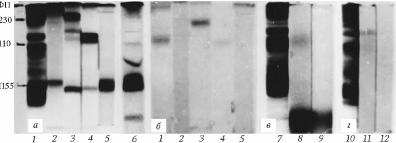
High mobility of dissociation-association processes in GFP and its isoforms is mostly expressed in ascites of patients with ovarian cancer (Fig.1, а): from one side, GFP, А230 and В110 dissociate in partial to final form CP55 (Fig.1, а 2-4), from the other side, CP55 reassociates partly to GFP (Fig.1, а 5, 6 ) that reflects pulsating form of protein bodies existence – the property characteristic for many enzymes. PA results for ascites GFP isoforms till and after incubation with l-Mab to antigen СА125 were absolutely equal (Fig.1b). the final form of GFP dissociation – CP55 does not demonstrate PA, but reassociated form of CP55 - GFP, does (Fig. 1, b 5).
After 7 months this form – CP55 represents up to 6 protein bandsin186the tube variant of n-PAGE, and one of which is a sloping band (Fig.1, а b). Such an inclined protein band was observed by group of Frаiles in the final form of dissociation of antigen СА125 with MW 55 kDa, but in plat variant of reducing PAGE [16].
Fig. 1. Electrophoresis under non-denaturing conditions of tube PAG and direct binding of l-MAB with antigen in PAG
а — electrophoretic mobility of ascites GFP and its isoforms in 7.5% n-PAGE: 1. ascites of a patient with ovary cancer № 305; 2. GFP; 3. А230; 4. В110, 5. CP55, isolated ex tempore according Davis (electrophoresis, dated 26.07.2005); 6 — dynamics of CP55 re-association (restoration of complexes)
after 7 months (electrophoresis dated 03.03.2006). Staining for proteins. b — peroxidase activity of ascites 305, ascites GFP and its isoforms with DAB substrate; c — direct binding of l-MAB to СА125 with antigen from serum of blood of female patient B. in 7.5% PAGE. 7 — staining for proteins; 8 — development with DAB after incubation with l-MAB to СА125; 9 — development with DAB only; d —
direct binding of I-MAB to antigen СА125 in ascites № 307. 10 — staining for proteins; 11 – development by substrate 3,3,5,5-tetramethylbenzidine (TMB) after incubation with m-MAB to СА125;
12 — development with TMB only.
Table 1. Some properties of main forms of the antigen CA125
Properties G500
ascites A230ascites B110ascites CP55ascites GFPserum IgGserum HSAserum Binding of l-MAB to
СА125 in ELISA + + + - - -
-Direct binding of l-MAB to CA125 in
PAG + + + - - -
-A dissociation form (kDa):
А. in SDS-PAGE B. in SDS-ME-PAGE
55
75 5575 5575 5575 5575 55; 1255 5575
Peroxidase activity + + + - - -
-Staining for Fe3+ + + + - + -
-Staining for
glycoproteins + + + - + -
b) and HSA permanently demonstrate. We did not observe such phenomenon with IgG (Fig. 2, а2) and AFP.
Fig. 2. Electrophoresis of serum proteins in reducing conditions of denaturing 10% PAG in plats
а – the “bowstring” effect: 1 – molecular weight markers (14,4 - 97 kDa); 2 – IgG; 3-5 – serum GFP; albumin). Lines 1, 5 contain 2% SDS and 5% ME; 2–4 - 2% SDS; 6-8 – serum CP55; 9-11 – BSA;
12-14 – rat albumin. 1, 2, 5, 8, 9, 12-14 lines contain 2% SDS and 5% - МE; 3, 4, 6, 7, 10 -13 lines contain 2% SDS; b – the spiral of albumin: 1 – thermostable BSA; 2-5 – renatured BSA: (in lines 2 and 4 – the
spiral of albumin (magnified by 3,5 times).
The transitional form from low to high level of molecular weight of a structure is presented by sloping line under angle ~ 30о that links different height levels (Fig. 3, а). Expected number of horizontal bands is absent; because the bladder bowstring in projection hides the number of horizontal grades by united sloping line. The «bowstring» effect was reproduced with GFP, antigen СА125, CP55, ТPС.А and all native, heated and renatured preparations of HSA and BSA and also with rat albumin, i.e. albumin in reducing conditions is “jumping up” (Fig. 2). Seemingly, this is a key stage in the mechanism of functional structures reconstruction on the basis of the stem complex «ТPС.А–albumin», in which amount ratio is equal to ~1:30, and molar - ~ 1:5 (seemingly, TPC.A has a molecular weight of 11,3 kDa).
PAGE (Fig. 2, а). In renatured BSA the «bowstring» line rises on closed helix from 2 to 75 kDa (Fig. 2, b 2,4), and a ghost/traces of a second helix in the upper levels (Fig. 2, b 2,4), do not allow excluding its possible double nature.
Revealing of antigen ТPС.А by reaction of precipitation in all serums and commercially available human preparations: HSA, IgG, and also in the antiserum to IGHC (Fig. 2, c), i.e. in places wher HSA- depot for ТPС.А evidences its dissemination. The effect of «bowstring», observed with BSA and rat albumin, indicates existence of functionally similar structure in animal proteins.Antibodiesto ТPС.А were revealed inall commercially available antiserums to human serum proteinsin a titer 1:8 (Sigmа,USA) and in a titer 1 : 40 (Binding Site Limited, UK; Russia). It is necessary to note that the titer of antibodies to TPC.A in antisera from different Russian and UK firms was stably high and was equal, as a rule, to 1:40 in a minimum. High titer of antibodies has been also revealed in all antisera against serum fractions or native sera of healthy donors and patients. Hence, а priori, we could expect high titer of antibodies to TPC.A in all anti-HSA sera after absorption of antibodies against HSA.
We suggest that extracellular N-domain of СА125 (1-1638 а.a.) may represent a full primary structure the isoform А230 of serum, and the primary structure of ТРС.А is accurately duplicated in its peptide motifs a. a. 421-524 and a. a. 641-742 [20]. The thermostable pair – «TPC.A-albumin», seemingly, is a stem structure if all CA 125-containing complexes up to supercomplexes with MW of 2700 kDa [18]. Number, weight and content of the complexes may depend on the number of bound toxic products of cell degradation (Fe ions, carbohydrates, heme groups etc.) that is clearly seen in ascites of ovary cancer patients. Seemingly, the ascites is a depot of toxins and a barrier for their penetration into the blood due to these complexes, which bind toxins and perform a sanitary role. For example, concentration of antigen СА125 in ascites (volume of 10 l) of the patient B. was equal to 9700 U/ml, and in the serum of the patient - 287 U/ml [2].
Hence, the final form of dissociation of all immunoreactive forms of the antigen CA125 is represented by a complex of polypeptides with MW of 55 kDa that includes IGCH, HSA and TPС.А, bud is not able to bind l-MAB to СА125. Ability of CP55 to re-associate and newly form peroxidase-active high molecular complexes with MW of 500 kDa (Fig.1, а5, b5) allows ranking CP55 among the stem structure of the antigen СА125. This unique structure is extremely interesting and it is impossible to name it as «dead»: it is always in movement. It is necessary to note the main difference of immunoreactive forms of ascitic CA125 from immunochemically similar serum forms – all immunoreactive forms of the ascites protein acquire peroxidase activity.
On the other side, identification of TPC.A highlights ambivalent nature of albumin, because it is difficult to explain heterogeneity, pulsating form of protein structure existence, reversible complexing and restoration of a denatured protein due to a helix principle only by «conformational adaptability» of a monovalent structure of albumin and this issue is of interest for further studies.
4. CONCLUSION
Ascites antigen СА125 and IgG-like serum glycoprotein represent a complex immunochemically identical structures that includes IgG, HSА and TPC.A .
The final form of dissociation of all isoforms of the ascitic antigen CA125 and IgG-like donor sera glycoferroprotein is presented in denaturing SDS-ME-PAGE by a single protein band with MW of 55 kDa that represents a complex of three polypeptides: HSА, IGHC and TPC.A. This structure demonstrates a form with MW of 75 kDa in reducing SDS-ME-PAGE.
Human, bovine and rat albumins demonstrate a bowstring effect in reducing SDS-ME-PAGE, and renatured BSA – bowstring twisted into a spiral.
TPC.A is not identical to HSA and IGHC, but is tightly coupled to HSA. The pair «ТРС.А-НSA», semingly, is a stem structure for reconstruction of chelating supercomplexes that bind toxic products of cells degradation.
CONSENT
Not applicable.
ETHICAL APPROVAL
All authors hereby declare that “Principles of laboratory animal care” (NIH publication No. 85-23, revised 1985) were followed as well as specific laws of the Russian Federation where applicable. All experiments have been examined and approved by the appropriate ethics committee.
ACKNOWLEDGEMENT
Authors are very grateful to Prof. G.I. Abelev and V.S.Poltoranila for consultance and real help in the present work implementation and understanding of the results.
COMPETING INTERESTS
Authors have declared that no competing interests exist.
REFERENCES
1. Bergmann J, Biclard JM, Georg M, et al. Evaluation of CA125 in patients with benign and malignant ascites. Cancer. 1987;59(2):213-217.
2. Prokopenko PG, Poltoranina VS, Jordania KI, et al. Problems and Perspectives in Diagnosis and Prevention of Ovarian Tumor Diseases. Brit. J. Med. & Medical Res. 2011; 1(4):182-197.
3. Timms TA, Smith JF, Devenyarov C, et al. Highly Accurate Detection of Ovarian Cancer Using CA125 but Limited improvement With Serum Matrix-Assisted Laser Desorption Time-of-Fight Mass Spectrometry Profiling. Int. J Gynecol. Cancer, 2010;20(9):1518-1524.
4. Weiland F, Fritz K, Oehler MK. Methods for identification CA125 from ovarian cancer ascites by high resolution mass spectrometry. Int. J. Mol. Sci. 2012;13(8):9942-58. 5. Prokopenko PG, Poltoranina VS, Shelepova VM, Terentiev AA. Serum IgG–like
glycoferroprotein: identification of its final dissociation form of thermostable protein coupled with albumin.Bul. Exp Biol. Med. 2012;153(1):43-48.
7. Wiggins, RC, Kshisagar B, Kelsch KC, Wilson, BS. Fragmentation and polymeric complexes of albumin in human urine. Clin. Chimica Acta. 1985;149(2):155-163. 8. Lloyd KO, Yin TWT, Synthesis and secretion of the ovarian cancer antigen CA125 by
the human cancer cell line NIH: OVCAR-3. Tumor Biology. 2001;22:77-82.
9. Borisenko SA, Prokopenko PG, Shelepova VM. Ovary cancer ascites proteins – 1: characterization of CA125 monoclonal antibody (OC125) binding proteins. Tumor Biology. 2007;28(1):90.
10. Buffe D, Rimbaut Ch. Alpha2H-glycoferroprotein: characterization and clinical Significance. Ann. N. Acad. Sci. 1975;259:417-426.
11. Prokopenko PG. Immunochemical identification of ferritin and its immunological analogs – beta-fetoprotein and alpha2H-globulin. Bull. Exp. Biol. Med. 1982;93(4):70-73.
12. Prokopenko PG, Terentiev AA. Determination of ferroproteins in biological fluids. Lab. Delo. 1975;6(259):115.
13. Sharma NC, Mohammad SF,Chuang HY. Albumin-IgG complexes In human serum and plasma. Prog. Nat. Acad. Sci. USA. 1981;78(12):7750-7753.
14. Silburn PA, Khoo SK, Hill R, Demonstration of tumor-associated immunoglobulin GIn ascites fluid of ovarian cancer. Diagn. Immunol. 1984;2(1):30-35.
15. Prokopenko PG, Moldogazieva N, Terentiev A. Peroxidase-activ glycoferroprotein ascites with ability to bind monocljnal antibody to CA125. Tumor Biology. 2010;b31^ 79.
16. Frailes MT, Stark S, Jaeger W, et al. Purification and Characterization of the CA125 Tumor-Associated Antigen from Human Ascites. Tumor Biol. 1993;14:18-29.
17. Davis HM, Zurawski VR, Bast RC, Klug TL. Characterization of CA125 antigen associated with human epithelial ovarian carcinomas. Cancer Res. 1986;46:6143-6148.
18. Nustad K, Lebedin Y, Lloyd KO, et al. Epitopes on CA125 Cervical Mucus and Ascites Fluid and Characterisation of Six New Antibody. Tumor Biol. 2002;23:5. 19. Poltoranina VS, Abelev GI, Iasova AK, Determination of monoclonal antibody
Specificity by mixed precipitation in gel. Bull. Exp. Biol. Med, 1985;4:492-494.
20. O,Brien TG, Beard JB, Underwood LJ, et al. The CA125 Gene: An Extracellular Superstructure Dominated by Repeat Sequences. Tumor Biology. 2001;22:348-366.
© 2014 Prokopenko et al.; This is an Open Access article distributed under the terms of the Creative Commons Attribution License (http://creativecommons.org/licenses/by/3.0), which permits unrestricted use, distribution, and reproduction in any medium, provided the original work is properly cited.
Peer-review history: