_____________________________________________________________________________________________________ *Corresponding author: E-mail: eradebamwen4real@yahoo.com;
(Past name: British Journal of Medicine and Medical Research, Past ISSN: 2231-0614, NLM ID: 101570965)
Prevalence of Clinically Concealed Prostatic
Diseases at Post Mortem; a Teaching Hospital
Experience in South-South, Nigeria
E. Imasogie Dele
1*and T. Azeke Akhator
21
Department of Morbid Anatomy, University of Benin Teaching Hospital, Benin City, Edo State, Nigeria.
2
Department of Anatomic Pathology, Irrua Specialist Teaching Hospital, Nigeria.
Authors’ contributions
This work was carried out in collaboration between both authors. Author EID designed the study, managed the literature searches, performed the statistical analysis, wrote the protocol, and wrote the first draft of the manuscript. Author TAA managed the analyses of the study, co-managed the literature searches and co- wrote the protocol. Both authors read and approved the final manuscript.
Article Information
DOI: 10.9734/JAMMR/2018/40271 Editor(s): (1) Thomas I. Nathaniel, University of South Carolina, School of Medicine-Greenville, Greenville, USA. Reviewers: (1) Serkan Degirmencioglu, Pamukkale University Medical Oncology Department, Turkey. (2)Nishith M. Paul Ekka, RIMS, Ranchi, India. (3)Iffat Raza, National University of Medical Sciences (NUMS), Karachi Institute of Medical Sciences, Pakistan. Complete Peer review History:http://www.sciencedomain.org/review-history/23852
Received 13th January 2018 Accepted 26th March 2018 Published 29th March 2018
ABSTRACT
Background: Most post-mortem studies on prostate gland are limited to malignant prostatic tumours. The possibilities of finding other histological types of prostatic diseases were worth exploring bearing in mind that Nodular hyperplasia and cancer are of epidemiological importance. Aim: This study aims to determine the pattern of prostatic diseases at post-mortem among individuals not previously diagnosed with prostate disease and who died from other causes. Materials and Methods: Prostate glands at post-mortem were obtained from individuals who died from non-prostate related causes, weighed, fixed in 10% neutral buffered formalin, processed and histologically analysed. Biodata and clinical diagnoses were obtained using clinical case notes and post-mortem register.
Results: The population under study were 86 adult males whose ages ranged from 30 to 85 years with a mean age of 52.71 ± 13.10 years. Ninety-three percent (93% / 80 cases) of the study
Dele and Akhator; JAMMR, 25(11): 1-13, 2018; Article no.JAMMR.40271
population were afflicted with prostatic diseases at post-mortem. The most common lesion was nodular hyperplasia. Latent or occult adenocarcinoma followed this, prostatic intraepithelial neoplasia and Schistosomiasis in decreasing order of frequency. The frequency of occult adenocarcinoma, prostatic intraepithelial neoplasia (PIN) and nodular hyperplasia increased significantly with age (P ≤ 0.05). The peak age for the development of occult adenocarcinoma, PIN and nodular hyperplasia was in the 6th, 8th and 6th decades respectively.
Conclusion: Clinically concealed prostatic diseases are common findings at post-mortem. There is a significant increase (P ≤ 0.05) in the frequency of covert prostatic diseases (nodular hyperplasia, PIN and adenocarcinoma) with advancing age. Convert prostatic diseases especially nodular hyperplasia and prostatic carcinoma have the potentials of contributing significantly to the burden of health care and the cost of receiving treatment if the life expectancy improves in our environment in particular and Nigeria in general.
Keywords: Concealed (latent); prostate; adenocarcinoma; Benign Prostatic Hyperplasia (BPH); schistosomiasis.
1. INTRODUCTION
Prostate gland diseases are common afflictions of men [1].Three pathological processes affect the prostate with sufficient frequency: inflammation, benign nodular hyperplasia (benign prostatic hyperplasia) and tumours [2]. Prostatitis can be broadly divided into acute prostatitis or chronic prostatitis. The latter can be bacterial, non-bacterial and special types of prostatitis [2,3]. Most of the post-mortem studies done on prostate gland focus primarily on malignant prostatic tumours, while some, of these studies, also looked at the premalignant lesion and nodular hyperplasia. The need to explore the possibilities of finding other histological types of prostatic diseases influenced our decision to study prostatic diseases instead of a single disease entity bearing in mind that Nodular hyperplasia and cancer are of epidemiological importance. However, both are commonly seen among middle age and elderly men [4]. Aside the clinical manifestation of lower urinary tract symptoms (LUTS) that reduces the patient’s quality of life, [5] Nodular hyperplasia has a significant health care problem due to its high prevalence and the cost associated with its treatment [6]. Several Hospital- and community- based epidemiological studies have documented the prevalence of nodular hyperplasia ranging from 30 to 50% and 18.1 to 25.3% respectively [7]. Prevalence studies using post-mortem population have not previously been done in this environment. It, therefore, follows that as the economy and health infrastructures improve in our environment in particular and in Nigeria in general, the life expectancy is also expected to increase. If this happens, the community – and hospital-based prevalence of the lesion would increase and ultimately contributes to the burden of healthcare delivery. Post-mortem studies are
invaluable in elucidating data that may help in planning to cushion the effect of the burden of increased prevalence of Nodular hyperplasia as life expectancy increases.
Prostate cancer can be found worldwide [8]. It has been labelled as a public health epidemic among blacks, [9] it occurs more often in blacks than whites and the mortality rate is also higher in the former [10]. It is among the leading causes of cancer deaths in the United States [8]. Post-mortem studies has a higher specificity and sensitivity than clinical studies in showing the prevalence of prostate carcinoma [11]. This can be ascribed to latent (occult) and incidental (unsuspected) prostate carcinoma [11].Prostatic intraepithelial neoplasia (PIN) is a premalignant lesion [12] and various studies have reported numerous associations between high grade prostatic intraepithelial neoplasia (HGPIN) and prostate cancer on epidemiologic clinical, genetic and molecular level [10]. Post-mortem studies have contributed immensely to the knowledge that is available to us today about the prevalence of prostate cancer in various parts of the world [8]. Post-mortem performed for epidemiological purposes offers an exact measure of the occurrence of prostate carcinoma [13]. There is, however, paucity of epidemiological data from post-mortem studies from Nigeria and other Africa population [8]. The aim of this study is, therefore, to determine the prevalence of prostatic diseases at post-mortem at the University of Benin Teaching Hospital.
2. MATERIALS AND METHODS
cause at the University of Benin Teaching Hospital, Benin City, Edo State, Nigeria. For each, the gland was removed intact, cleaned of any non-prostate tissue, weighed and fixed in 10% neutral buffered formalin. The partial (limited) sampling method of the prostate gland as documented by Bostwick and Meiers (2009) was employed during cut-up of each prostate gland in this study [14]. Haematoxylin and eosin (H&E) was employed to stain the paraffin-embedded sections. These sections were examined for prostate diseases. The clinical case note of each individual, as well as the mortuary/ post-mortem register, was consulted for details of the age and clinical diagnosis. The data were analysed using the Statistical Package for Social Sciences, version 16 (SPSS 16, SPSS Inc. Chicago, Illinois, United States of America). Ethical clearance was sought and obtained from the University of Benin Teaching Hospital Ethics and Research Committee.
3. RESULTS
3.1 Study Population
The population under study was 86 patients. Of these, 98.8% (85 cases) were Africans of Nigerian extraction while 1.2% (one case) was an Asian of Chinese extraction. These were adult males 30 years and above who died of causes unrelated to prostatic diseases. The most common cause of death in the population under study was Cardiac diseases (33.7%). Next in frequency is accident/unnatural death (32.6%). Others causes of death are as shown in Fig. 1.
3.2 Age Distribution of the Study
Population
The ages of the patients ranged from 30-85 years. While the mean age was 52.71 ± 13.10,
the median and modal ages were 53.48 and 50 years respectively Fig. 2.
3.3 Distribution of Prostatic Weight
The weight of the prostate gland ranged from 10 to 130g. The mean weight of the prostate glands was 30.87±15.76 g. There was a significant increase in the mean weight of the prostate gland with age. The mean age of patients with prostate glands weighing 30 g or less (47 ± 10.78 years) was significantly less than that of patients with prostate glands weighing more than 30g (63 ± 10.71 years), p < 0.001. The maximum mean weight (51g) was seen in the 8th decade, Fig. 3 and Table 2.
3.4 Prevalence of Prostatic Disease
Of the 86 patients in this study, 80 had histologically diagnosed prostatic diseases accounting for 93% of the study population as shown in Table 1. The remaining 6 patients had no prostatic disease. Prostatic diseases peaked in the 6th decade (50-59 years) which accounted for 27 cases (31.4%), Table 1 and Fig. 2.
The prevalence of prostatic intraepithelial neoplasia (PIN) was 5.8% of the study population. It was seen in the 4th to 6th and 8th decades. It peaked in the 8th decade which accounted for 40% (2 cases), Table 2.The prevalence of latent (occult or concealed) adenocarcinoma was 8.1% of the study population. It peaked in the 6th decade accounting for 3 cases of those who had the disease. The 7th, 8th and 9th decades accounted for those who had the disease in 14% (one case), 29% (2 cases) and 14% (one case) respectively, Table 2. There was a significant increase in the frequency of occult adenocarcinoma with age, P = 0.02.
Table 1. Age distribution of prostatic diseases. There is an increase in the incidence of prostatic disease with age, with a peak in the 6th decade of life and thereafter, a fall in the
incidence of prostatic diseases
Age group (years) Prostatic disease(s) no of case[s] (%)
Normal prostate gland no of case[s] (%)
Total no of case[s] (%)
30-39 12 (13.95) 3(3.5)
40-49 16 (18.61) 3(3.5) 19 (22.11)
50-59 27 (31.40) 0(0.0) 27 (31.40)
60-69 14 (16.28) 0(0.0) 14 (16.28)
70-79 10 (11.63) 0(0.0) 10 (11.63)
80-89 1 (1.16) 0(0.0) 1 (1.16)
Fig. 1. Pie chart showing percentage distribution of the cause of death in the study population. Cardiac-related death (33.7%) and accident/unnatural death (32.6%)) were the most common causes of death in the study population (both responsible for 66.3% of death). Other causes of
death are as depicted in the colour coded pie chart
Fig. 2. A histogram is showing age distribution of patients in the population under study. There is an increase in the incidence of prostatic disease with age, with a peak in the 6
decade of life and after that, a fall in the
8.10% 8.10%
Cardiac causes Neoplastic disease Gastrointestinal disease Endocrinology/metabolic disease Haematological disease
Dele and Akhator; JAMMR, 25(11): 1-13, 2018; Article no.
Pie chart showing percentage distribution of the cause of death in the study population. related death (33.7%) and accident/unnatural death (32.6%)) were the most common causes of death in the study population (both responsible for 66.3% of death). Other causes of
death are as depicted in the colour coded pie chart
gram is showing age distribution of patients in the population under study. There is an increase in the incidence of prostatic disease with age, with a peak in the 6
decade of life and after that, a fall in the incidence of prostatic diseases
33.70%
32.60% 8.10%
8.10% 7.00%
3.50% 3.50%
2.30% 1.20%
CAUSE OF DEATH
Cardiac causes Accident/unnatural cause Neoplastic disease Infectious disease Gastrointestinal disease Respiratory disease Endocrinology/metabolic disease Renal disease Haematological disease
13, 2018; Article no.JAMMR.40271
Pie chart showing percentage distribution of the cause of death in the study population. related death (33.7%) and accident/unnatural death (32.6%)) were the most common causes of death in the study population (both responsible for 66.3% of death). Other causes of
gram is showing age distribution of patients in the population under study. There is an increase in the incidence of prostatic disease with age, with a peak in the 6th
Fig. 3. A graph showing the distribution of mean weight of prostate gland (y axis) with age group (x axis). The mean weight of the prostate gland increases with age. The highest mean
weight (51 g) is seen in the 8th decade
Table 2. Histopathological pattern of the different diagnostic categories and mean weight of prostate in relation to various age groups seen in the population under study. The mean weight of the prostate gland increases with age from 4th to 8th decade of life. There is a decline
in the mean weight in the 9th decade of life
Age group (years)
BPH AdenoCa HGPIN SCH N.
prostate
Total (%) Mean weight(g)
30-39 11 0 1 0 3 15(17.4) 19.33
40-49 14 0 1 1 3 19(22.1) 26.05
50-59 23 3 1 0 0 27(31.4) 29.07
60-69 13 1 0 0 0 14(16.3) 38.21
70-79 6 2 2 0 0 10 (11.6) 51.00
80-89 0 1 0 0 0 0 (1.6) 40.00
Total 67 7 5 1 6 86 (100) 30.87
BPH= Benign prostatic hyperplasia or nodular prostatic hyperplasia. AdenoCa = Adenocarcinoma HGPIN = High grade prostatic intraepithelial neoplasia. SCH = Schistosomiasis
N. PROSTATE = Normal Prostate
The prevalence of benign prostatic hyperplasia (BPH) was 77.9% of the study population, thus making it the most common prostatic lesion observed in this study. Benign prostatic hyperplasia was seen from 4th to 8th decade with a peak age in the 6th decade. There was a steady increase in its frequency up to the 6th decade and there after a decline was noted in the 7th and 8th decades. The 4th to 8th decades accounted for 12.8% (11 cases), 16.3% (14
cases), and 26.7% (23 cases), 15.1% (13 cases) and 7.0% (6 cases) of the study population respectively Table 2. Histological nodular prostatic hyperplasia was seen in 73.3% of men in their 4th decade. This figure increases to 73.7% by the 5th decade, 85.2% by the 6th
Dele and Akhator; JAMMR, 25(11): 1-13, 2018; Article no.JAMMR.40271
Table 3. The frequency of BPH and its percentage within each age group. The frequency of BPH increases with age, with a peak in the 6th decade of life. There is a progressive fall in the
frequency from the 7th decade of life
Age group (years) BPH Total
Yes No
30 – 39 (Frequency / % within Age group) 40 – 49 (Frequency / % within Age group) 50 – 59 (Frequency / % within Age group) 60 – 69 (Frequency / % within Age group) 70 – 79 (Frequency / % within Age group) 80 – 89 (Frequency / % within Age group)
11(73.3%) 14(73.7%) 23(85.2%) 13(92.9%) 6(60.00%) 0(0.00%)
4(26.7%) 5(26.3%) 4(14.8%) 1(7.1%) 4(40%) 1(100%)
15(100%) 19(100%) 27(100%) 14(100%) 10(100%) 1(100%) Total (Frequency / % within Age group) 67(77.9%) 19(22.1%) 86(100%)
BPH = Benign prostatic hyperplasia (nodular prostatic hyperplasia).
Table 4. The mean age, mean weight, frequency and the corresponding percentages of the different diagnostic categories. There is a progressive increase in the mean age and mean weight of prostate gland in those patient with NPG, SCH, BPH, HGPIN and AdenoCa in this
order
Frequency (%) Mean age (± SD) years Mean weight (±SD) g Diagnostic category
BPH 67(77.9) 52.60 ± 12.02 29.85 ± 10.97
AdenoCa 7(8.1) 65.29 ± 14.28 48.57 ± 37.72
PIN 5(5.8) 55.40 ± 16.94 36.00 ± 16.36
SCH 1(1.2) 40.00 ± 00.00 25.00 ± 00.00
NPG 6(7) 39.17 ± 7.30 18.33 ± 6.06
Total 86(100) 52.71 ± 13.10 30.87 ± 15.76
BPH= Benign prostatic hyperplasia or nodular prostatic hyperplasia. AdenoCa = Adenocarcinoma, PIN = prostatic intraepithelial neoplasia, SCH = Schistosomiasis , NPG= normal prostate gland
histological BPH had significantly less mean age (44.44 ± 6.55 years) and rate of occurrence (25 cases/37%) when compared to those who were 50 years and above with a mean age of 59.83 ± 7.97 years (42 cases/63%). These statistical figures were obtained at p < 0.001. Only one case of Schistosomiasis was noted in the 5th decade, Table 2.
The mean age in decreasing order of frequency for patients diagnosed of adenocarcinoma (65.29 ± 14.28 years), PIN (55.40 ± 16.94 years), nodular hyperplasia (52.60 ± 12.02 years), Schistosomiasis (40.00 ± 00.00years) and normal prostate glands (39.17 ± 7.3 years), Table 4.
4. HISTOPATHOLOGICAL PATTERN OF PROSTATIC DISEASE
Nodular hyperplasia was the most common lesion accounting for 67 cases (77.9%). Adenocarcinoma was next in frequency accounting for 7 cases (8.1%). PIN and Schistosomiasis accounted for 5 cases (5.8%) and a case (1.2%) respectively, Table 4. Figs. 4
to 7 show photographs of some of the lesions seen.
5. FOCAL HISTOLOGICAL CHANGES
SEEN IN ASSOCIATION WITH
NODULAR PROSTATIC HYPERPLASIA
Cystic change, chronic inflammation and acute inflammation were present in association with nodular hyperplasia in 73%, 51% and 3% of cases respectively, Table 5.
6. DISCUSSION
Okani et al. [15] reported that the prevalence of prostatic diseases at post-mortem in the University College Hospital, Ibadan was 90%.
This finding was collaborated in this study where 93% of the study population had prostatic
incidence of prostatic diseases at post-mortem and the disparity created (i.e. from studies done on one or two prostatic diseases and that on all prostatic diseases) does not allow for comparison unlike the study done by Okani et al15 and in this study that looked at prostatic diseases.
Benign prostatic hyperplasia (BPH) of the prostate was observed to be the most common lesion in this study. Okani et al. [15] in their study identified BPH as the most common prostatic lesion at post-mortem accounting for 64 cases (81%). Paucity of data on the incidence of prostatic diseases at post-mortem greatly limits the comparison of this finding in this study with other studies. In this study, 73.3%, 73.7%, 85.2%, 92.9% and 60% of men in their 4th, 5th, 6th, 7th
and 8th decades respectively developed histological BPH. From this, it is seen that there is an increase in its frequency with age from the 4th to 7th decade, and thereafter, there was a drop in frequency. Despite this drop, there is a significant increase in the frequency of histological BPH as age increases. This is supported by the fact that its frequency in patients whose ages were between 30 and 49 years with a mean age of 40.44 ± 6.55 years was 37% (25 cases), which was significantly less than those patient whose ages were 50 years and above with a mean age of 59.83 ± 7.97 years (42 cases/ 63%), at P < 0.001. Thus, this study is in
agreement with those of Lepor, [16] Okani et al. [15] and Berry et al. [17] in that there is a significant increase in the frequency of histological BPH as age increases.
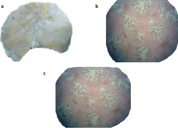
Fig. 4. From a 33 year old man with chronic liver disease who died from hypovolaemic shock secondary to ruptured esophageal varice. (a) Cut section of the prostate gland showing multiple sieve like spaces. (b and c) Nodular prostatic hyperplasia. Haematoxyllin and Eosin: b
and c photomicrographs taken with x 40 and x 100 magnification respectively
Table 5. Histological changes associated with nodular prostatic hyperplasia
Histological change Frequency Percentage (%)
Cystic change 49 73
Chronic inflammation 34 51
Acute inflammation 3 5
a b
Dele and Akhator; JAMMR, 25(11): 1-13, 2018; Article no.JAMMR.40271
Fig. 5. A 76 year old male who died from congestive heart failure secondary to hypertensive heart disease. (a) Transverse cut section of the prostate gland show multiple predominantly
solid nodules. (b) Grade 2 score 4 adenocarcinoma of the prostate. The right of the photomicrograph (short arrow) show well circumscribed nodule of malignant glands, while the
left (long arrow) show uninvolved prostate glands. Haematoxyllin and Eosin, x 40 magnification. (c) Closely packed malignant prostatic glands with straight luminal borders and
a single layer of epithelial lining with prominent nucleolus (arrow). Haematoxyllin and Eosin, x 200 magnification
Fig. 6. A 42 year old man who died from severe head injury secondary to a close range shot from a Shotgun. (a) Prostate gland weighing 50g and (b) Cribriform PIN in which the epithelial
lining of pre-existing ducts show architectural atypia. Haematoxyllin and Eosin; x100 magnification
a
b c
a
Fig. 7. These figures are from a 40 year old man who died of intracranial haemorrhage secondary to severe head injury following vehicular road traffic accident. (a) Prostate gland (25g). (b,c,d) Ova of schistosoma haematobium and adjacent prostatic glands. Haematoxyllin
and Eosin: b, c and d photomicrographs taken with x40, x100 and x200 magnification respectively
The reason for the drop is probably because the life expectancy of Nigerians which, according to World Health Organization [18,19] is less than 55 years (i.e. 53 years for males and 55 for females). Furthermore, 60%, 20% and 20% of the lesions observed in the 8th decade were BPH, prostatic intraepithelial neoplasia and adenocarcinoma respectively, while 100% of the lesion in the 9th decade was adenocarcinoma. These three lesions has been hitherto found to have an increase frequency with increasing age, hence prostatic intraepithelial neoplasia and adenocarcinoma competing with BPH for diagnosis and frequency in the setting of a reduced sample size might be responsible for the decline in the frequency of nodular prostatic hyperplasia in the 8th and 9th decades. In prostate specimen removed surgically, it is common to find histological evidence of acute or chronic inflammation in men with no clinical symptoms of acute or chronic prostatitis [20]. In these instances, aetiological infectious agents have not yet been identified [20]. To eliminate confusion with the clinical syndromes of acute
and chronic prostatitis, these prostate specimens are instead diagnosed in descriptive terms as showing “acute inflammation” or “chronic inflammation” and not as prostatitis.2 Acute and chronic inflammations were present in association with nodular hyperplasia in this study.
Prostatic adenocarcinoma was next in frequency after benign prostatic hyperplasia, thus making it the second most common lesion in this study. There was a significant increase in its frequency with age (P = 0.02). This is consistent with the
findings of other post-mortem studies [11,15,21-24]. The peak age of development of concealed adenocarcinoma at post-mortem in this study was in the 6th decade. This is similar to the finding of Okani et al. [15] unlike in the studies done by Franks, [25] Akpolat et al. [11] and Sakr et al. [23] where it was reported that occult adenocarcinoma observed at post-mortem in Caucasians peaked in the 8th decade. It is important to note that the human knowledge on the aetiology of prostate cancer is limited, [2]
a b
Dele and Akhator; JAMMR, 25(11): 1-13, 2018; Article no.JAMMR.40271
however several factors including but not limited to age and environmental influences may play a role [2]. The precise role of aging as an aetiological factor for prostate cancer is debatable. Kovi et al. [26] reported that the aging process per se does not increases susceptibility to cancer. To come to this conclusion, they studied the age-related changes of vessels, glands, and stroma in 795 unselected and consecutive autopsy prostates of black men from the Washington, DC, area and from Ibadan, Nigeria, and Accra, Ghana, West Africa. They did not find any correlation between histologic changes related to aging and the presence of
carcinoma. Miller DG [27] reported that the aging process provides the time necessary for
the cumulative effects of nonlethal genetic injury from environmental insults (i.e. internal or
external) over a prolonged period of time. This results in clonal expansion of a single precursor
cell that has incurred the genetic damage [2], which may cumulate in time depended increase susceptibility to cancer development.
[27] Perhaps an aging population vis-à-vis the
development of prostatic adenocarcinoma with increasing age may be responsible for the difference noted in Caucasians and Africans as seen in the index study, and
reinforced by the report of Okani et al. [15] in their study.
Prostatic intraepithelial neoplasia (PIN) was the third most common lesion in this study. It was first detected in the 4th decade. Its frequency was noted to increase with increasing age with a peak (40%) in the 8th decade. Akpolat et al. [11] reported that PIN was first detected in the 3rd decade at post-mortem. This finding is contrary to an earlier study by Billis [28] Billis [28]noted that prostatic intraepithelial neoplasia appear at a much younger age in African-Brazilian when compared to their Caucasian counterparts.
Prostatic adenocarcinoma and its precursor lesion are located preferentially at the peripheral zone of the gland in close proximity to each other in a majority of cases [2,29]. Many of the molecular changes seen in invasive cancers are present in PIN thus giving credence to the argument that PIN is a precursor lesion of the invasive prostate cancer [2]. There is an inverse relationship between the increasing tumour frequency and the decreasing percentage of PIN foci in the prostate [24]. This implies that the earlier developed PIN foci may show: spontaneous regression; or may slowly undergo consumption through progression toward a
malignant transformation from one of the DNA-damaged dysplastic cells of PIN as the age is advancing; or as the age is advancing, the senescent (atrophized) glandular epithelial cells have a decreasing capacity to regenerate with active cell cycle resulting in a steady decline in chances for acquiring sequential DNA damages required for dysplastic cell formation [24]. What remains unclear is whether PIN inevitably progresses to cancer, or instead sometimes remains latent or even regresses [2]. It, however, remains clear from previous studies that an increasing frequency of histologic prostate carcinoma and its premalignant lesion (PIN) is a function of advancing age, [29-31] a position upheld by the findings of the index study. Perhaps the ageing process provides the time necessary for the growing effects of nonlethal genetic injury from environmental insults over a prolonged period of time, which is sufficient to cause PIN but fall short of causing cancer. On the other hand, the insult may be enough to cause cancer only in the presence of other factors that include but not limited to race and geographical location. Soos et al. [24] opined that further investigations using cell and molecular biology techniques are invaluable to an understanding of the course of PIN with advancing age.Prostatic intraepithelial neoplasm was not found co-existing with the determined cases of occult carcinomas in this study, though it has been reported to have a high predictive value as a marker of adenocarcinoma [12]. Okani et al. [15] reported that there was no case of isolated PIN or that co-existing with occult adenocarcinoma in their study. These observations are contrary to the report of previous studies done by Akpolat et al. [11] and Stamatiou et al. [13] where a relationship between PIN and prostatic adenocarcinoma was established. While Akpolat et al. [11] Okani et al. [15] and this study employed a partial sampling method, Stamatiou et al. [13] sectioned the whole prostatic tissue. Despite these disparities in sampling method and outcome, the partial sampling method of the prostate gland adopted in this study might be responsible for the inability to establish a relationship between PIN and adenocarcinoma.
(blastomycosis) [32-34]. The non-specific granulomatous prostatitis occurs mostly in patients over 50 years of age whose prostate is affected by nodular hyperplasia [35]. It is associated with the escape of prostatic secretions into the surrounding stroma. This elicits a granulomatous inflammation [32,36]. Prostatic Schistosomiasis has been reported in previous autopsy studies that were done in the 70’s in a much higher frequency. Edington et al. [37] (Ibadan, Nigeria) and Gelfand et al. [38] (Rhodesia now present day Zimbabwe) reported prostatic schistosomiasis in 8.1% and 20.7% respectively from their respective autopsy study populations. Okani et al. [15] reported 3.8% (3 cases) of prostatic schistosomiasis at autopsy. Schistosomiasis was seen in 1.2% (one case) of the population under study. These shows a decrease in the frequency of schistosomiasis at autopsy which might be related to disease awareness and improved health care with resultant change in the disease trend from the 70’s to present time bearing in mind that Edo state where this study was carried out and neighbouring Delta state are endemic areas for Schistisomiasis infection in Nigeria.
7. CONCLUSION
In conclusion, clinically concealed prostatic diseases are common findings at post-mortem. There is a significant increase (p ≤ 0.05) in the frequency of covert prostatic diseases (nodular hyperplasia, PIN and adenocarcinoma) with advancing age. Convert prostatic diseases have the potentials of contributing significantly to the burden of health care and the cost of receiving treatment if the life expectancy improves in our environment in particular and Nigeria in general. Data generated thus from this study are invaluable in coming to this realisation as well as serving as a basis for allocating resources (human and capital), and also for planning to carter for an eventual surge in the frequency of middle aged and elderly that will eventually afflicted with prostatic diseases especially nodular hyperplasia and prostatic carcinoma.
CONSENT
It is not applicable.
ETHICAL APPROVAL
As per international standard or university standard, written approval of Ethics committee
has been collected and preserved by the authors.
COMPETING INTERESTS
Authors have declared that no competing interests exist.
REFERENCE
1. Humphrey PA. The prostate gland. In: Silverberg SG, editor. Silverberg's Principle and Practice of Surgical Pathology and Cythopathology, 4th ed. Philadelphia: Churchill Livingstone Elsevier. 2006;1791-1830.
2. Epstein JI. The lower urinary tract and male genital system. In: Kumar V, editor. Robbins and Cotran Pathologic Basis of Disease, 8th ed. Philadelphia: Saunders Elsevier; 2006;993-1002.
3. Attah EB. Nonspecific inflammatory lesions of the prostate. International Surgery. 1975; 60(3):158-62.
4. Forae G, Obaseki DE, Aligbe JU, Ekanem VJ. Morphologic patterns of prostatic lesions in Benin City, Nigeria: A twenty year retrospective study. Ann Trop Pathol. 2011;2(1):23-8.
5. Oesterling JE, Jacobsen SJ, Chute CG, Guess HA, Girman CJ, Panser LA, et al. Serum prostate-specific antigen in a community-based population of healthy men. Establishment of age-specific reference ranges. J Am Med Assoc. 1993; 270(7):860–4.
6. Saigal CS, Joyce G. Economic cost of benign prostatic hyperplasia in the private sector. J Urol. 2005;173(4):1309-13. 7. Ojewola RW, Oridota ES, Balogun OS,
Alabi TO, Ajayi AI, Olajide TA, et al. Prevalence of clinical benign prostatic hyperplasia amongst community-dwelling men in a South-Western Nigerian rural setting: A cross-sectional study. Afr J Urol. 2017;23:109–15.
8. Haas GP, Delongchamps N, Otis W, Brawley OW, Wang CY, de la Roza G. The worldwide epidemiology of prostate cancer: Perspectives from autopsy studies. Can J Urol. 2008;15(1):3866–71.
Dele and Akhator; JAMMR, 25(11): 1-13, 2018; Article no.JAMMR.40271
10. Haas GP, Sark WA. Epidemiology of prostate cancer. CA Cancer J Clin. 1997; 47:273-87.
11. Akpolat N, Buyuk Y, Uzun I, Gecit I, Kurnaz G. Prevalence of latent prostate cancer and prostatic intraepithelial neoplasia in Istanbul, Turkey: An autopsy study. Turk J Med Sci. 2012;42(3):449-56. 12. Botswick DG, Brawer MK, Lina L, Junqi Q.
High grade prostatic intraepithelial neoplasia. Rev Urol. 2004;6(4):171-9. 13. Stamatiou K, Alevizos A, Perimeni D,
Sofras F, Agapitos E. Frequency of impalpable prostate adenocarcinoma and precancerous conditions in Greek male population: An autopsy study. Prostate Cancer and Prostatic Diseases. 2006; 9:45–9.
14. Bostwick DG, Meiers I. Prostate, In: Weidner N, editor. Modern Surgical Pathology, 2nd edition. Philadelphia: Saunders Elsevier. 2009;1121–1158. 15. Okani C, Akang E, Ogunbiyi O. Incidence
of Sub-clinical prostatic disease at autopsy in the university college hospital, Ibadan. Open Journal of Urology. 2013;33.
DOI: 10.4236/oju
(Last viewed on March 14, 2015)
16. Lepor H. Pathophysiology, epidemiology, and natural history of benign prostatic hyperplasia. Rev Urol. 2004;6(9):3–10. 17. Berry SJ, Coffey DS, Walsh PC, Ewing LL.
The development of human prostatic hyperplasia with age. J Urol. 1984; 132:474–9.
18. This Day Live. WHO Report: Nigeria, Angola, Others' Life Expectancy Stagnant. Available:www.thisdaylive.com/articles/wh o-report-nigeria-angola-others-life-expectancy-stagnant/178808 (Last viewed on May 18, 2014)
19. World Health Organization Global Health Observatory Data Repository. Life expectancy: Life expectancy Data by country.
Available:www.apps.who.int/gho/data/node .688?lang=en
(Last viewed on July 1, 2014)
20. Kohnen PW, Drach GW. Patterns of inflammation in prostatic hyperplasia: A histologic and bacteriologic study. J Urol. 1979;121(6):755-60.
21. Zare-Mirzaie A, Balvayeh P, Ali Imamhadi M, Lotfi M. The frequency of latent prostate carcinoma in autopsies of over 50 years old males, the Iranian experience. Medical
Journal of Islamic Republic of Iran. 2012; 26(2):73-77.
22. Rich AR. On the frequency of occurrence of occult carcinoma of the prostate. Int J Epidemiol. 2007;36:274–77.
23. Sakr WA, Grignon DJ, Haas GP, Heilbrun LK, Pontes JE, Crissman JD. Age and racial distribution of prostatic intraepithelial neoplasia. Eur Urol. 1996;30:138–44. 24. Soos G, Tsakirisa I, Szantob J, Turzoa C,
Haasc PG, Dezsoa B. The prevalence of prostate carcinoma and its precursor in hungary: An autopsy study. European Urology. 2005;48:739–44.
25. Franks LM. Latent Carcinoma of the Prostate. J Pathol Bacteriol. 1954;68:603-16.
26. Kovi J, Jackson MA, Rao MS, Heshmat MY, Akberzie ME, Williams AO, et al. Cancer of the prostate and aging: An autopsy study in black men from Washington, DC, and selected African cities. Prostate. 1982;3(1):73-80.
27. Miller DG. On the nature of susceptibility to cancer: The presidential address. Cancer. 1980;46:1307-18.
28. Billis A. Age and race distribution of high grade prostatic intraepithelial neoplasia (HGPIN): An autopsy study in Brazil (South America). J Urol Pathol. 1996;5:1-7. 29. Sánchez-Chapado M, Olmedilla G,
Cabeza M, Donat E, Ruiz A. Prevalence of prostate cancer and prostatic intraepithelial neoplasia in Caucasian Mediterranean males: An autopsy study. Prostate. 2003; 54:238-47.
30. Breslow N, Chan CW, Dhom G, Drury RAB, Franks LM, Gellei B, et al. Latent carcinoma of the prostate at autopsy in seven areas. Int J Cancer. 1977;20:680–8. 31. Sakr WA, Grignon DJ, Crissman JD,
Heilbrun LK, Cassin BJ, Pontes EJ, et al. High grade prostatic intraepithelial neoplasia (HGPIN) and prostatic adenocarcinoma between the ages of 20-69: An autopsy study of 249 cases. In vivo.
1994;8:439–44.
32. Weidner W, Brunner H, Krause W. Quantitative culture of Ureaplasma urealyticum in patients with chronic prostatitis or prostatosis. J Urol. 1980;124: 622-5.
33. Akang EEU, Aligbe JU, Olisa EG. Prostatic tumours in Benin city, Nigeria. WAJM. 1996;15(1):56-60.
(“Warm”) autopsy study for procurement of metastatic prostate cancer. Clinical Cancer Research. 2000;6:1038–45.
35. Rosai J. Rosai and ackerman's surgical pathology, 10th ed. Edinburgh London: Mosby Elsevier. 2011;1287-1314.
36. Sakr WA, Grignon DJ, Haas GP, Schomer KL, Heilbrun LK, Cassin BJ, et al. Epidemiology of high grade prostatic intraepithelial neoplasia. Pathol Res Pract. 1995;19:838-41.
37. Edington GM, Nwabuebo I, Junaid TA. The pathology of schistosomiasis in Ibadan,
Nigeria with special reference to the appendix, Brain, pancreas and genital organs. Transactions of the Royal Society of Tropical Medicine & Hygiene.1975; 69(1):153-156.
38. Gelfand M, Ross CM, Blair DM, Castle WM. Schistosomiasis of the male pelvic organs. Severity of infection as determined by digestion of tissue and histologic methods in 300 cadavers. American Journal of Tropical Medicine and Hygiene. 1970; 19(5): 779-784.
_________________________________________________________________________________ © 2018 Dele and Akhator; This is an Open Access article distributed under the terms of the Creative Commons Attribution License (http://creativecommons.org/licenses/by/4.0), which permits unrestricted use, distribution, and reproduction in any medium, provided the original work is properly cited.
Peer-review history: