_____________________________________________________________________________________________________
*Corresponding author: E-mail: [email protected];
Journal of Advances in Medicine and Medical Research
26(11): 1-6, 2018; Article no.JAMMR.42244
ISSN: 2456-8899
(Past name: British Journal of Medicine and Medical Research, Past ISSN: 2231-0614, NLM ID: 101570965)
Evaluation of Co-Relation between Philtral Width
and Maxillary Central Incisors Width
Abhinav Agrawal
1, Kaushik Kumar Pandey
1*, A. K. Verma
1, Mariyam Ali
1,
Pratibha Katiyar
1and Abhishek Gaur
11Department of Prosthodontics, Career Post Graduate Institute of Dental Sciences, Lucknow, India.
Authors’ contributions
This work was carried out in collaboration between all authors. Authors AA, KKP and AKV designed the study, performed the statistical analysis, wrote the protocol and wrote the first draft of the manuscript. Authors MA and PK managed the analyses of the study. Author AG managed the literature searches. All authors read and approved the final manuscript.
Article Information
DOI: 10.9734/JAMMR/2018/42244
Editor(s):
(1) Sandra Aparecida Marinho, Professor, Paraíba State University (Universidade Estadual da Paraíba - UEPB), Campus, Brazil.
Reviewers:
(1) G. H. Sperber, University of Alberta, Canada. (2)Fatima Betul Basturk, Marmara University, Istanbul. (3) F. Armando Montesinos, National Autonomous University of Mexico, Mexico. Complete Peer review History:http://www.sciencedomain.org/review-history/25179
Received 26th March 2018 Accepted 11th June 2018 Published 18th June 2018
ABSTRACT
Aim and Objective: To evaluate the co-relation between philtral and maxillary combined central incisor width in north Indian population.
Materials and Methods: Measurement of philtral width was done by measuring the width of philtrum with digital vernier calipers (to the fraction of 1/10th of a millimeter). Measurement of width of central incisor was done intraorally with a digital vernier caliper.
Results: There was a mild significant correlation of philtral width with central incisor width (r=0.27, p=0.003).
Conclusion: There is a mild significant coorelation between the philtrum width and central incisor width, so we can use the philtrum width to determine the combined central incisor width in north indian population in cases of no pre- extraction records available. Using this result we can easily provide esthetically pleasant maxillary central incisors with proper tooth width and tooth form according to philtrum width and face form.
1. INTRODUCTION
The restoration of natural and pleasing lip support is one of the prime requisites of an esthetic denture. Denture esthetics is defined as ‘the cosmetic effect produced by a dental prosthesis which affects the desirable beauty, attractiveness, character, and dignity of the individual’ [1].
Determination of width of maxillary anterior teeth is an important criteria for selection and arrangement of teeth in completely and partially edentulous patients to enhance aesthetics and function [2].
Artificial tooth selection in anterior maxilla is
currently done by numerous guidelines
introduced by authors. The reliability of these guidelines, however, becomes questionable when the predicted size for anterior teeth leads to fabrication of a prosthesis which does not meet the esthetic, functional and
demands of the patient [3]. John H. Lee co related the combined mesio-distal width of Maxillary Central Incisors with philtrum. He has stated that the width of these two teeth is equal to the width of the philtrum. This test was done in English subjects [4]. Based on William’s theory; many studies have attempted to evaluate the correlation between the upside down facial form and the form of the maxillary central incisor. Also, measuring devices such as the Trubyte Tooth Indicator, Trubyte Teleform gauge, and Tooth selector have been used for determining the form of an artificial tooth. To date, William’s classification is the most universally accepted method of determining maxillary central incisor tooth form [5].
The literature of Prosthodontics is based mostly on study populations outside India, and there is an apparent lack of information about the selection of Maxillary Central Incisor forms in subjects of Indian ethnicity [5].
Hence this study is framed to evaluate the co relation between philtral width and combined
Fig. 2. Maxillary perforated dentulous metal stock tray
Agrawal et al.; JAMMR, 26(11): 1-6, 2018; Article no.
The restoration of natural and pleasing lip support is one of the prime requisites of an denture. Denture esthetics is defined as ‘the cosmetic effect produced by a dental prosthesis which affects the desirable beauty, attractiveness, character, and dignity of the
Determination of width of maxillary anterior teeth is an important criteria for selection and arrangement of teeth in completely and partially edentulous patients to enhance aesthetics and
Artificial tooth selection in anterior maxilla is
currently done by numerous guidelines
introduced by authors. The reliability of these guidelines, however, becomes questionable when the predicted size for anterior teeth leads to fabrication of a prosthesis which does not meet the esthetic, functional and phonetic John H. Lee
co-distal width of Maxillary Central Incisors with philtrum. He has stated that the width of these two teeth is equal to the width of the philtrum. This test was done in Based on William’s theory; many studies have attempted to evaluate the correlation between the upside down facial form and the form of the maxillary central incisor. Also, measuring devices such as the Trubyte Tooth gauge, and Tooth selector have been used for determining the form of an artificial tooth. To date, William’s classification is the most universally accepted method of determining maxillary central incisor
is based mostly on study populations outside India, and there is an apparent lack of information about the selection of Maxillary Central Incisor forms in
Hence this study is framed to evaluate the co-iltral width and combined
maxillary central incisor width in North Indian population. If positive co-relation is found, this study will be helpful to clinicians in the selection of maxillary anterior teeth for fabrication of partial and complete denture prosthesis.
2. MATERIALS AND METHODS
This study has been conducted in the Department of Prosthodontics and Crown & Bridge, Career Post Graduate Institute of Dental Sciences and Hospital, Lucknow. The study involved 120 dentulous subjects including male & female of Indian origin. All dentulous subjects were in-between 20-35 year age, group. Written consent was taken from subjects after explaining the procedure in detail. Approval of ethical committee of the institution was taken for the study. The inclusion criterions of the study were; dentulous subjects without any missing teeth and with fully erupted second molars, Angles Class I occlusion without any deformity. The exclusion criteria of the study were; carious, fractured, nonvital maxillary central incisor, Scarring in the upper lip, Cleft lip, Nasal infection, Previous orthognathic surgery or orthodontic treatment, congenital or surgical facial defects and any anomalies of the teeth.
2.1 Instruments and Materials Study
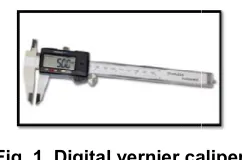
Instruments: Digital Vernier caliper. (Fig. 1)
Fig. 1. Digital vernier caliper
Materials: Maxillary perforated dentulous metal stock tray of varying sizes, Rubber bowl (flexible) and curved spatula, Rubber bowl and plaster spatula, Base former, Cast trimmer
marker,Type III Dental stone (Kalabhai) (Fig. 2, 3, 4, 5).
perforated dentulous Fig. 3. Alginate – zelgan
Fig. 4. Dental type
; Article no.JAMMR.42244
maxillary central incisor width in North Indian relation is found, this study will be helpful to clinicians in the selection of maxillary anterior teeth for fabrication of partial
2. MATERIALS AND METHODS
This study has been conducted in the Department of Prosthodontics and Crown & Bridge, Career Post Graduate Institute of Dental Sciences and Hospital, Lucknow. The study involved 120 dentulous subjects including male & female of Indian origin. All dentulous subjects 35 year age, group. Written consent was taken from subjects after explaining the procedure in detail. Approval of ethical committee of the institution was taken for the n criterions of the study were; dentulous subjects without any missing teeth and with fully erupted second molars, Angles Class I occlusion without any deformity. The exclusion criteria of the study were; carious, fractured, sor, Scarring in the upper lip, Cleft lip, Nasal infection, Previous orthognathic surgery or orthodontic treatment, congenital or surgical facial defects and any
ts and Materials Used in
Vernier caliper. (Fig. 1).
Digital vernier caliper
Maxillary perforated dentulous metal stock tray of varying sizes, Rubber bowl (flexible) and curved spatula, Rubber bowl and plaster
spatula, Base former, Cast trimmer, Point
Type III Dental stone (Kalabhai) (Fig. 2,
Fig. 5. Rubber bowl and spatula
2.2 Methodology
Fig. 7. Measuring philtrum width with vernier caliper
2.3 Measurement of Philtral Width
Patients were seated in upright position and lips lightly touching each other. It was ensured that lip was completely in relaxed position. Two most prominent points were marked at base of philtrum. These points were marked by drawing a line along the vertical ridge of philtrum and marking a point where they meet the vermillion border of upper lip. Radiographic pen (0.4 was used to mark these lines and points. The width between these two points was
Fig. 8. Measuring combined central incisor width with digital Vernier
Caliper
Agrawal et al.; JAMMR, 26(11): 1-6, 2018; Article no.
Fig. 6. Cast trimmer
philtrum width with vernier
2.3 Measurement of Philtral Width
Patients were seated in upright position and lips lightly touching each other. It was ensured that lip was completely in relaxed position. Two most prominent points were marked at base of philtrum. These points were marked by drawing a cal ridge of philtrum and marking a point where they meet the vermillion pen (0.4 mm) used to mark these lines and points. The width between these two points was measured
by digital vernier calipers to the fraction of of a millimeter (Fig. 7).
2.4 Measurement of Width of Central Incisor
Measurements of the maxillary central incisor were made intraorally with a Digital Vernier Caliper. The external edges of the vernier was a direct facilitating proper fit in the embrasures, the internal measuring edges of the beak remained unaltered. The mesio measurements were recorded at the widest dimension (contact areas). (Fig. 8,
After that an alginate impression was taken of all subjects and type III improved dental stone cast were prepared and the mesio distal width was again cross-checked using a digital vernier caliper and the result was compared with the previously measured result for greater accuracy. Mean of both the reading was tabulated.
All tabulated data was send for statistical analysis. Chi-square test was used to compare categorical variables. The Unpaired t
used to compare continuous variables. Pearson correlation coefficient was calculated. The p value<0.05 was considered significant. All the analysis was carried out on SPSS 16.0 version (Chicago, Inc., USA).
combined central incisor width with digital Vernier
Caliper
Fig. 9. Measuring the width of central incisors with digital
Vernier Caliper on model
; Article no.JAMMR.42244
by digital vernier calipers to the fraction of 1/10th
Measurement of Width of Central
Measurements of the maxillary central incisor were made intraorally with a Digital Vernier Caliper. The external edges of the vernier caliper was a direct facilitating proper fit in the embrasures, the internal measuring edges of the beak remained unaltered. The mesio-distal recorded at the widest
, 9)
After that an alginate impression was taken of all subjects and type III improved dental stone cast were prepared and the mesio distal width was checked using a digital vernier caliper and the result was compared with the sult for greater accuracy. Mean of both the reading was tabulated.
All tabulated data was send for statistical square test was used to compare categorical variables. The Unpaired t-test was used to compare continuous variables. Pearson lation coefficient was calculated. The p-value<0.05 was considered significant. All the analysis was carried out on SPSS 16.0 version
Agrawal et al.; JAMMR, 26(11): 1-6, 2018; Article no.JAMMR.42244
3. RESULTS
In this study, there was mild significant correlation of Philtral width with Central Incisor width seen. (r=0.27, p=0.003). (Table 1 and Scattered Dig. 1).
Table 1. Correlation of Philtral width with central incisor width
Philtral width with central incisor width
Correlation coefficient 0.27
p-value 0.003*
4. DISCUSSION
The proper selection of artificial teeth is the prime concern of clinicians and lab technicians for the success of the prosthesis. Various authors have suggested that artificial teeth should have same position, shape and color as natural teeth. Teeth selection is not simply a mechanical procedure, but requires dexterity and knowledge of biology. Selection of teeth forms is an important step before teeth arrangement. Prime objective of teeth selection is to create a dentofacial harmony [1].
Daniel et al. [6] reported that mesio-distal width of all maxillary six anteriors is equal to bizygomatic width / 3.3. John H. Lee [4] reported that combined mesio-distal width of maxillary Central Incisor to be equal to width of Philtrum. Bell. R.A [7] studied photographs of dental casts, x-rays of Central Incisors and photographs of face and he did not support Williams method [8].
Mavroskoufis et al. [9] studied the nasal width and incisive papilla as guide of selection of maxillary anterior teeth. Vuttiparum et al. [10] studied the relationship between width of the maxillary Central Incisors and the philtrum and found a direct correlation between the width of the Central Incisor and philtrum. Al wazzan [11] reported intercanthal distance can be used as the preliminary method of determining width of maxillary anterior teeth in edentulous patients. Abdullah [12] reported intercanthal distances and geometric progression as a predictor of maxillary Central Incisor width [2].
The relation between width of philtrum and mesio-distal width of the Central Incisor as proposed by J. H. Lee was found to be valid in English population, Sandhu Navreet et al. [4] found that the philtrum can be used to make a provisional or initial size selection of maxillary Central Incisors, Ganapathy Dhanaraj et al. [2]
also found that philtrum width can be used in predicting and selecting the width of maxillary Central Incisor.
Many authors did studies for suggesting a guideline for calculation of size and shape of maxillary anterior teeth. Only few Indian authors had worked on selecting the size and shape of maxillary Central Incisor for edentulous patients. Keeping all above things in mind, this study was framed to evaluate the relationship between philtral width and combined maxillary Central Incisor width in male and female subjects [13,14].
For measuring philtrum width, a digital vernier caliper was used. The mesio-distal width of the maxillary Central Incisors was measured using the digital vernier caliper intra-orally and on the cast. The mean of both the combined Central Incisor width were taken as final reading. Digital Vernier Caliper has been used to standardize the study. Sandhu Navreet et al. [4], Ahmed Naseer [15], Ganapathy Dhanraj et al. [2], Bansal Deepak et al. [16] have used digital vernier caliper in their studies.
Philtrum is the anatomical depression present in the facial midline inferior to the base of the nasal septum and superior to the upper labia. The shape is blunt pyramidal with the apex at the nose and the base on the centre of the superior border of the upper lip. Philtrum is the concave depression and bounded by elevation on the right and left side termed philtral ridges, with
corresponding medial and lateral slopes.
Philtrum is taken for this study as it is a clear and demarcated landmark on face and being in the midline is one of the easiest to measure. The advantage of using philtrum includes easy
location, non-invasive technique with no
discomfort to the patient [2].
In this study 89 patients were in between 20-25 year age group and 31 patients were of more than 25 year age. Majority of patients were between 20-25 years (74.2%). The mean age of patients was 24.01±2.72 years ranging from 20-32 years. In the present study 62 male and 58 female subjects had participated. About half of patients were males (51.7%).
Agrawal et al.; JAMMR, 26(11): 1-6, 2018; Article no.JAMMR.42244
Scatter Dia. 1. Showing correlation of Philtral width with Central Incisor width
5. CONCLUSION
Within the limitation of the study, the following conclusions were drawn:-
1 – There is a mild significant correlation between the philtrum width and Central Incisor width as indicated by the correlation coefficient which is 0.27 and p-value which is 0.003. So we can use the philtrum width to determine the combined Central Incisor width in north Indian population in cases of no pre extraction records available.
The findings of this study can be used for selection of size of maxillary central incisors.
CONSENT
Consent was taken individually from all the patients.
ETHICAL APPROVAL
Ethical approval was taken from institutional ethical research cell committee.
COMPETING INTERESTS
Authors have declared that no competing interests exist.
REFERENCES
1. Winkler S. Essentials of complete denture prosthodontics. Second Edition. A.I.T.B.S. 2009;202.
2. Dhanraj Ganapathy, Janani Nandakumar,
Prathap Sekhar. Philtral width and width of maxillary central incisor - A correlational analysis in dentate adult human subjects. Innovative Journal of Medical and Health Science. 2015;5(4):133–135.
3. Jafari HJ, Orkideh Radmehr. The analysis
of correlation between the facial width and mesio-distal width of the maxillary anterior teeth. J Res Dent Sci Spring. 2011;11(1): 49-53.
4. Sandhu Navreet, et al. Role played by soft
tissue landmarks such as philtrum in selecting the width of artificial maxillary central incisors. Indian Journal of Dental Sciences. 2012;4(1).
5. Koralakunte Pavankumar R, et al. A
clinical study to evaluate the correlation between maxillary central incisor tooth form and face form in an Indian population. Journal of Oral Science. 2012;54(3):273-278.
6. Daniel HG, Drasen OM. Complete Denture
Prosthesis 4th Edition. 1958;138.
7. Bell Ra. The geometric theory of selection of artificial teeth: Is it valid? J Am Dent Assoc. 1978;97:637-640.
8. Mavroskoufis F, Ritchie GM. The face-form
as a guide for the selection of maxillary
central incisors. J Prosthet Dent.
1980;43(5):501-5.
9. Mavroskoufis F, Ritchie Gm. Nasal width
Agrawal et al.; JAMMR, 26(11): 1-6, 2018; Article no.JAMMR.42244
10. Vuttiparum N, Benjakul C. Relationship
between the width of maxillary central incisors and philtrum. J Dent Assoc Thai. 1989;39;233-9.
11. Al Wazzan Ka. The relationship between
intercanthal dimension and the widths of maxillary anterior teeth. J Prosthet Dent. 2001;86:608-12.
12. Abdullah Ma. Inner canthal distance and
geometric progression as a predictor of maxillary central incisor width. J Prosthet Dent. 2002;88:16-20.
13. Madhu Ranjan, Ravi Raj, Rohit Chetan,
Ritu Kumara. Correlation between
mesiodistal width of maxillary anterior teeth and interhamular notch distance amongst dakshin kannada population. International
Journal of Contemporary Medical
Research. 2017;4(7):1471-1474.
14. Raghavendra N, Nayana R. Somayaji,
Venkatesh V. Kamath. The correlation
between permanent maxillary central
incisor crown length, facial height and body height and weight. An allometric analysis of 100 individuals. RRJDS. 2014;2(2): 127.
15. Naseer Ahmed, Muhammad Abbas, Asma
Naz, Afsheen Maqsood. Correlation
between innercanthal distance and the mesiodistal width of the maxillary central incisors. Isra Medical Journal. 2015; 7(3).
16. Deepak Bansal, Shruti Sharma, Manjit
Kumar, Amrit Khosla. A clinical study to correlate the facial form and maxillary central incisor tooth form in males and females of davangere population. Djas. 2016;4(Iii):156-164.
_________________________________________________________________________________ © 2018 Agrawal et al.; This is an Open Access article distributed under the terms of the Creative Commons Attribution License
(http://creativecommons.org/licenses/by/4.0), which permits unrestricted use, distribution, and reproduction in any medium,
provided the original work is properly cited.
Peer-review history: