_____________________________________________________________________________________________________ *Corresponding author: E-mail: [email protected];
ISSN: 2231-0614, NLM ID: 101570965
SCIENCEDOMAIN international www.sciencedomain.org
Evaluation of a New Stapler with Unique Surface
Gripping Technology
Masahiro Kimura
1*, Hironori Tanaka
1, Motoki Hato
1, Satoshi Taniwaki
1,
Yasuyuki Shibata
1, Kotaro Mizuno
1, Nobuo Ochi
1, Yoichiro Mori
1,
Takaya Nagasaki
1, Shuhei Ueno
1and Yuki Eguchi
11
Department of Surgery, Nagoya City East Medical Center, 2-23 Wakamizu 1, Chikusa-ku, Nagoya 464-8547, Japan.
Authors’ contributions
This work was carried out in collaboration between all authors. Author MK designed the study, wrote the protocol and wrote the first draft of the manuscript. Authors HT, MH, ST, YS, KM, NO, YM and TN managed the literature searches, analyses of the study, performed the spectroscopy analysis and authors SU and YE managed the experimental process. All authors read and approved the final manuscript.
Article Information
DOI: 10.9734/BJMMR/2016/30153 Editor(s): (1) Syed Faisal Zaidi, Department of Basic Medical Sciences, College of Medicine, King Saud Bin Abdulaziz University-HS, National Guard Health Affairs, King Abdulaziz Medical City, Kingdom of Saudi Arabia. Reviewers: (1) Gulzar Ahmad Bhat, King Khalid University, Kingdom of Saudi Arabia. (2)Bhupesh H. Tirpude, Maharashtra University of Health Sciences, India. Complete Peer review History:http://www.sciencedomain.org/review-history/16866
Received 21st October 2016 Accepted 8th November 2016 Published 10th November 2016
ABSTRACT
Background: Staplers make it possible to create a gastrointestinal anastomosis quickly, easily, and securely. Staplers have undergone several improvements. We herein evaluate the effect of a new stapler with unique surface gripping technology that provides a superior tissue grip without trauma during firing.
Methods: Porcine small bowel was used. The stapling devices compared were the ECHELON FLEXTM with White (E) and GST System White (G). The number of total malformed staples, severely malformed staples, staples malformed to the cutting side, and the absolute value of the degree of malformation were evaluated.
Results: The number of malformed staples and the absolute value of the degree of malformation were significantly lower in group G. The number of occurrences of total malformations <0 was greater in group G. Comparing the inner, middle, and outer staple rows, the number of occurrences
of malformations were lower on the outer row in group E. Comparing the front, middle, and back parts malformation was lower in the front.
Conclusions: We found that a new stapler is superior to a standard stapler with regard to a reliable “B” shape formation of sta
staple malformation and the strength of the suture line.
Keywords: Staple; malformation; tissue slippage
1. INTRODUCTION
Stapling devices are gaining popularity and have undergone many technical improvements Staplers make it possible to create a gastrointestinal anastomosis quickly, easily, and securely. Mechanical stapling devices are being used more frequently in gastrointestinal and thoracic surgery. One of the issues to be improved upon is the movement of and trauma to the tissue at the time of firing. This movement is likely to affect the formation of the staples. Recently, a new stapler has been introduced as an alternative to the standard stapler. This new stapler is designed to have less tissue slippage during firing. However, there are no data on the outcomes of this new stapler in the literature. Hence, we conducted this experiment to evaluate the efficacy of this new stapler.
2. MATERIALS AND METHODS
Fresh porcine small bowel was used for all experiments. The specimens were obtained from animals that had been sacrificed for use in
(b)
a. Comparison of surface of two cartridges
ations were lower on the outer row in group E. Comparing the front, middle, and back parts malformation was lower in the front.
We found that a new stapler is superior to a standard stapler with regard to a reliable “B” shape formation of staples. Future directions include studying the correlation between staple malformation and the strength of the suture line.
tissue slippage; gripping surface technology.
Stapling devices are gaining popularity and have undergone many technical improvements [1]. Staplers make it possible to create a gastrointestinal anastomosis quickly, easily, and securely. Mechanical stapling devices are being rointestinal and thoracic surgery. One of the issues to be improved upon is the movement of and trauma to the tissue at the time of firing. This movement is likely to affect the formation of the staples. Recently, a new stapler has been introduced as ternative to the standard stapler. This new stapler is designed to have less tissue slippage during firing. However, there are no data on the outcomes of this new stapler in the literature. Hence, we conducted this experiment to evaluate
MATERIALS AND METHODS
Fresh porcine small bowel was used for all experiments. The specimens were obtained from animals that had been sacrificed for use in
approved non-gastrointestinal research studies. The specimens were used within
sacrifice. Each segment of the intestinal tract was 20 cm in length. The front and rear walls of the small intestine segments were stapled in the longitudinal direction using a linear surgical stapler. To match the thickness of the human small intestine, the two pieces of intestine were overlapped in the experiments. The stapling devices used were the ECHELON FLEX Ethicon Echelon Stapler Reloads White (ECR60W, Ethicon, Japan) and the GST System White (GST60W) (Fig. 1). To observe the s in the original sequence, we performed the stapling procedure shown in previous experiments, we wrapped a plastic bag on the cartridge side of the stapler, and then we stapled the intestine. Each device was clamped on the tissue for 1 minute. Firings were performed automatically (Powered ECHELON FLEX Five staplings were completed in each stapler. After stapling, the intestine with a plastic bag was put in the sodium hydroxide to dissolve the intestine [2].
(a)
(c) Fig. 1. Shape of stapler
Comparison of surface of two cartridges; b. Step of cartridge side; c. Step of anvil side
ations were lower on the outer row in group E. Comparing the front, middle, and back
We found that a new stapler is superior to a standard stapler with regard to a ples. Future directions include studying the correlation between
gastrointestinal research studies. The specimens were used within 24 hours after sacrifice. Each segment of the intestinal tract was 20 cm in length. The front and rear walls of the small intestine segments were stapled in the longitudinal direction using a linear surgical stapler. To match the thickness of the human ll intestine, the two pieces of intestine were overlapped in the experiments. The stapling devices used were the ECHELON FLEXTM with Ethicon Echelon Stapler Reloads White (ECR60W, Ethicon, Japan) and the GST System 1). To observe the staples in the original sequence, we performed the stapling procedure shown in previous experiments, we wrapped a plastic bag on the cartridge side of the stapler, and then we stapled the intestine. Each device was clamped on the s were performed automatically (Powered ECHELON FLEXTM). Five staplings were completed in each stapler. After stapling, the intestine with a plastic bag was put in the sodium hydroxide to dissolve the
Kimura et al.; BJMMR, 18(9): 1-6, 2016; Article no.BJMMR.30153
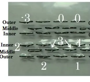
Observation was carried out in a state in which all of the staples were attached to a plastic bag. We divided the specimens into +/- by the malformation direction of the hook portion. We gave a negative score when the hook portion was malformed to the knife side and a positive score when the hook portion was malformed to the outside. The degree of malformation was categorized into four scores (Fig. 2):
0: Well-formed staples.
1: The degree of malformation is so small that the hook portion is in contact with the linear portion.
2: The distance of the hook portion from the linear portion is less than twice the diameter of the staple.
3: The degree of malformation is larger than score 2.
The degree of malformation was evaluated by the total of the two hooks. The number of malformed staples, strongly malformed staples, staples malformed to the knife side, and the absolute value of the malformation degree were compared for each group. Furthermore, the 88 staples were divided by columns (inner, middle, outer) and location (front, middle, back), and they were compared with each other.
Fig. 2. Example of the score
The number of malformed staples, strongly malformed staples, and staples malformed to the cutting side were expressed as percentages. The absolute value of the degree to which the staples were malformed was expressed as the average per staple. Engineer unrelated to the authors measured and scored using the optical microscope.
2.1 Statistical Analysis
Discrete variables were analyzed by the Mann-Whitney test and significance was indicated at p<0.05.
3. RESULTS
In all experiments, 88 staples could be observed while attached to the plastic bag. The number of malformed staples and the absolute value of the degree of malformation were significantly lower in group G compared with group E (Table 1). On the contrary, the number of occurrences of <0 staple malformations was significantly greater in group G. Comparing the inner, middle, and outer staple rows, the number of occurrences of 2≦
staple malformations and the absolute value of the malformation degree were both
significantly lower in the outer row of staples in
group E (Table 2). Unlike in group E, there was no difference in any of the measured
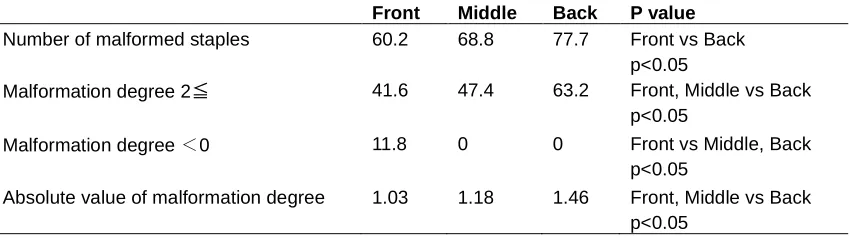
parameters in group G (Table 3). Comparing the front, middle, and back parts of the stapler, with the exception of staples with a malformation degree <0, malformation was significantly lower in the front part of the stapler in both groups (Tables 4, 5).
4. DISCUSSION
Development of and improvements in the automatic stapler have changed surgery dramatically. Staples built into staplers are designed to be a “B” shape when firing has been completed, which is believed to be the optimal shape [3-6]. However, there are no studies comparing the shape of staples as they relate to postoperative complications including leaks and bleeding. Rodeheaver compared staple formation and the resistance to leak with various staplers. He found that the leak pressure of the stapler with a smaller degree of malformation is lower than that with a large degree of malformation. This is the only experiment to our knowledge that mentions the "B" staple formation and its clinical relevance. This data has not been published scientifically, but rather is included in the information booklet as published by Covidien. To this end, various improvements have been made in order to form “B” shape staples.
There are various factors involved in the formation of staples including:
・Tissue thickness
・Movement of tissue at the time of firing ・Tissue firmness
Table 1. Malformed staples in each group
ECR GST P value
Number of malformed staples 74.2±3.1 68.7±4.4 p<0.05
Malformation degree 2≦ 51.4±7.4 50.2±4.3 ns
Malformation degree <0 0.7±0.7 4±2.5 p<0.05
Absolute value of malformation degree 1.56±0.18 1.22±0.04 p<0.05
Table 2. Comparison in each column with ECR stapler
Inner Middle Outer P value
Number of malformed staples 74.6 76.2 71.4 ns
Malformation degree 2≦ 59.2 64 31.4 Inner, Middle vs Outer
p<0.05
Malformation degree <0 0.6 0 1.7 ns
Absolute value of malformation degree 1.74 1.78 1.11 Inner, Middle vs Outer p<0.05
Table 3. Comparison in each column with GST stapler
Inner Middle Outer P value
Number of malformed staples 66.6 72 67.4 ns
Malformation degree 2≦ 52.8 51.8 45.7 ns
Malformation degree <0 5.8 2.6 3.4 ns
Absolute value of malformation degree 1.26 1.26 1.14 ns
Table 4. Comparison in each location with ECR stapler
Front Middle Back P value
Number of malformed staples 70 77.4 74.9 Front vs Middle
p<0.05
Malformation degree 2≦ 36.2 59.8 58.9 Front vs Middle, Back
p<0.05
Malformation degree <0 2.1 0 0 Front vs Middle, Back
p<0.05 Absolute value of malformation
degree
1.21 1.8 1.71 Front vs Middle, Back
p<0.05
Table 5. Comparison in each location with GST stapler
Front Middle Back P value
Number of malformed staples 60.2 68.8 77.7 Front vs Back
p<0.05
Malformation degree 2≦ 41.6 47.4 63.2 Front, Middle vs Back
p<0.05
Malformation degree <0 11.8 0 0 Front vs Middle, Back
p<0.05
Kimura et al.; BJMMR, 18(9): 1-6, 2016; Article no.BJMMR.30153
Regarding tissue thickness, staples are ideally compatible with the tissue being stapled, which varies in thickness, from thin blood vessels to the thick stomach. In order to make the stapler more robust, more staple rows can be added and a changing staple height can be employed. In any case, it is important to select the staple of appropriate height that matches the thickness of the tissue.
Nakayama and colleagues reported that a sufficient amount of pre-compression time is necessary for achieving a secure staple formation [7]. However, the effect of pre-compression cannot be observed in our previous experimental system. One stapling method that was significantly different, though, was the power stapling method. With this method, good results were obtained on all measures of malformation.
Movement of the tissue during firing is likely to affect the formation of the staple. It is a possibility that the legs of the staples are distracted along with the tissue. A new staple cartridge has been introduced to potentially limit tissue extrusion during multiple staple fires. The unique gripping surface technology on each reload provides a superior tissue grip without additional trauma during firing. In this study, we compared this new staple cartridge (GST60W) to the existing staple cartridge (ECR60W) (Fig. 1a).
There was no movement of the tissue at the time of firing with ECR. However, the malformation of the inner row was clearly greater than that of the outer row. This may have to do with the movement of the blade on the cutting side which lies along the inner row. If the blade runs down the center, the tissue is forced to the outer rows. There is a step in both the cartridge and the anvil (Fig. 1b,c). Therefore, these staplers can grip tissue strongly. However, in the space of each of the two inner rows, the tissue is forced to the outside by the cutting blade. As a result, as shown in Fig. 1, malformation degree 2≦and absolute value of malformation degree was significantly greater in the inner and middle rows. On the other hand, there was no difference by site in any deformation item in GST60W. These results are likely due to the strength of the gripping force on the tissue in all columns (inner, middle, outer).
With respect to the location along the length of the stapler (front, middle, back), deformity tends to be small towards the front. In both staplers, the gap between the cartridge and anvil has
been designed to be large. This gap may cause tissue movement during the firing. From the shape of the gripping surface, the gripping forces might be strong enough for longitudinal distraction.
5. CONCLUSION
In our experiment, the new stapler (GST) is superior to the standard stapler (ECR) with regard to a consistent “B” shape formation of staples. However, the relationship of the malformation of staples and postoperative complications is not clear. That being said, there is no doubt that the “B” is the best shape.
As a next step, we would measure the correlation between staple malformation and the strength of the suture line using our experimental model.
CONSENT
It is not applicable.
ETHICAL APPROVAL
It is not applicable.
COMPETING INTERESTS
Authors have declared that no competing interests exist.
REFERENCES
1. Chekan E, Whelan RL. Surgical stapling device-tissue interactions: What surgeons need to know to improve patient outcomes. Med Devices. 2014;7:305-318.
2. Kimura M, Terashita Y. Superior staple formation with powered stapling devices. Surg Obes Relat Dis. 2016;12:668-72. 3. Kawasaki K, Fujino Y, Kanemitsu K, Goto
T, Kamigaki T, Kuroda D, et al. Experimental evaluation of the mechanical strength of stapling techniques. Surg Endosc. 2007;21:1796-9.
4. Mery CM, Shafi BM, Binyamin G, Morton JM, Gertner M. Profiling surgical staplers: Effect of staple height, buttress, and overlap on staple line failure. Surg Obes Relat Dis. 2008;4:416-422.
challenging tissue applications. Surg Obes Relat Dis. 2013;9:417-421.
6. Baker RS, Foote J, Kemmeter P, Brady R, Vroeqop T, Serveld M. The science of stapling and leaks. Obes Surg. 2004; 14:1290-8.
7. Nakayama S, Hasegawa S, Nagayama S, Kato S, Hida K, Tanaka E, et al. The importance of precompression time for secure stapling with a linear stapler. Surg Endosc. 2011;25:2382-2386.
_________________________________________________________________________________ © 2016 Kimura et al.; This is an Open Access article distributed under the terms of the Creative Commons Attribution License (http://creativecommons.org/licenses/by/4.0), which permits unrestricted use, distribution, and reproduction in any medium, provided the original work is properly cited.
Peer-review history: