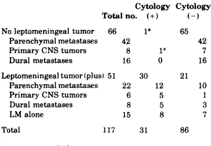
cytology) and pathologic findings at autopsy. The purpose was to discover: (1) the incidence of negative CSF cytology in patients with CNS malignancy, (2) the incidence of false-positive cytology, and (3) the relationship between a true-positive cytology and the distribution of malignant tumor a t autopsy. Of 117 patients with CNS tumor and premortem cytologic examination of the CSF, 3 1 (26 percent) were positive and 86 (74 percent) were negative. Only 1 of 66 patients with tumor t h a t did not reach the leptomeninges had a positive cytology. Of 51 patients with leptomeningeal tumor at autopsy, cytology was positive in 30 (59 percent) and negative in 21 (41 percent). Five potentially “false-positive” cytologies were encountered: Three patients were treated, and tumor may have been eradicated; in two patients with lymphoma, inflammatory cells associated with infection were apparently mistaken for malignant cells. These data indicate that a positive CSF cytology is a reliable indicator of CNS malignancy and almost always reflects leptomeningeal tumor.
NEUROLOGY 29: 1369-1375, October 1979
Malignant
cells
i
n
cerebrospinal
fluid
(CSF‘):
The
meaning
of
a
positive
CSF
cytology
J.
Peter Glass, M.D., Myron Melamed, M.D., Norman L. Chernik, M.D., and Jerome B. Posner, M.D.Examination of the cerebrospinal fluid (CSF) for malignant cells is a time-honored and widely used technique. Since DuFour’ first identified malig- nant cells in the CSF in 1904, many reports attest to the usefulness of CSF cytology in assisting in the diagnosis of primary and metastatic central nervous system (CNS) tumors.2-11 In most series, a “positive cytology”-i.e., malignant cells in CSF sampled from the lumbar sac, cistern, or lateral ventricles-was encountered in 20 to 40 percent of
patients harboring malignant tumors of the CNS. Most of these reports dealt with only small num- bers of metastatic tumors and did not address three major clinical considerations: (1) the inci- dence of negative CSF cytology in patients with histologically proved brain or, especially, lep- tomeningeal tumors, (2) the incidence of false- positive cytology, and (3) the relationship between positive cytology and distribution of the tumor at
autopsy. Since all of these clinical considerations have therapeutic implications, we reviewed our experience with cytologic examination of the CSF in patients subsequently examined at autopsy. Be- cause of the nature of Memorial Sloan-Kettering
Cancer Center’s (MSKCC) hospital population, this report deals largely with CNS metastases rather than with primary brain tumors.
Materials
andmethods.
The records of the Cytology Laboratory and the Autopsy Service of MSKCC for the years 1976 and 1977 were com-pared. We identified all patients who had cytologic examination of the CSF prior t o death, whether positive or negative, and who subsequently underwent autopsy that included the CNS. The pathologic examination of the nervous system was performed by one neuropathologist
(N.L.C.)
using a standardized coding system12 that emphasizes the extent and distribution of disease. Because of our interest in leptomeningeal metastases, special attempts were made t o identify this neurologic complication of systemic cancer.13 Patients har- boring CNS neoplasms were classified into three groups: (1) those with dural metastases, including invasion of the cavernous space, other dural sinuses or the subdural surface of brainor
spinal cord; (2) those with parenchymal tumors within the substance of the brain or spinal cord; and (3)From the Departments of Neurology and Pathology, Memorial Sloan-Kettering Cancer Center, and the Departments of Neurology and Pathology, Cornell University Medical College, New York, NY.
Accepted for publication March 19, 1979.
Malignant cells in CSF
Results.
Autopsy findings. During 1976 and 1977 there were 777 autopsies of patients who died with malignant neoplasms and whose brains and spinal cords were examined; 363 of these patients were consideredto
be “neuropathology cases,” requiring more careful examination because the patient had been evaluated neurologically during life or be- cause a suspicious CNS lesion was encountered by the prosector on gross or microscopic examination. In 210 patients, tumor was found in CNS (table 1). One hundred forty-two patients had malignant tumors within the parenchyma of the brain or spi- nal cord. In 98 of these patients, there was no evidence of leptomeningeal or dural involvement; in the other44
there was additional tumor in the leptomeninges as well as the brain or spinal cord. These 142 parenchymal lesions include 114 meta- static lesions and 28 primary CNS tumors (table 1).Fifty patients had dural lesions. All were meta- static; 33 had no additional leptomeningeal in- volvement, and in 17 leptomeningeal metastases accompanied the dural involvement. Seventy- three patients had histologic evidence of lep- tomeningeal tumor, either metastases from a sys- temic cancer or direct spread from a primary brain tumor.
In 18
patients, leptomeningeal tumor was unaccompanied by parenchymal or dural neo- plasia; in 41 patients there Were also parenchymal lesions, and in 14 there were dural tumors.CSF cytology. During 1976 and 1977,2775 indi-
‘SF. These 182 Of the 363 “neuropathol- ogy cases” and 117 patients with histologically proved CNS malignancy (table 2). The interval
Figure 1 . CSF cytology from a patient with malignant lymphoma. The spinal fluid is very cellular, and is composed of immature and abnormal lymphoreticular cells that are characteristically single rather than in
cytoplasm, and have irregular and convoluted nuclei with visible nucleoli.
groups, The cells vary in size, with little or no visible viduals examination Of the
those with leptomeningeal metastases. This last group was subdivided into patients with only a single focus of neoplasia in the leptomeninges and patients with diffuse or widespread multifocal seeding of the leptomeninges.
The findings on CSF examination were taken from the official reports. Only those reports in which malignant cells were definitely identified were considered positive (figures 1 t o 3). “Suspi- cious” or “equivocal” findings were considered negative. The microscopic slides of the CSF re- ported as positive for malignant cells from patients with no evidence of CNS tumor at autopsy (false- positive cytology) were examined again by one of the authors
(M.M.).
The usual CSF sample submit- ted t o the Cytology Laboratory was obtained by lumbar puncture and contained approximately 4cc of CSF immediately fixed with 4 cc of 50 percent alcohol. Smears were prepared from the cen- trifuged sediment or by direct centrifugation onto glass slides, and subsequently stained with the Papanicolaou technique. We do not use filter prep- arations.
.
Figure 2. CSF cytology from a patient with breast carcinoma. The clustering of these tumor cells readily identifies them as carcinoma rather than lymphoma. T h e cells of carcinoma are usually larger than lymphoma, with more abundant but variable amounts
patient with malignant melanoma. This specimen shows great variation in cell size and staining
characteristics with many mitotic features. The
cytoplasm of the larger cells is more deeply stained and contains finely granular melanin pigment. ___
Table 1. CNS tumors encountered at autopsy (1976-1977)
T u m o r type
Primary brain tumor Malignant astrocytoma Medulloblastoma Meningioma
Breast Lung Melanoma
Gynecologic malignancies Testicular malignancies Neuroblastoma
Renal carcinoma Osteogenic sarcoma Others
Solid systemic tumor
Hematologic malignancies Leukemia
Hodgkin disease Non-Hodgkin lymphoma Myeloma
Site of CNS involvement
Parenchymal D u a l Leptomeningeal
Totals 142 (98) 50 (33) 73 (18)
Malignant cells in CSF
Table 2. CSF cytology in patients with CNS malignancies
Cytology Cytology
Total no. (+) ( - )
No leptomeningeal tumor 66 1* 65
Parenchymal metastases 42 42
Dural metastases 16 0 16
Primary CNS tumors 8 1* 7
Leptomeningeal tumor (plus) 51 30 21
Parenchymal metastases 22 12 10
Primary CNS tumors 6 5 1
Dural metastases 8 5 3
LM alone 15 8 7
Total 117 31 86
* ( + ) to ( - ) with therapy.
between the last antemortem CSF examination and death varied, but in most patients it was less than 30 days. Of the 117 patients with CNS tumor and premortem cytologic examination of the CSF, 31 (26 percent) were positive at least once and 86 (74 percent) were negative. The CSF cytology was negative in all except 1 of the 66 patients with parenchymal or d u r a l lesions without lep- tomeningeal involvement. Of the 22 patients with both parenchymal disease and leptomeningeal disease, there were 12 positive cytologies and 10
negative cytologies. In the eight patients with lep- tomeningeal involvement in addition to dural tumor, there were five positive and three negative cytologies. Thus, in only one instance were malig- nant cells found in the CSF of patients whose meninges were not invaded by tumor at autopsy, and that patient’s cytology reverted
to
negative with treatment, which may have eradicated previ- ously present leptomeningeal disease.The CSF was examined before death in 51 of the
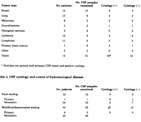
73 patients with autopsy-proved leptomeningeal tumor. These include metastases from a variety of primary tumors (table 3); in 30 patients (59 per- cent), the cytology was positive and in 21 it was negative. No primary tumor appeared
to
exude cells into the CSF more consistently than any other. The incidence of positive cytologies corre- lated with the extent of disease (table 4). Of the 73 patients with leptomeningeal metastases, the dis- ease was focal in 19 and multifocal or disseminated in 54. Among the 13 patients with focal lep- tomeningeal seeding and CSF examination, 5cytologies were positive (38 percent) and 8 nega- tive. Of the 38 patients with disseminated lep- tomeningeal tumor and CSF examination, 25 were positive (66 percent) and 13 negative (figure 4). When viewed the other way, 25 of 30 patients with a positive CSF cytology had disseminated lep-
tomeningeal disease (83 percent); only 5 of the 30
positive CSFs occurred with focal involvement. In 13 of the 21 patients with a negative CSF cytology the leptomeningeal disease was disseminated, and in 8 it was focal.
‘?False-positive cytology.” Five patients with malignant cells in CSF had no evidence of CNS malignancy a t autopsy. These included three pa- tients with lymphoma, one with acute myelocytic leukemia, and one with medulloblastoma (table 5 ) . The patients with medulloblastoma and acute myelocytic leukemia both had clinical evidence of
leptomeningeal involvement during life, and both were treated for that disorder. Both responded to treatment clinically, and malignant cells disap- peared from the CSF before death. Thus, the dis- ease was probably eradicated from the CNS in these patients prior to death, and they were not really false-positives. The same may have been true for one patient with lymphoma (patient 3)
who received radiation therapy to the brain to treat multiple cranial nerve dysfunction. Cytology was positive prior to radiation therapy, and there was no follow-up CSF cytology. Patient 1 received intrathecal chemotherapy as prophylaxis against meningeal metastasis in September and October
1975. In November 1976, CSF cytology was read as positive, but there was no clinical evidence of either leptomeningeal tumor or meningitis. CSF cultures for bacteria and fungi were negative, and all other CSF measurements were normal. There
was no known cause for the positive cytology. Pa- tient 2 was also a true false-positive. Apparently malignant cells appeared in the CSF in association with disseminated herpes zoster and symptoms of
herpes zoster meningitis. No therapy was given to the CNS for malignant lymphoma, and when the generalized zoster cleared the CSF cytology be- came negative.
Discussion. We undertook this retrospective analysis of cytologic examinations of the CSF for three reasons: (1) I t had been our clinical impres- sion that positive cytology was strong evidence that a malignant brain tumor had not only reached the ventricular or leptomeningeal surface but had seeded the leptomeninges, at least in focal areas. Although it had been recognized that tumor cells could not appear in the CSF unless they reached
the leptomeningeal or ventricular s u ~ f a c e , ~ * ~ , ~ * few attempts had been made to correlate the find-
Tumor type Breast Lung Melanoma Neuroblastoma Osteogenic sarcoma Leukemia Lymphoma
Primary brain tumors
Other Totals No. patients 14 10 6 3 3 14 1 1 7 5 73
No. CSF samples examined 10 6 3 2 2 9 8 6 5 51
Cytology (+)
6 4 3 1 0 4 5 5 2 30*
* Excludes one patient with primary CNS tumor and positive cytology.
Table 4.
CSF
cytology and extent of leptomeningeal diseaseNo. CSF samples
No. patients examined Cytology (+)
Focal seeding 19
Primary Metastatic
1 18
MultifocaUdisseminated seeding 54
Primary Metastatic
6
48
have encountered an increasing number of posi- tive cytologic diagnoses, and we occasionally con- sidered some to be falsely positive. There have been several reported cases of false-positive
CSF
cytology in malignant lymphomasB at times as- sociated with
CNS
i n f e c t i ~ n s . * * ~ ~ * l ~A
significant incidence of false-positive cytologic diagnoses would, of course, have major clinical implications. (3) Finally, we have been concerned about the pa- tients with clinical (or pathologic) evidence of widespread leptomeningeal involvement in whom malignant cells are not found in theCSF.
Since most physicians require a finding of malignant cells in the CSF to make a diagnosis of lep- tomeningeal tumor, we wanted t o know how often cytologic examination was negative in patients with proved disseminated leptomeningeal tumor. This survey appearsto
answer all three ques- tions. At least in our hands, malignant cells in theCSF imply that the leptomeninges have been
13 5
1 0
12 5
38 25
5 5
33
Cytology (- 1
4 2 0 1 2 5 3 1 3 21
Cytology (-)
8
1 7
13
0
seeded by viable tumor. Moreover, tumor is usu- ally not j u s t present i n a focal area of lep- tomeninges, but has seeded the leptomeninges rather widely in a multifocal or disseminated fash- ion. This statement applies not only to metastatic
CNS neoplasms but also to primary tumors. Sec- ond, false-positive cytologic diagnoses appear to be rare, at least in our experience, and occurred only in patients with lymphoma who were suffering from meningitis in which reactive or immature lymphocytes were mistaken for malignant lym-
phoid cells. With experience, even most of these cases could be correctly recognized. Thus, in one report of a false-positive cytologic diagnosis,15 lymphocytes from the CSF of a patient suffering from cryptococcal meningitis were interpreted as lymphoma; we believe from the photomicrographs in that article that the cells can be identified as
reactive.
Malignant cells in C-SF
Figure 4. T h e cauda equina from a patient with a negative CSF cytology. T hi s patient suffered from adenocarcinoma of the lung with clinical signs of widespread leptomeningeal involvement by tumor. Three lumbar punctures were performed 10 clays prior to her death. I n each there were a n increased number of lymphocytes, an elevated protein level, and a depressed glucose level, but in all three instances the cytologic examindion for malignant cells was negative. At autopsy, the day after the third lumbar puncture, the. cauda equina was grossly infiltrated by tumor, and microscopic sections revealed large numbers o f tumor cells surrounding each of the roots.
Table 5. Positive CSF cytology not confirmed at autopsy
Patient Primary
no. tumor
1 Lymphoma
2 Lymphoma
3 Lymphoma
4 Leukemia (AML)
5 Medulloblastoma
Positive cytology
11/17/76
4/19/76
1/16/76
9/21/76
6/24/76
Treatment
None following cytologic exam
None
Radiation therapy to the brain
Radiation therapy
tq base of skull Intraventricular
methotrexate
Radiation therapy to spinal cord
Negative C y t o l O g Y
follow-up No
6/24/76
High-dose intravenous methotrexate
No follow-up
10/11/76
9/2/76
Date of death
11/21/76
11/21/77
2/3/76
10129176
6/3/77
Comments
Probable false-posi tive associated with sepsis
False-positive effect of herpes zoster
Tumor m a y have Good clinical
responded to treatment
evidence of CNS
involvement prior to treatment
evidence of CNS
is still a significant problem.
It
is well to remember that in a patient with appropriate clinical or other evidenceof
leptomeningeal tumor, absence ofmalignant cells in the CSF does not rule out that diagnosis.
Our findings differ from other surveys which suggested that parenchymal metastases without leptomeningeal involvement can be identified by examination of CSF. There are several possible reasons for this discrepancy: (1) Our techniques may be less sensitive than other^,^.^^ resulting in excessive false-negative cytologies unless the lep- tomeninges are widely seeded and large numbers
of cells are actually present in the CSF. (2) Previ- ous reports of positive cytologies in parenchymal tumors may not have examined the entire nervous system carefully enough to identify leptomenin- geal spread of tumor, which was, in fact, responsible for the positive cytology. (3) We may be “under- reading” malignant cells or others may be over- reading malignant cells in the CSF. We believe that leptomeningeal metastases may be over- looked a t autopsy if not specifically sought by meticulous examination of the entire CNS in pa- tients with positive cytologies, a practice we have consistently followed. Overall, about 25 percent of
our patients with autopsy-proved CNS involve- ment by malignant tumor had a positive cytology, a figure similar to that encountered in most other large series. Furthermore, when considered by in- dividual tumor type, the percentage of cases with positive cytology seen by us was similar to that of other studies. Thus, the difference between our studies and others is not in the technique of isolat- ing and identifying malignant cells in the CSF, but in the meticulous examination of the
CNS at
au- topsy, revealing a much higher incidence of lep- tomeningeal seeding than was previously appar- ent.Conclusions. We draw several conclusions from these data and our review of the literature: (1) Malignant cells in the CSF mean that there is
malignant tumor in the CNS. In rare instances of patients with systemic lymphoma and CNS infec- tion, immature or reactive lymphocytes in the CSF may mimic lymphoma and lead to errors in diag- nosis. Occasional patients with systemic leukemia in blastic crisis may seed a few malignant cells into the CSF, but the implications of that are not en- tirely clear. (2) Malignant cells appear in the CSF
most commonly when the leptomeninges a r e widely seeded by tumor, less often when they are focally seeded by tumor, and almost never when tumor is limited to the brain and the pial surface has not been breached. The clinical implication of this finding is that if malignant cells are present in the CSF, cure by ablative surgery is highly un- likely, and treatment must be directed to the en-
the CSF does not exclude the diagnosis of lep- tomeningeal seeding when clinically suspected. An exception is acute lymphoblastic leukemia, which will almost always yield cells in the CSF if the meninges are seeded.
(4)
Cytologic examina- tion of the CSF may be useful in assessing the results of therapy. Three of our patients with un- equivocal clinical evidence of leptomeningeal seed- ing and positive CSF cytology reverted t o negative during the course of treatment, and no tumor was found in their CNS at autopsy.References
1. DuFour MH: Meningite sarcomateuse diffuse avec encahis- sement de la moelle et des racines: Cytologie positive et special du liquide cephalorachidien. Rev Neurol (Paris)
2. Gondos B, King EB: Cerebrospinal fluid cytology: Diagnos- tic accuracy and comparison of different techniques. Acta
3 . Wertlake PT, Markovits BA, Stellar S: Cytologic evalua- tion of cerebrospinal fluid with clinical and histologic corre- lation. Acta Cytol 16:224-239, 1972
4. McMenemey WH, Cumings JN: The value of the examina- tion of the cerebrospinal fluid in the diagnosis of intracra- nial tumours. J Clin Pathol 12:400-411, 1959
5. Balhuizen JC, Bots GTAM, Schaberg A, e t al: Value of cerebrospinal fluid cytology for the diagnosis of malignan- cies in the central nervous system. J Neurosurg 48:747-753, 1978
6. Kline TS: Cytological examination of the cerebrospinal fluid. Cancer 15:591-597, 1962
7. Rich JR: A survey of cerebrospinal fluid cytology. Bull Los Angeles Neurol SOC 34:115-131, 1969
8. Naylor B: The cytologic diagnosis of cerebrospinal fluid. Acta Cytol 8:141-149, 1964
9. McCormack LJ, Hazard JB, Gardner WJ, e t al: Cere- brospinal fluid changes i n secondary carcinoma of
meninges. Am J Clin Pathol23:470-478, 1953
10. Wilkins RH, Odom GL: Cytological changes in cere- brospinal fluid associated with resections of intracranial neoplasms. J Neurosurg 2594-34, 1966
11. El-Batata M Cytology of cerebrospinal fluid in the diag- nosis of malignancy. J Neurosurg 28:317-326, 1968 12. Chernik NL, McDowell LB: Work book Neuropathology. In
Gilbert HA, Posner JB, Weiss L (Editors). Brain Metas- tasis. Boston, GK Hall, 1979
13. Olson ME, Chernik NL, Posner JB: Infiltration of the lep- tomeninges by systemic cancer: A clinical and pathologic study. Arch Neurol 30:122-137, 1974
14. McGarry P, Holmquist ND, Carmel S A A postmortem study of cerebrospinal fluid with histologic correlation. Acta Cytol 13:48-52, 1969
15. Davies SF, Gormus BJ, Yarchoan R, et al: Cryptococcal meningitis with false-positive cytology in the CSF: Use of T-cell rosetting to exclude meningeal lymphoma. JAMA
16. Rawlinson DG, Billingham ME, Berry PF, e t al: Cytologyof the cerebrospinal fluid in patients with Hodgkin’s disease or malignant lymphoma. Acta Neuropathol (Berlin) [Suppl
12:104-106, 1904
Cyt01 201542-547, 1976
23912369-2370, 1978
_ .
v11:187-191, 1976
DOI 10.1212/WNL.29.10.1369
1979;29;1369-1375
Neurology
J. Peter Glass, Myron Melamed, Norman L. Chernik, et al.
cytology
Malignant cells in cerebrospinal fluid (CSF): The meaning of a positive CSF
This information is current as of October 1, 1979
Services
Updated Information &
http://n.neurology.org/content/29/10/1369.full
including high resolution figures, can be found at:
Citations
les
http://n.neurology.org/content/29/10/1369.full##otherartic
articles:
This article has been cited by 19 HighWire-hosted
Permissions & Licensing
ssions
http://www.neurology.org/about/about_the_journal#permi
(figures,tables) or in its entirety can be found online at: Information about reproducing this article in parts
Reprints
http://n.neurology.org/subscribers/advertise
Information about ordering reprints can be found online:
Communications, Inc.. All rights reserved. Print ISSN: 0028-3878. Online ISSN: 1526-632X. ©1979Advanstar continuously since 1951, it is now a weekly with 48 issues per year. Copyright
® is the official journal of the American Academy of Neurology. Published