Serological Diagnosis of
Toxoplasma gondii
Infection in Humans
Pavlo Maksimov,aJohannes Zerweck,bAline Maksimov,aAndrea Hotop,cUwe Groß,cUwe Pleyer,dKatrin Spekker,eWalter Däubener,e Sandra Werdermann,fOlaf Niederstrasser,gEckhardt Petri,hMarc Mertens,iRainer G. Ulrich,iFranz J. Conraths,aand Gereon Scharesa
Friedrich-Loeffler-Institut, Federal Research Institute for Animal Health, Institute of Epidemiology, Wusterhausen, Germanya
; JPT Peptide Technologies GmbH, Berlin, Germanyb
; Department of Medical Microbiology and National Consulting Laboratory for Toxoplasma, University Medical Center Göttingen, Göttingen, Germanyc ; Department of Ophthalmology, Campus Virchow Klinikum, Charité-Universitätsmedizin Berlin, Berlin, Germanyd
; Institute of Medical Microbiology and Hospital Hygiene, Heinrich-Heine-University Düsseldorf, Düsseldorf, Germanye
; Institut für Arbeits- und Sozialhygiene Stiftung, Kyritz, Germanyf
; Helios Kliniken, Bad Saarow, Germanyg ; Novartis Vaccines and Diagnostics, Marburg, Germanyh
; and Friedrich-Loeffler-Institut, Federal Research Institute for Animal Health, Institute for Novel and Emerging Infectious Diseases, Greifswald-Insel Riems, Germanyi
Toxoplasma gondii
infections occur worldwide in humans and animals. In immunocompromised or prenatally infected
hu-mans,
T. gondii
can cause severe clinical symptoms. The identification of specific epitopes on
T. gondii
antigens is essential for
the improvement and standardization of the serological diagnosis of toxoplasmosis. We selected 20 peptides mimicking linear
epitopes on GRA1, GRA2, GRA4, and MIC3 antigenic
T. gondii
proteins
in silico
using the software ABCpred. A further 18
pep-tides representing previously published epitopes derived from GRA1, SAG1, NTPase1, and NTPase2 antigens were added to the
panel. A peptide microarray assay was established to prove the diagnostic performance of the selected peptides with human
se-rum samples. Seropositive human sese-rum samples (
n
ⴝ
184) were collected from patients presenting with acute toxoplasmosis
(
n
ⴝ
21), latent
T. gondii
infection (
n
ⴝ
53), and inactive ocular toxoplasmosis (
n
ⴝ
10) and from seropositive forest workers (
n
ⴝ
100). To adjust the cutoff values for each peptide, sera from seronegative forest workers (
n
ⴝ
75) and patients (
n
ⴝ
65) were
used. Univariate logistic regression suggested the significant diagnostic potential of eight novel and two previously published
peptides. A test based on these peptides had an overall diagnostic sensitivity of 69% (100% in ocular toxoplasmosis patients, 86%
in acutely infected patients, 81% in latently infected patients, and 57% in seropositive forest workers). The analysis of
seronega-tive sera performed with these peptides revealed a diagnostic specificity of 84%. The results of our study suggest that the use of a
bioinformatic approach for epitope prediction in combination with peptide microarray testing is a powerful method for the
se-lection of
T. gondii
epitopes as candidate antigens for serological diagnosis.
T
oxoplasma gondii
is a widespread protozoan parasite. The
se-roprevalence of
T. gondii
infection in humans varies
depend-ing on the region or country and is reported to usually range from
30% to 60% in women of child-bearing age (
17
,
56
). The infection
is associated with a large spectrum of clinical diseases in both
humans and animals. The progression and severity of
toxoplas-mosis are variable, presumably due to a combination of host and
parasite factors (
37
,
55
).
Infection of immunocompromised individuals or prenatally
infected persons with the parasite can have severe consequences
(
43
). Congenital transmission from a nonimmune mother to the
fetus may cause abortion or may result in the birth of a prenatally
infected child. Clinical signs of toxoplasmosis may be evident at
birth (e.g., neurological disorders) but can also develop later (e.g.,
retinochorioiditis). In immunocompetent individuals,
T. gondii
infection often occurs unnoticed, eventually being associated with
lymphadenitis or other flu-like symptoms, but may also cause
infectious retinochorioiditis, accounting for 30% to 50% of all
cases of posterior uveitis (
15
,
30
).
Serological methods play a major role in the diagnosis of
toxo-plasmosis. Assays for the detection of
T. gondii-
specific antibodies
are often based on crude antigen extracts (e.g., tachyzoite lysates)
or purified native antigenic proteins such as the major surface
antigen SAG1 (
12
,
18
,
20
,
38
). Production of these native antigens
is limited, and amounts are difficult to standardize. To avoid these
limitations, recombinant antigens were produced and successfully
applied in diagnostic assays (
32
). Potential contaminations with
proteins of the organisms used for the production of recombinant
antigens (e.g.,
Escherichia coli
) may be a constraint for the use of
such preparations (
34
). Moreover, some recombinant antigens
show lower reactivity with specific antibodies than the
corre-sponding native antigens, mainly because of the differences in
protein folding that can result in altered epitope presentation (
9
,
19
). Variations in sensitivity and specificity were also observed
with respect to the level of recognition of recombinant antigens by
human serum samples. This effect might be the result of
differ-ences in the methods of production of recombinant proteins in
various studies (
32
).
Several studies have shown that synthetic peptides can be used
for the serological diagnosis of viral and bacterial as well as
para-sitic diseases (
1
,
31
,
36
,
44
,
46
,
54
). Large amounts of synthetic
peptides can be produced with a high level of purity within a
relative short period of time. However, to achieve a satisfactory
Received21 February 2012 Returned for modification19 March 2012 Accepted4 April 2012
Published ahead of print11 April 2012
Address correspondence to Pavlo Maksimov, pavlo.maksimov@fli.bund.de, or Gereon Schares, gereon.schares@fli.bund.de.
Supplemental material for this article may be found athttp://cvi.asm.org/. Copyright © 2012, American Society for Microbiology. All Rights Reserved.
doi:10.1128/CVI.00119-12
on August 17, 2020 by guest
http://cvi.asm.org/
diagnostic sensitivity and a high specificity, it is necessary to use
optimized peptide combinations, mimicking reactive epitopes on
natural antigens (
36
,
53
). Peptides can be arranged in microarrays,
thus allowing simultaneous analysis of up to several thousands of
peptides with a single serum sample. With this technique, global
patterns of antibody responses to various infectious agents were
studied (
3
,
35
,
36
,
42
,
44
).
Several experimental approaches have been used to identify
protein regions and epitopes suitable for the serological diagnosis
of
T. gondii
, including phage display of cDNA libraries, epitope
mapping, and reactivity with monoclonal antibodies (
5
,
11
,
26
,
29
,
41
,
49
). For this study, a bioinformatic approach was used for
in
silico
prediction of 20 B-cell linear epitopes on the
T. gondii
anti-genic proteins GRA1, GRA2, GRA4, and MIC3. These 20 novel
peptides and 18 peptides reported in studies previously published
by others (
6
,
11
,
25
,
26
) were printed into a peptide microarray
and validated by analyzing a large number of well-characterized
human serum samples.
MATERIALS AND METHODS
Patient sera.A total of 84T. gondii-positive human patient serum samples were provided by the Institute of Medical Microbiology and Hospital Hygiene, Heinrich-Heine-University, Düsseldorf, Germany, and the De-partment of Medical Microbiology and the National Reference Center for Systemic Mycoses, University Medical Center, Göttingen, Germany. Of these, 21 originated from patients with acute toxoplasmosis, 53 from pa-tients with latentT. gondiiinfection, and 10 from patients with latent ocular toxoplasmosis showing typical lesions in the retina. These institu-tions also provided 65 samples from serologicallyT. gondii-negative pa-tients for the study. Detailed information about the serological status of patients with acute or latentT. gondiiinfection andT. gondii-seronegative patients was published previously by Maksimov et al. (39). Sera collected from serologicallyT. gondii-positive (n⫽100) or -negative (n⫽75) forest workers were also used and were described in detail previously (39,40). The presence or absence ofT. gondii-specific antibodies in the sera was confirmed by a latex agglutination test (LAT; see Table S1 in the supple-mental material) andT. gondiisurface antigen 1 (SAG1) immunoblot analysis (39).
Ethical considerations.The study reported here was a collaborative work of the Toxonet01 and Toxonet02 projects of the National Research Platform for Zoonoses and was approved by the respective ethical com-mittees of the medical faculties of the University of Düsseldorf (3174; 20 January 2009) and the University of Göttingen (8 June 2009) and by the State Medical Association of Brandenburg (19 April 2010). Serum sam-ples were collected using approved protocols. For the anonymized patient sera provided by the Institute of Medical Microbiology and Hospital Hygiene, Heinrich-Heine-University Düsseldorf, Düsseldorf, Germany, and the Department of Medical Microbiology and the National Reference Center for Systemic Mycoses, University Medical Center Göttingen, Göt-tingen, Germany, informed consent was obtained verbally that was in agreement with the ethical committee’s approved guidelines. All volun-teers (forest workers) were included in the study on the basis of written informed consent as described in detail in reference40.
In silicoprediction ofT. gondiilinear B-cell epitopes by the use of ABCpred.For the prediction of linear B-cell epitopes onT. gondiiprotein sequences, the bioinformatic tool ABCpred (http://www.imtech.res.in /raghava/abcpred) was used. This software has been developed with a combination of recurrent neural networks (RNN) and standard feed-forward networks (52). FourT. gondiiantigens, i.e., GRA1, GRA2, GRA4, and MIC3, were analyzed in ABCpred for the presence of linear epitopes. Protein sequences were obtained either from the immune epitope data-base (http://www.immuneepitope.org) or from GenBank.
For the GRA1 protein, a sequence at the amino-terminal region between amino acid positions 160 and 189 was used (linear sequence
source: GenBank accession no.ADK88943.1). This region was de-scribed as immunogenic for humanT. gondii-specific B cells (6).
The GRA2 protein sequence (57) was analyzed for linear epitopes within the signal peptide (amino acid positions 1 to 23) and the amino-terminal region (amino acid positions 24 to 94). This protein sequence was described previously as a region containing major epitopes (29) (lin-ear sequence source:http://www.immuneepitope.org/assayId/8305).
The GRA4 protein sequence, including amino acids 297 to 345 (linear sequence source: http://www.immuneepitope.org/assayId/1244669) at the carboxy-terminal region, was used for selecting specific epitopes. This region has been reported to be well recognized by IgG antibodies from T. gondii-infected humans, mice, and sheep (41).
The entire MIC3 protein sequences (linear sequence source: GenBank accession no.CAB56644andEEB02693) were also analyzed for potential linear epitopes.
For the prediction of potential epitopes, a threshold value of 0.6 and a window length of 16 amino acids with overlapping filters were used (48). Published peptide sequences.A total of 18 previously described pep-tides representing four different antigens, i.e., SAG1, GRA1, NTP1, and NTP2, were also included in the study.
Two peptides derived from NTPase1 (length⫽20 amino acids) and from NTPase2 (length⫽18 amino acids) (25) were selected (Table 1).
A peptide mimicking a GRA1-specific epitope (named “epi24”) de-rived from protein residues 172 to 186 of GRA1 and published by Be-ghetto et al. was also included in the peptide panel (6) (Table 1).
Finally, a total of 15 SAG1-specific epitopes, which have previously been described by other workers (11,26), were also selected for the pep-tide panel (Table 1).
Preparation of peptide microarray slides.Peptides were synthesized and printed on peptide microarray slides by JPT Peptide Technologies GmbH, Berlin, Germany, essentially as described by Maksimov et al. (39). Examination of sera by peptide microarray.Each array block on the slides was first framed with a “Pap Pen” (Kisker Biotech GmbH & Co. KG, Steinfurt, Germany). Arrays were then incubated (60l/well) with block-ing solution (phosphate-buffered saline [PBS], 0.05% Tween 20, 0.2% I-Block [Applied Biosystems, Bedford, MA]) for 30 min.
Human serum samples (60l/well), diluted 1:200 in blocking solution, were incubated at 37°C for 1 h and washed seven times for 3 min each time with PBS-T (PBS [pH 7.2], 0.5% Tween 20) at room temperature. Conjugate (Cy5-AffiniPure donkey anti-human IgG; Fc␥fragment specific; and mini-mal cross reactions to bovine, horse, and mouse serum proteins) (Jackson ImmunoResearch Laboratories, West Grove, PA) diluted 1:1,000 (1g/ml) was added to the wells (60l/well), incubated at 37°C for 30 min, and washed as indicated above, followed by three additional washing steps (1 min each) with sterile filtered Milli-Q water. Afterwards, the slides were spun dry for 10 s using a slide spinner (DW-41MA-230; Qualitron Inc./Eppendorf, Berzdorf, Germany).
Scanning and measurement of spot signal intensities and data ex-traction.Scanning and evaluation of microarray slides as well as data extraction were performed essentially as described previously (39,44,45). Peptide microarray data analysis.To analyze the raw data (median of signal intensity) in GPR files, index values (IVs) were recovered for each peptide spot as the log2of the quotient of the medians of foreground and
background values (39,44,45). Each serum sample was analyzed on a single block, with the peptides printed in triplicate on each block. To obtain the serum-specific spectrum for each peptide, the means of the IVs for each peptide spot per block (mean sample index value [MSIV]) were calculated using the “corrected mean” formula (Microsoft Office Excel 2003) to exclude artifacts, i.e., false-positive and -negative signals within the replicas in each block (39). The peptide microarrays used in this study failed to meet the criteria required for submission to MIAME-based pub-lic databases, as the data types of biomolecular interactions and parame-ters studied and the protocols, as well as the types of information extracted from the microarray experiment, are different from those of standard DNA microarray experiments (47,58). Therefore, the MSIVs for all sera
on August 17, 2020 by guest
http://cvi.asm.org/
and peptides are presented as supplemental material (see Table S1 in the supplemental material).
To ensure the specificity of all of the peptides used, we established an individual cutoff value for each peptide to classify the result of the reaction performed with each particular peptide as positive or negative. The cutoff value was selected for each peptide separately by using the MSIVs ob-tained for a panel of 140T. gondii-seronegative human serum samples and accepting a maximum of 4% false-positive reactions.
Statistical analysis.Fisher’s exact test and logistic regression (LR) were computed with R, version 2.8.1 (R Foundation for Statistical Com-puting, Vienna, Austria; ISBN 3-900051-07-0; http://www.R-project .org). Linear regression analysis and the Wilcoxon signed-rank test were performed with STATISTICA 8 (StatSoft, Tulsa, OK). This program was also used to produce figures.Pvalues⬍0.05 were regarded as statistically significant. Univariate LR analysis (R, package “epicalc”) was performed to assess the diagnostic potential of tested peptides. APvalue (Wald’s test)
⬍0.05 was regarded as statistically significant (13). The R-package “stats” was used for analysis of variance (ANOVA) in calculations of peptide and serum reactivity.
To perform multiple comparisons of means determined from all index values, a post hoc test (LSD [least significant difference]-Bonferroni test) of ANOVA results was applied using the R package “agricolae.” The dif-ferences between means of positive peptide reaction values determined in the analysis of peptide and serum reactivities within tested groups were accepted as significant when the differences were equal to or higher than the LSD values as corrected by the Bonferroni method (10).
RESULTS
Diagnostic specificity and peptide-specific reactivity of sera.
Twenty candidate B-cell linear epitopes (
Table 1
) derived from the
antigenic proteins GRA1, GRA2, GRA4, and MIC3 were selected
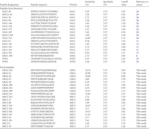
TABLE 1Peptides used in this study and their diagnostic propertiesaPeptide designation Peptide sequence Protein
Sensitivity (%)
Specificity (%)
Cutoff value
Reference or source
Peptides from literature
SAG1-48 SDPPLVANQVVTCPDKKS SAG1 4.35 3.57 1.87 26
dSAG1-62 DKKSTAAVILTPTENHFT SAG1 3.26 3.57 1.62 26
SAG1-76 NHFTLKCPKTALTEPPTLA SAG1 2.72 3.57 2.81 26
SAG1-90 PPTLAYSPNRQICPAGTTS SAG1 3.26 3.57 2.03 26
SAG1-104 AGTTSSCTSKAVTLSSLIP SAG1 1.63 3.57 1.29 26
SAG1-146 PIEKFPVTTQTFFWGCIKG SAG1 3.26 3.57 1.30 26
SAG1-273 AFPAESKSVIIGCTGGSPE SAG1 2.17 3.57 1.84 26
SAG1-287 GGSPEKHHCTVKLEFAGAA SAG1 5.43 3.57 0.54 26
dSAG1-301 FAGAAGSAKSAAGTASHVS SAG1 1.09 3.57 2.28 26
SAG1-315 ASHVSIFAMVIGLIGSIAACVA SAG1 16.85 3.57 1.13 26
SAG1-61 DKKSTAAVILTPTENHFTL SAG1 9.78 3.57 1.53 11
SAG1-181 SVVNNVARCSYGADSTLGP SAG1 1.63 3.57 2.06 11
SAG1-241 SFKDILPKLTENPWQGNAS SAG1 2.72 3.57 2.60 11
SAG1-261 DKGATLTIKKEAFPAESKS SAG1 2.17 3.57 1.48 11
SAG1-301 AGAAGSAKSAAGTASHVSI SAG1 0.54 3.57 1.71 11
GRA1-100 EEVIDTMKSMQRDED GRA1 2.72 3.57 2.81 6
NTP1 CKAPMIVTGGGMLAA INTLK NTP1 4.35 3.57 1.82 25
NTP2 APMFITGREMLASIDTLK NTP2 3.26 3.57 1.91 25
Novel peptides
GRA1-162 MKVIDDVQQLEKDKQQ GRA1 34.24 3.57 2.83 This study GRA2-61 DERQQEPEEPVSQRAS GRA2 30.98 3.57 1.28 This study GRA2-28 GVVNQGPVDVPFSGKP GRA2 50.00 3.57 0.98 This study GRA2-67 PEEPVSQRASRVAEQL GRA2 4.89 3.57 3.39 This study GRA2-38 PFSGKPLDERAVGGKG GRA2 9.24 3.57 1.71 This study GRA4-300 GGTRTSTAPAEAGKTE GRA4 9.24 3.57 1.06 This study GRA4-316 LDDGYRPPPFNPRPSP GRA4 4.35 3.57 0.95 This study GRA4-309 PAEAGKTELDDGYRPP GRA4 33.15 3.57 1.13 This study GRA4-321 PPPFNPRPSPYAELLK GRA4 13.04 3.57 2.33 This study MIC3-219 GETLVNLPEGGQGCKR MIC3 2.72 3.57 3.19 This study MIC3-15 SGAVWMCTPAEALPIQ MIC3 0.00 3.57 3.08 This study MIC3-88 RQLHTDNGYFIGASCP MIC3 1.09 3.57 2.82 This study MIC3-324 GSEGSLSEKMNIVFKC MIC3 10.33 3.57 1.37 This study MIC3-238 HAFRENCSPGRCIDDA MIC3 2.17 3.57 2.64 This study MIC3-282 GVEVTLAEKCEKEFGI MIC3 19.57 3.57 2.82 This study MIC3-191 SKRGNAKCGPNGTCIV MIC3 31.52 3.57 1.22 This study MIC3-26 ALPIQKSVQLGSFDKV MIC3 2.17 3.57 1.66 This study MIC3-291 CEKEFGISASSCKCDN MIC3 7.61 3.57 1.73 This study MIC3-8 LLHALTFSGAVWMCTP MIC3 1.63 3.57 2.25 This study MIC3-39 DKVVPSREVVSESLAP MIC3 2.17 3.57 1.26 This study a
Peptide sequences were either previously published (n⫽18) or selected afterin silicoepitope prediction (n⫽20). The number at the end of the peptide names indicates the position of the first amino acid of the peptide in the protein sequence. The individual cutoff value for each peptide was selected using the mean index values obtained for a panel of 140T. gondii-seronegative human sera and accepting a maximum of 4% false-positive reactions.
on August 17, 2020 by guest
http://cvi.asm.org/
in silico
using the computer program ABCpred. The respective
peptides containing these epitopes (referred to here as novel
pep-tides) and 18 additional peptides with published sequences were
synthesized.
All sera of seronegative patients (
n
⫽
65) and 75 randomly
selected sera of seronegative forest workers were used to establish
cutoff values for each peptide. Individual application of these
pep-tide-specific cutoff values resulted in a diagnostic specificity for
T.
gondii
-seronegative sera of 96.6% for each individual peptide.
Sera of patients with acute signs of
T. gondii
infection
recog-nized statistically significantly more peptides than the sera of
se-ropositive forest workers (Wilcoxon signed-rank test,
P
⫽
0.0003;
Fig. 1
). Similarly, sera of patients with latent
T. gondii
infection or
ocular signs of toxoplasmosis recognized a significantly higher
number of peptides than sera of serologically positive forest
work-ers (Wilcoxon signed-rank test;
P
⬍
0.000 or
P
⫽
0.001,
respec-tively) (
Fig. 1
). The differences between the results determined for
the groups of patients with ocular, acute, or latent toxoplasmosis
were not statistically significant (Wilcoxon signed-rank test,
P
⬎
0.6) (
Fig. 1
).
The sera of 72 of 84 (86%)
T. gondii
-seropositive patients with
acute, latent, or ocular toxoplasmosis and 66 of 100 (66%)
T.
gondii-
seropositive forest workers recognized at least 1 of the 38
peptides. The remaining 46 of 184 (25%) sera failed to react with
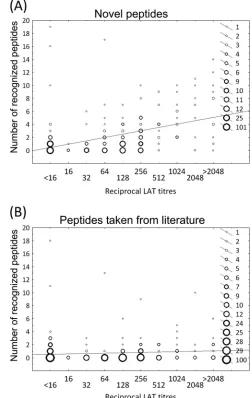
any of the 38 peptides. Linear regression analysis revealed a
signif-icant positive correlation between reciprocal LAT titers and the
number of novel peptides recognized by the individual sera (
r
2⫽
0.24,
P
⬍
0.0001;
Fig. 2A
). No significant correlation was found
between the reciprocal LAT titers and the number of peptides
taken from the literature that were recognized by the individual
sera (
r
2⫽
0.008,
P
⫽
0.1;
Fig. 2B
).
Individual diagnostic sensitivities of novel peptides
pre-dicted
in silico
.
Fourteen of the 20 novel peptides were recognized
by at least one serum sample from the patients with acute and
ocular toxoplasmosis. Individual peptide sensitivities in these two
serum panels (acute and ocular) ranged from 0% to 86% and from
0% to 100%, respectively (
Fig. 3A
and
B
).
Sera from patients with latent
T. gondii
infection recognized 19
of the 20 novel peptides. The individual diagnostic sensitivities of
the peptides ranged from 0% to 60% (
Fig. 3C
). The lowest number
of peptides (13 of 20) was recognized by sera from forest workers,
with a range of individual peptide sensitivities of 1% to 30% (
Fig.
3D
). Taken together for all sera (
n
⫽
184), the individual
diagnos-tic sensitivities determined for each novel peptide ranged from 0%
to 49% (
Table 1
).
The GRA2–28 peptide was the most reactive of the novel
pep-tides and was recognized by 100% of the serum samples from the
ocular toxoplasmosis patients, 85.7% of the serum samples from
patients with acute infections, 60.3% of the serum samples from
latently infected patients, and 30% of the serum samples from
T.
gondii
-seropositive forest workers (
Fig. 3A
,
B
,
C
, and
D
,
respec-FIG 1Number of peptides recognized by individualT. gondii-positive sera.Sera from forest workers recognized a significantly lower number of peptides per serum sample compared to sera from acutely or latently infected patients or patients with ocular toxoplasmosis (OT patients) (Wilcoxon signed-rank test;Pⱕ0.001). No significant differences were observed within the three patient groups (acute, latent, and ocular toxoplasmosis) concerning the num-ber of recognized peptides per serum sample.
FIG 2Latex agglutination test (LAT) titers and the numbers of recognized peptides in all seronegative and seropositive human serum samples (n⫽324) were analyzed for potential correlations by linear regression analysis. (A) Novel peptides: statistically significant correlation (r2⫽0.24;P⬍0.0001). (B)
Peptides taken from the literature: no significant correlation (r2⫽0.008;P⫽
0.1).
on August 17, 2020 by guest
http://cvi.asm.org/
tively). GRA-derived peptides were statistically significantly more
often recognized than MIC3-derived peptides (Wilcoxon
signed-rank test,
P
⬍
0.0001). One peptide (MIC3–15) failed to show a
positive reaction with any of the analyzed sera (
Table 1
).
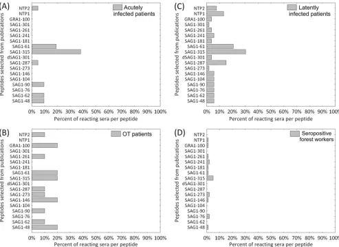
Individual diagnostic sensitivities of peptides taken from the
literature.
All 18 peptides taken from the literature were
recog-nized by sera from latently infected patients, with peptide-specific
sensitivities of 2% to 30% (
Fig. 4C
). Sera from ocular
toxoplas-mosis patients and forest workers recognized 11 of these 18
pep-tides, with individual peptide sensitivities of 0% to 20% (
Fig. 4B
and
D
). The lowest number of peptides (7 of 18) was recognized
by sera from patients with acute signs of toxoplasmosis, with
in-dividual peptide sensitivities ranging from 0% to 38% (
Fig. 4A
).
Among the 18 previously published peptides, SAG1–315 was
the peptide most often recognized in all tested serum groups. The
diagnostic sensitivities seen with this peptide ranged from 5%
(forest workers) to 38% (acutely infected patients). The individual
diagnostic sensitivities of each peptide with respect to all sera (184
sera per peptide) ranged between 1% and 17%.
SAG1-derived peptides showed reactivity that was statistically
significantly higher than the reactivity shown by the
NTPase-de-rived peptides (Wilcoxon signed-rank test,
P
⬍
0.0001;
Table 1
).
Diagnostic sensitivity and specificity of an optimized peptide
array.
The diagnostic potential of all 38 tested peptides was
ana-lyzed in relation to LAT seropositivity by univariate logistic
re-gression (LR). LR revealed a statistically significant association of
the results obtained for eight novel and two published peptides in
relation to the serological status of sera in LAT (
Table 2
). For all
serologically positive individuals, the peptide-specific diagnostic
sensitivity within this panel of 10 peptides ranged from 9.78%
(SAG1– 60) to 50% (GRA2–28) (
Table 1
).
An optimized array using a combination of these 10 peptides
had a diagnostic sensitivity of 100% in ocular toxoplasmosis
pa-tients, 86% in acutely infected papa-tients, 81% in latently infected
patients, and 57% in seropositive forest workers when the array
was regarded as positive when at least one of the peptides was
recognized.
The overall sensitivity mounted to 69% when sera were not
grouped.
Analysis of seronegative sera (
n
⫽
140) revealed a specificity of
84% for this optimized peptide array.
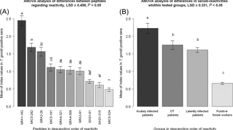
Reaction intensity of peptides with diagnostic potential.
Dif-ferences in the reaction intensities within the group of the 10
pep-tides with diagnostic potential were analyzed by ANOVA and an
FIG 3Diagnostic sensitivity of 20 novel peptides predictedin silicoin this study in groups of human serum samples (A) from patients with acute toxoplasmosis (n⫽21), (B) from patients with nonactive ocular toxoplasmosis (OT patients) (n⫽10), (C) from patients with latent toxoplasmosis (n⫽53), and (D) from serologically positive forest workers (n⫽100).on August 17, 2020 by guest
http://cvi.asm.org/
LSD post hoc test. Reactions performed with GRA1–162 showed
the highest statistically significant intensities, i.e., the highest
MSIVs, followed by MIC3–282 and GRA2–28 (LSD
ⱖ
0.458).
Peptides SAG1– 61, SAG1–315, and MIC3–324 displayed the
low-est means (LSD
ⱖ
0.458) of the reaction intensities (
Fig. 5A
).
ANOVA and LSD test analyses further showed that sera from
acutely infected patients recognized the 10 peptides with the
high-est statistically significant MSIVs (LSD
ⱖ
0.331) compared to the
sera from the other groups (
Fig. 5B
). Patients presenting with
latent and ocular
T. gondii
infection revealed MSIVs (LSD
ⱖ
0.331) that were significantly higher than those of serologically
positive forest workers. No statistical differences were observed
between patients presenting with ocular and latent toxoplasmosis
(LSD
ⱖ
0.331) (
Fig. 5B
).
One novel peptide (GRA2–28) was recognized with higher
sta-tistically significant MSIVs (LSD
ⱖ
1.09) by sera of acutely
in-fected patients compared to those determined for the members of
the other groups (
Fig. 6
). Two further novel peptides (MIC3–282
and MIC3–191) were recognized with significantly higher MSIVs
by sera of acutely infected patients compared to latently infected
patients and seropositive forest workers (LSD
ⱖ
1.09 for MIC3–
282; LSD
⬎
0.95 for MIC3–191) (
Fig. 6
). Sera of patients with
ocular toxoplasmosis showed no statistically significant
differ-ences in their reactivity with these two peptides compared to sera
of acutely infected patients and of patients with latent
toxoplas-mosis (LSD
ⱖ
1.09 for MIC3–282; LSD
⬎
0.95 for MIC3–191)
(
Fig. 6
). For all 10 peptides with diagnostic potential, the sera from
forest workers showed the lowest mean MSIV index values relative
to those from the members of the other groups.
DISCUSSION
A number of
T. gondii
proteins, including SAG1, NTPase1,
NT-Pase2, GRA1, GRA2, and GRA4, have long been known to possess
high antigenicity and are suitable antigens for the serological
di-agnosis of
T. gondii
infection (
2
,
7
,
11
,
16
,
21
,
24
–
27
,
33
,
41
). We
therefore selected these proteins to identify novel diagnostic
pep-tides for the serological diagnosis of human toxoplasmosis.
To identify
T. gondii
epitopes with diagnostic potential, we
predicted potentially suitable epitopes by the use of the
bioinfor-matic software ABCpred. A variety of computing methods for the
prediction of epitopes (e.g., PREDITOPE, PEOPLE, BEPITOPE,
BcePred, AAP, BepiPred, the Hopp and Woods method, the
Jame-son-Wolf antigenicity index, etc.) have been developed in the past
(
14
,
50
). Most of them utilize the physical and chemical properties
FIG 4Diagnostic sensitivity of 18 peptides taken from the literature in groups of human serum samples (A) from patients with acute toxoplasmosis (n⫽21), (B) from patients with nonactive ocular toxoplasmosis (OT patients) (n⫽10), (C) from patients with latent toxoplasmosis (n⫽53), and (D) from serologically positive forest workers (n⫽100).on August 17, 2020 by guest
http://cvi.asm.org/
of individual amino acids for the prediction of structural and
functional properties of the peptide chain suggesting the
localiza-tion of epitopes. However, the proporlocaliza-tion of correct prediclocaliza-tions
made with these tools was only marginally better than that seen
with random epitope selection (
8
).
Saha and Raghava (
52
) developed ABCpred, a software
pro-gram designed to predict linear B-cell epitopes, using a
combina-tion of recurrent and artificial neural networks. This software was
trained with a data set of 700 experimentally detected B-cell
epitopes from the Bcipep database and 700 random peptides from
the Swiss-Prot database. Thereafter, ABCpred showed a better
prediction rate than other tools, and the results obtained were
significantly better than those obtained by random selection (
48
,
52
). Our study supports these findings, as almost all (19 of 20)
ABCpred-selected
T. gondii
peptides were recognized by various
groups of serologically
T. gondii
-positive human serum samples,
and 40% of 20 ABCpred-predicted linear B-cell epitopes appeared
to possess diagnostic potential. However, considerable variation
in the respective diagnostic sensitivities was observed that
de-pended on the individual peptide and the serum panel analyzed.
The moderate success in predicting such epitopes was expected,
since the ABCpred developers described a prediction accuracy of
65.93% by this method (
52
). Since we analyzed the protein
se-quences without prior knowledge of the linear B-cell epitopes and
nonepitopes, we cannot directly compare our data on the
propor-tion of correctly predicted epitopes with the predicpropor-tion accuracy
reported for ABCpred.
All previously described peptides, which included mainly
SAG1-derived peptides, were recognized by the human serum
samples with lower diagnostic sensitivity. This was unexpected, as
a large proportion of anti-
T. gondii
antibodies in infected humans
and animals are directed against SAG1. However, it is known that
most B-cell epitopes on the SAG1 antigen are conformation
de-pendent (
9
,
28
,
33
,
51
), whereas we analyzed only linear epitopes
in this study.
This could therefore be one explanation for the low reactivity
of SAG1-derived peptides in our study. However, SAG1 peptide
sequences had diagnostic sensitivity in our hands that was lower
than that reported previously (
11
). Since Cardona et al. (
11
)
con-ducted their study in South America, differences between South
America and Europe in the prevailing clonal types of
T. gondii
appear to be the most likely reason for this discrepancy. Moreover,
the results might also be biased by differences in the major
histo-compatibility complex (MHC) haplotype distributions in the
hu-man populations from South America and Europe (
22
,
23
,
37
).
We also observed lower reactivity of peptide GRA1–100 (also
called “epi-24”), which was previously described as highly
immu-noreactive by Beghetto and colleagues (
6
). In our hands, GRA1–
100 had a diagnostic sensitivity of only 2.7% (5/184), while 29 of
40
T. gondii
-positive sera reacted with GRA1–100 in the study of
Beghetto et al. (
6
). This discrepancy with respect to the diagnostic
sensitivity of GRA1–100 might be explained by differences in the
methods by which the antigens were produced. We applied
syn-thetic peptides, whereas Beghetto and colleagues used a
recombi-nant protein (
6
). The oligopeptide produced in an expression
vec-tor may be folded in a fashion different from that of synthetic
peptides or might carry secondary modifications, which could
also explain differences in antigenicity.
The diagnostic sensitivities of individual peptides analyzed in
our study were limited, not exceeding 50% within the group of all
serologically positive (i.e., all LAT-positive) human serum
sam-ples. Furthermore, we observed that each
T. gondii
-positive serum
recognized an individual peptide pattern. Thus, our results
sug-gest that it is necessary to use several peptides at the same time, i.e.,
a peptide array, for the serological diagnosis of
T. gondii
infections
to achieve a level of sensitivity similar to the sensitivity of
conven-tional serological diagnostic tests. Similar effects were also
ob-served when synthetic peptides were used as diagnostic antigens
for human echinococcosis and tuberculosis studies (
36
,
44
,
53
).
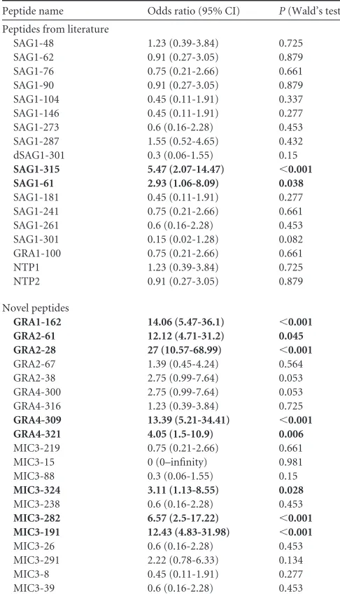
Univariate LR analysis identified 10 peptides for which the
se-rological results showed a statistically significant concordance
with those obtained with established serological tests. These
pep-tides contain epitopes from the
T. gondii
antigenic proteins SAG1,
GRA1, GRA2, and GRA4. The peptide-specific diagnostic
sensi-TABLE 2Univariate logistic regression analysis to examine potentialrelationships between seropositivity in the latex agglutination test (LAT) and positive reactions with individual peptidesa
Peptide name Odds ratio (95% CI) P(Wald’s test)
Peptides from literature
SAG1-48 1.23 (0.39-3.84) 0.725 SAG1-62 0.91 (0.27-3.05) 0.879 SAG1-76 0.75 (0.21-2.66) 0.661 SAG1-90 0.91 (0.27-3.05) 0.879 SAG1-104 0.45 (0.11-1.91) 0.337 SAG1-146 0.45 (0.11-1.91) 0.277 SAG1-273 0.6 (0.16-2.28) 0.453 SAG1-287 1.55 (0.52-4.65) 0.432 dSAG1-301 0.3 (0.06-1.55) 0.15
SAG1-315 5.47 (2.07-14.47) ⬍0.001
SAG1-61 2.93 (1.06-8.09) 0.038
SAG1-181 0.45 (0.11-1.91) 0.277 SAG1-241 0.75 (0.21-2.66) 0.661 SAG1-261 0.6 (0.16-2.28) 0.453 SAG1-301 0.15 (0.02-1.28) 0.082 GRA1-100 0.75 (0.21-2.66) 0.661 NTP1 1.23 (0.39-3.84) 0.725 NTP2 0.91 (0.27-3.05) 0.879
Novel peptides
GRA1-162 14.06 (5.47-36.1) ⬍0.001
GRA2-61 12.12 (4.71-31.2) 0.045
GRA2-28 27 (10.57-68.99) ⬍0.001
GRA2-67 1.39 (0.45-4.24) 0.564 GRA2-38 2.75 (0.99-7.64) 0.053 GRA4-300 2.75 (0.99-7.64) 0.053 GRA4-316 1.23 (0.39-3.84) 0.725
GRA4-309 13.39 (5.21-34.41) ⬍0.001
GRA4-321 4.05 (1.5-10.9) 0.006
MIC3-219 0.75 (0.21-2.66) 0.661 MIC3-15 0 (0–infinity) 0.981 MIC3-88 0.3 (0.06-1.55) 0.15
MIC3-324 3.11 (1.13-8.55) 0.028
MIC3-238 0.6 (0.16-2.28) 0.453
MIC3-282 6.57 (2.5-17.22) ⬍0.001
MIC3-191 12.43 (4.83-31.98) ⬍0.001
MIC3-26 0.6 (0.16-2.28) 0.453 MIC3-291 2.22 (0.78-6.33) 0.134 MIC3-8 0.45 (0.11-1.91) 0.277 MIC3-39 0.6 (0.16-2.28) 0.453 aStatistically significant (P⬍0.05) associations are marked (bold). CI, confidence interval.
on August 17, 2020 by guest
http://cvi.asm.org/
tivities of the individual peptides ranged from 9.78% to 50%.The
diagnostic sensitivity of an array based on the 10 peptides ranged
between 79% and 100%, depending on the group of sera analyzed,
while the diagnostic specificity was 84%. These values are
accept-able, though far from optimal. In any case, the 10 peptides may in
the future be included in a larger multiple antigenic peptide array
for detecting
T. gondii
infection in patients. Further suitable
pep-tides need to be identified to improve sensitivity. The specificity of
a future peptide microarray could be increased by applying more
stringent thresholds, i.e., by increasing the cutoffs for individual
peptides or by increasing the number of peptides that must be
recognized by an individual serum for it to be classified as testing
positive. To increase the number of peptide candidates for an
optimized peptide microarray, additional immunoreactive
anti-genic proteins, such as rhoptry (ROP1, ROP2), microneme
(MIC2, MIC4, MIC5), dense granule (GRA1, GRA2, GRA4,
GRA6, GRA7, GRA8), and surface antigens (SAG2A), should be
taken into consideration for the identification of linear B-cell
epitopes. If such an optimized array can be established, it may
prove superior to conventional serological tests, because the
epitopes recognized by sera from individual patients are
unambig-uously defined. The peptide recognition pattern may allow further
conclusions regarding the status of the patient, e.g., differentiation
of the acute stage versus chronic stage of infection. A
T. gondii
peptide array may also represent a powerful tool to compare
se-rological reactions, e.g., reactions of a mother and her child, to
find evidence for congenital transmission. Another field of
appli-cation could be the differential examination of serum and
intra-ocular fluids of patients with suspected intra-ocular toxoplasmosis to
confirm or rule out active infections of the eye.
Even within our small array, certain antipeptide reactions
could be associated with the acute stage of infection. Sera from
acutely infected patients generally recognized peptides with
signif-icantly higher index values than sera from other groups.
More-over, we found that particular peptides (e.g., GRA2–28, MIC3–
282, MIC3–191) were recognized with a significantly higher
intensity by sera from acutely infected patients than by sera from
others, indicating that these peptides may be candidates for a
pre-dictive peptide panel for the diagnosis of acute toxoplasmosis in
humans.
The present report demonstrates that ABCpred is an
appropri-ate bioinformatical tool for use in the selection of potential
epitope candidates. However, we also observed the prediction of
epitopes that show low reactivity in the analysis with
T. gondii
-positive human serum samples. False--positive epitope prediction
was also reported by the developers of the tool and can be
ex-plained, e.g., by the use of a fixed amino acid length in predicting
epitopes (
52
). Moreover, most epitopes analyzed in this study
were continuous, whereas the majority of naturally recognized
epitopes are discontinuous (
4
). Because of this, it may also make
sense to search for discontinuous epitopes, e.g., by using
bioinfor-matical software for the analysis of the three-dimensional (3D)
X-ray structure of an antigen of interest. Unfortunately, there are
only limited 3D X-ray protein structure data for
T. gondii
anti-genic proteins available. Nevertheless, our report shows that the
use of a bioinformatic prediction method in combination with a
FIG 5Intensity index values for peptides with diagnostic potential among testedT. gondii-seropositive human serum samples. The differences between the means of the sample index values (MSIVs) for intensity for single peptides and those for human groups (acutely infected patients, latently infected patients, patients with ocular toxoplasmosis [OT], and seropositive forest workers) determined with 10 peptides with diagnostic potential were regarded as statistically significant when the differences were equal to or higher than the LSD values. Different letters above the whiskers indicate significant differences between the mean intensities in the post hoc LSD test ([A] LSDⱖ0.458; [B] LSDⱖ0.331). Whiskers in bar plots represent 95% confidence intervals of the MSIVs. (A) MSIVs of the reactions performed with 10 peptides with diagnostic potential observed for seropositive humans. (B) MSIVs for sera from different groups of humans determined with 10 peptides with diagnostic potential.on August 17, 2020 by guest
http://cvi.asm.org/
peptide microarray assay is a powerful tool for selection and
anal-ysis of
T. gondii
epitopes as candidate antigens for serological
di-agnosis.
ACKNOWLEDGMENTS
This work was supported by the German Federal Ministry of Educa-tion and Research (Toxonet01 and Toxonet02) by funds to G.S. (01KI0765 and 01KI1002F), W.D. (01KI0764 and 01KI1002E), U.P. (01KI1002G), and U.G. (01KI0766 and 01KI1002B).
We acknowledge the excellent technical assistance of Andrea Bärwald, Lieselotte Minke, and Robert Carus. We thank Andreas Fröhlich for dis-cussions about the statistical analysis of our study results.
REFERENCES
1.Alcaro MC, Peroni E, Rovero P, Papini AM.2003. Synthetic peptides in the diagnosis of HIV infection. Curr. Protein Pept. Sci.4:285–290. 2.Altcheh J, et al.2006. Kinetic analysis of the humoral immune response
against 3Toxoplasma gondii-recombinant proteins in infants with sus-pected congenital toxoplasmosis. Diagn. Microbiol. Infect. Dis.56:161– 165.
3.Bacarese-Hamilton T, Mezzasoma L, Ardizzoni A, Bistoni F, Crisanti A. 2004. Serodiagnosis of infectious diseases with antigen microarrays. J. Appl. Microbiol.96:10 –17.
4.Barlow DJ, Edwards MS, Thornton JM.1986. Continuous and discon-tinuous protein antigenic determinants. Nature322:747–748.
5.Beghetto E, et al.2003. Use of an immunoglobulin G avidity assay based
on recombinant antigens for diagnosis of primaryToxoplasma gondii in-fection during pregnancy. J. Clin. Microbiol.41:5414 –5418.
6.Beghetto E, et al. 2001. Identification of a human immunodominant B-cell epitope within the GRA1 antigen ofToxoplasma gondiiby phage display of cDNA libraries. Int. J. Parasitol.31:1659 –1668.
7.Beghetto E, et al. 2003. Molecular dissection of the human B-cell re-sponse againstToxoplasma gondiiinfection by lambda display of cDNA libraries. Int. J. Parasitol.33:163–173.
8.Blythe MJ, Flower DR.2005. Benchmarking B cell epitope prediction: underperformance of existing methods. Protein Sci.14:246 –248. 9.Burg JL, Perelman D, Kasper LH, Ware PL, Boothroyd JC. 1988.
Molecular analysis of the gene encoding the major surface antigen of Toxo-plasma gondii.J. Immunol.141:3584 –3591.
10. Cabral HJ.2008. Multiple comparisons procedures. Circulation117: 698 –701.
11. Cardona N, de la Torre A, Siachoque H, Patarroyo MA, Gomez-Marin JE.2009.Toxoplasma gondii: P30 peptides recognition pattern in human toxoplasmosis. Exp. Parasitol.123:199 –202.
12. Chaves-Borges FA, Souza MA, Silva DA, Kasper LH, Mineo JR.1999. Detection ofToxoplasma gondiisoluble antigen, SAG-1(p30), antibody and immune complex in the cerebrospinal fluid of HIV positive or nega-tive individuals. Rev. Inst. Med. Trop. Sao Paulo41:329 –338.
13. Domínguez-Almendros S, Benítez-Parejo N, Gonzalez-Ramirez AR. 2011. Logistic regression models. Allergol. Immunopathol. (Madr.)39: 295–305.
14. El-Manzalawy Y, Honavar V.2010. Recent advances in B-cell epitope prediction methods. Immunome Res.6(Suppl. 2):S2. doi:10.1186/1745-7580-6-S2-S2.
FIG 6Differences between various groups of humans regarding the reactivity of sera with peptides suitable forT. gondiidiagnosis. The differences between the MSIVs among groups of humans (acutely infected patients, latently infected patients, patients with ocular toxoplasmosis [OT], and seropositive forest workers) for each peptide with diagnostic potential were regarded as statistically significant when the differences were equal to or higher than the LSD values. Different letters on the bars indicate statistically significant differences between the mean intensities in the post hoc LSD-Bonferroni test. Whiskers in bar plots represent 95% confidence intervals of the MSIVs. Peptide GRA2-28 was recognized by the sera from acutely infected patients with a statistically significant higher intensity than by sera from other groups. MIC3–282 and MIC3–191 peptides were recognized with statistically significant higher index values by sera from acutely infected patients than by sera from ocular toxoplasmosis patients and seropositive forest workers. For all 10 peptides with diagnostic potential, the sera from the forest workers displayed the lowest MSIVs relative to the sera from other groups.
on August 17, 2020 by guest
http://cvi.asm.org/
15. Garweg JG, et al.2005. Congenital ocular toxoplasmosis— observations on the outcome after an early diagnosis. Klin. Monbl. Augenheilkd.222: 721–727.
16. Golkar M, et al.2007. The dense granule protein GRA2, a new marker for the serodiagnosis of acuteToxoplasmainfection: comparison of sera col-lected in both France and Iran from pregnant women. Diagn. Microbiol. Infect. Dis.58:419 – 426.
17. Groß U.2004. Prevalence and public-health-aspects of toxoplasmosis. Bundesgesundheitsblatt Gesundheitsforschung Gesundheitsschutz47: 692– 697. (In German.)
18. Gross U, Roos T, Appoldt D, Heesemann J.1992. Improved serological diagnosis ofToxoplasma gondiiinfection by detection of immunoglobulin A (IgA) and IgM antibodies against P30 by using the immunoblot tech-nique. J. Clin. Microbiol.30:1436 –1441.
19. Harning D, Spenter J, Metsis A, Vuust J, Petersen E.1996. Recombinant Toxoplasma gondiisurface antigen 1 (P30) expressed inEscherichia coliis recognized by humanToxoplasma-specific immunoglobulin M (IgM) and IgG antibodies. Clin. Diagn. Lab. Immunol.3:355–357.
20. Hosseininejad M, Azizi HR, Hosseini F, Schares G.2009. Development of an indirect ELISA test using a purified tachyzoite surface antigen SAG1 for sero-diagnosis of canineToxoplasma gondiiinfection. Vet. Parasitol. 164:315–319.
21. Ismael AB, Sekkai D, Collin C, Bout D, Mévélec MN.2003. The MIC3 gene ofToxoplasma gondiiis a novel potent vaccine candidate against toxoplasmosis. Infect. Immun.71:6222– 6228.
22. Jamieson SE, et al.2009. Host genetic and epigenetic factors in toxoplas-mosis. Mem. Inst. Oswaldo Cruz104:162–169.
23. Jamieson SE, et al.2008. Genetic and epigenetic factors at COL2A1 and ABCA4 influence clinical outcome in congenital toxoplasmosis. PLoS One 3:e2285. doi:10.1371/journal.pone.0002285.
24. Jiang T, et al.2008. Evaluation of a recombinant MIC3 based latex ag-glutination test for the rapid serodiagnosis ofToxoplasma gondiiinfection in swines. Vet. Parasitol.158:51–56.
25. Johnson MS, Broady KW, Johnson AM.1999. Differential recognition of Toxoplasma gondiirecombinant nucleoside triphosphate hydrolase iso-forms by naturally infected human sera. Int. J. Parasitol.29:1893–1905. 26. Kato M, et al.2007. Reactivity of synthetic SAG1 (p30) peptide sequences
with RH, S273 and Beverley strain-induced anti-Toxoplasma gondii anti-bodies. Pathobiology74:50 –56.
27. Kato M, et al.2005.Toxoplasma gondiiantigens GRA1 (p24) and SAG1 (p30): a comparison of their stimulatory influence on T-cell activation and cytokine expression in in vitro cultures. Pathobiology72:160 –164. 28. Kim K, Bülow R, Kampmeier J, Boothroyd JC.1994. Conformationally
appropriate expression of theToxoplasmaantigen SAG1 (p30) in CHO cells. Infect. Immun.62:203–209.
29. Klaren VN, Peek R.2001. Evidence for a compartmentalized B cell re-sponse as characterized by IgG epitope specificity in human ocular toxo-plasmosis. J. Immunol.167:6263– 6269.
30. Kodjikian L, et al.2006. Ocular manifestations in congenital toxoplas-mosis. Graefes Arch. Clin. Exp. Ophthalmol.244:14 –21.
31. Kong JT, Grigg ME, Uyetake L, Parmley S, Boothroyd JC.2003. Sero-typing ofToxoplasma gondiiinfections in humans using synthetic pep-tides. J. Infect. Dis.187:1484 –1495.
32. Kotresha D, Noordin R.2010. Recombinant proteins in the diagnosis of toxoplasmosis. APMIS118:529 –542.
33. Lekutis C, Ferguson DJ, Grigg ME, Camps M, Boothroyd JC.2001. Surface antigens ofToxoplasma gondii: variations on a theme. Int. J. Para-sitol.31:1285–1292.
34. Li S, et al.2000. Serodiagnosis of recently acquiredToxoplasma gondii infection with a recombinant antigen. J. Clin. Microbiol.38:179 –184. 35. Liang L, et al.2011. Identification of potential serodiagnostic and subunit
vaccine antigens by antibody profiling of toxoplasmosis cases in Turkey. Mol. Cell. Proteomics10:M110.006916. doi:10.1074/mcp.M110.006916. 36. List C, et al.2010. Serodiagnosis ofEchinococcusspp. infection:
explor-ative selection of diagnostic antigens by peptide microarray. PLoS Negl. Trop. Dis.4:e771. doi:10.1371/journal.pntd.0000771.
37. Lüder CG, Seeber F.2001.Toxoplasma gondiiand MHC-restricted anti-gen presentation: on degradation, transport and modulation. Int. J. Para-sitol.31:1355–1369.
38. Maksimov P, et al.2011. Serological survey and risk factors for Toxo-plasma gondiiin domestic ducks and geese in Lower Saxony, Germany. Vet. Parasitol.182:140 –149.
39. Maksimov P, et al.2012. Analysis of clonal type-specific antibody reac-tions inToxoplasma gondiiseropositive humans from Germany by pep-tide-microarray. PLoS One7:e34212. doi:10.1371/journal.pone.0034212. 40. Mertens M, et al.2011. Seroprevalence study in forestry workers of a non-endemic region in eastern Germany reveals infections by Tula and Dobrava-Belgrade hantaviruses. Med. Microbiol. Immunol.200:263–268. 41. Mévélec MN, et al.1998. Mapping of B epitopes in GRA4, a dense granule antigen ofToxoplasma gondiiand protection studies using recombinant proteins administered by the oral route. Parasite Immunol.20:183–195. 42. Mezzasoma L, et al.2002. Antigen microarrays for serodiagnosis of
in-fectious diseases. Clin. Chem.48:121–130.
43. Montoya JG, Liesenfeld O.2004. Toxoplasmosis. Lancet363:1965–1976. 44. Nahtman T, et al.2007. Validation of peptide epitope microarray
exper-iments and extraction of quality data. J. Immunol. Methods328:1–13. 45. Ngo Y, et al.2009. Identification and testing of control peptides for
antigen microarrays. J. Immunol. Methods343:68 –78.
46. Noya O, Patarroyo ME, Guzmán F, Alarcón de Noya B.2003. Immu-nodiagnosis of parasitic diseases with synthetic peptides. Curr. Protein Pept. Sci.4:299 –308.
47. Pamelard F, et al.2009. PASE: a web-based platform for peptide/protein microarray experiments. Methods Mol. Biol.570:413– 430.
48. Reineke U, Schutkowski M.2009. Epitope mapping protocols. Preface. Methods Mol. Biol.524:v–vi.
49. Reineke U.2009. Antibody epitope mapping usingde novogenerated synthetic peptide libraries. Methods Mol. Biol.524:203–211.
50. Roggen EL.2006. Recent developments with B-cell epitope identification for predictive studies. J. Immunotoxicol.3:137–149.
51. Saeij JP, Arrizabalaga G, Boothroyd JC.2008. A cluster of four surface antigen genes specifically expressed in bradyzoites, SAG2CDXY, plays an important role inToxoplasma gondiipersistence. Infect. Immun.76:2402– 2410.
52. Saha S, Raghava GP.2006. Prediction of continuous B-cell epitopes in an antigen using recurrent neural network. Proteins65:40 – 48.
53. Shen G, Behera D, Bhalla M, Nadas A, Laal S. 2009. Peptide-based antibody detection for tuberculosis diagnosis. Clin. Vaccine Immunol. 16:49 –54.
54. Sousa S, et al.2009. Selection of polymorphic peptides from GRA6 and⬘ GRA7 sequences ofToxoplasma gondiistrains to be used in serotyping. Clin. Vaccine Immunol.16:1158 –1169.
55. Suzuki Y.2002. Host resistance in the brain againstToxoplasma gondii.J. Infect. Dis.185(Suppl. 1):S58 –S65.
56. Tenter AM, Heckeroth AR, Weiss LM.2000.Toxoplasma gondii: from animals to humans. Int. J. Parasitol.30:1217–1258.
57. Travier L, et al. 2008. Functional domains of theToxoplasmaGRA2 protein in the formation of the membranous nanotubular network of the parasitophorous vacuole. Int. J. Parasitol.38:757–773.
58. Vigil A, et al.2010. Identification of the feline humoral immune response to Bartonella henselaeinfection by protein microarray. PLoS One 5:e11447. doi:10.1371/journal.pone.0011447.
![FIG 6 Differences between various groups of humans regarding the reactivity of sera with peptides suitable forMSIVs among groups of humans (acutely infected patients, latently infected patients, patients with ocular toxoplasmosis [OT], and seropositive for](https://thumb-us.123doks.com/thumbv2/123dok_us/7730174.1265431/9.585.46.543.65.370/differences-regarding-reactivity-formsivs-latently-patients-toxoplasmosis-seropositive.webp)
