Cells via Caveolin-1
Bing Wu,aShuang Geng,bYanmin Bi,aHu Liu,aYanxin Hu,cXinqiang Li,aYizhi Zhang,aXiaoyu Zhou,bGuoxing Zheng,dBin He,d Bin Wanga,b
State Key Laboratory for Agro-Biotechnology, College of Biological Science, China Agricultural University, Beijing, People’s Republic of Chinaa
; Key Laboratory of Medical Molecular Virology of MOH and MOE, Fudan University Shanghai Medical College, Shanghai, People’s Republic of Chinab
; College of Veterinary Medicine, China Agricultural University, Beijing, People’s Republic of Chinac
; Department of Microbiology and Immunology, College of Medicine, University of Illinois, Chicago, Illinois, USAd
Caveolin-1 (Cav-1), the principal structural protein of caveolae, has been implicated as a regulator of virus-host interactions.
Several viruses exploit caveolae to facilitate viral infections. However, the roles of Cav-1 in herpes simplex virus 1 (HSV-1)
infec-tion have not fully been elucidated. Here, we report that Cav-1 downregulates the expression of inducible nitric oxide synthase
(iNOS) and the production of nitric oxide (NO) in dendritic cells (DCs) during HSV-1 infection. As a result, Cav-1 deficiency led
to an accelerated elimination of virus and less lung pathological change following HSV-1 infection. This protection was
depen-dent on iNOS and NO production in DCs. Adoptive transfer of DCs with Cav-1 knockdown was sufficient to confer the
protec-tion to wild-type (WT) mice. In addiprotec-tion, Cav-1 knockout (KO) (Cav-1
ⴚ/ⴚ) mice treated with an iNOS inhibitor exhibited
signif-icantly reduced survival compared to that of the nontreated controls. We found that Cav-1 colocalized with iNOS and HSV-1 in
caveolae in HSV-1-infected DCs, suggesting their interaction. Taken together, our results identified Cav-1 as a novel regulator
utilized by HSV-1 to evade the host antiviral response mediated by NO production. Therefore, Cav-1 might be a valuable target
for therapeutic approaches against herpesvirus infections.
H
erpes simplex virus 1 (HSV-1) is a double-stranded DNA
(dsDNA) virus belonging to the
Alphaherpesvirus
family,
which causes oral herpes, encephalitis, keratitis, neonatal herpes,
and pneumonia disease, establishing latency in the neurons after
acute infection of mucosal tissues (
1–3
). Notably, HSV-1 can be
isolated from the respiratory tract of immunosuppressed patients
and newborn infants, where it induces pneumonitis, resulting in
remarkable morbidity and mortality (
4
). Recent studies have
sug-gested that HSV-1-induced bronchopneumonitis is common in
nonimmunocompromised persons who are undergoing
continu-ous mechanical ventilation (
5
). Currently, the mechanisms of
HSV-1-induced pneumonia and obstructive pulmonary disease
are not fully understood, although intranasal (i.n.) infection with
HSV-1 in mice can be used as a model to investigate these
mech-anisms (
4
,
6
,
7
). Such investigations might reveal a valuable
ther-apeutic approach for HSV-1-induced pneumonia.
Innate defense cells and inflammatory factors serve as the
first-line of host defense against viral infections. DCs can be recruited
to the lungs and in the cornea of the eye, where they contribute to
host defense (
8
,
9
). Studies have shown that diphtheria toxin
(DT)-induced depletion of DCs in CD11c-DTR mice (in which
the DT receptor [DTR] is expressed under the control of the
CD11c promoter) inhibited the migration of natural killer cells
and neutrophils to locally infected cornea, resulting in severe
pa-thology (
10
,
11
). Moreover, involvement of the free radical nitric
oxide (NO) has been indicated. This is a powerful vasodilator
factor and cell signaling molecule, with a short half-life of 3 to
⬃
4
s in the blood, and it is synthesized by nitric oxide synthase (NOS)
in epithelial cells, macrophages, DCs, and other myeloid cells (
12
,
13
). NOS has three isoenzymes: endothelial NOS (eNOS),
neuro-nal NOS (nNOS), and inducible NOS (iNOS) (iNOS is induced by
a single stimulus, like lipopolysaccharide [LPS] or gamma
inter-feron [IFN-
␥
]). Induction of iNOS and NO production
consti-tutes a critical component of the innate antiviral host response to
HSV-1, influenza A virus, and other intracellular parasites (
14–
16
) and is potent in clearing the invading pathogens. Early
inhibi-tion of NO by i.n. administrainhibi-tion of aminoguanidine (AG) was
found to increase HSV-1 infection in the eyes and lungs of mice
(
17
). Conversely, pretreatment with an NO donor, sodium
nitro-prusside (SNP), decreased the titer of Sindbis virus (
18
). Despite
the general importance of DCs and NO in antiviral responses, it is
unknown whether this is applicable to HSV-1 infection in the
lungs.
Caveolin-1 (Cav-1), a scaffolding protein found in most types
of cells, is the major coating protein of caveolae (with 50- to
100-nm plasma membrane invaginations) (
19
,
20
). A deficiency
in Cav-1 leads to disruption of the caveolae structure. Although
best known in lipid metabolism, roles for Cav-1 in the
internal-ization of pathogens, signal transduction, host defenses, and
sup-pression of inflammatory responses have also been indicated by
numerous studies (
20–22
). Viral entry into cells occurs by
clath-rin, caveolae, or receptor-mediated pathways (
23–25
). However,
recent studies revealed that simian virus 40 (SV40) enters cells via
Received24 March 2015Returned for modification10 April 2015 Accepted20 May 2015
Accepted manuscript posted online27 May 2015
CitationWu B, Geng S, Bi Y, Liu H, Hu Y, Li X, Zhang Y, Zhou X, Zheng G, He B, Wang B. 2015. Herpes simplex virus 1 suppresses the function of lung dendritic cells via caveolin-1. Clin Vaccine Immunol 22:883–895.doi:10.1128/CVI.00170-15.
Editor:H. F. Rosenberg
Address correspondence to Bin Wang, [email protected].
Copyright © 2015, American Society for Microbiology. All Rights Reserved.
doi:10.1128/CVI.00170-15
on August 17, 2020 by guest
http://cvi.asm.org/
Madin-Darby canine kidney (MDCK) cells (
29
). By using the
mu-tant Cav-1 protein, hepatitis B virus (HBV) was found to require
intact Cav-1 to initiate a productive infection in HepaRG cells
(
30
). In addition, Cav-1 has been implicated in cell signaling and
inflammation (
31
). Reports show that caveolin-1 might
down-regulate iNOS/NO via a proteasome pathway and mediate the
posttranscriptional regulation of iNOS (
32
,
33
). Through the
abil-ity of Cav-1 to regulate NO, Cav-1
⫺/⫺mice exhibited attenuation
of lung injury and less edema formation in response to LPS (
34
).
These observations suggested to us that Cav-1 might be involved
in HSV-1 infection.
We hypothesized that Cav-1 might suppress host antiviral
im-munity during HSV-1 infection. In the present study, we used a
murine model of HSV-1-induced pneumonia to demonstrate for
the first time that Cav-1
⫺/⫺mice are resistant to fatal HSV-1
in-fection and that the increased antiviral activity is due to increased
expression of iNOS/NO in DC cells in Cav-1
⫺/⫺mice. In addition,
the protective effect of Cav-1 deficiency was largely abolished by
iNOS inhibition in Cav-1
⫺/⫺mice or by DC depletion in
CD11c-DTR/Cav-1
⫺/⫺mice. Furthermore, we observed that Cav-1
colo-calized with iNOS and HSV-1 in caveolae in the virus-infected
DCs. Thus, these findings indicate that HSV-1 exploits Cav-1 to
downregulate the expression of iNOS in DCs. The corollary is that
a deficiency in Cav-1 can restore antiviral immune responses,
which has implications for the design of drugs against herpes
in-fection.
MATERIALS AND METHODS
Ethics statement.The use of laboratory animals in our study was ap-proved by the Beijing Association for Science and Technology (approval ID SYXK [Beijing] 2007-0023), and all animal procedures were conducted according to the guidelines of Beijing Laboratory Animal Welfare and Ethics of the Beijing Administration Committee of Laboratory Animals. All animal research was also carried out in accordance with the China Agricultural University Institutional Animal Care and Use Committee guidelines (ID: SKLAB-B-2010-003) and approved by the animal welfare committee of China Agricultural University.
Mice.Female C57BL/6 mice at 6 to 8 weeks of age were purchased from the Animal Institute of Chinese Medical Academy (Beijing, China). Cav-1⫺/⫺mice (stock Cav1tm1Mls/J, C57BL/6 background) and CD11c-DTR-GFP mice (B6.FVB-Tg [Itgax-DTR/GFP] 57Lan/J) (GFP, green flu-orescent protein) were purchased from the Jackson Laboratory (Bar Harbor, ME). NOS2 (iNOS) KO mice (B6, 129P2-NOS2tm1Lau/J) were purchased from the China Institute of Laboratory Animal Sciences, Chi-nese Academy of Medical Sciences (CAMS), and Peking Union Medical College (PUMC).
Reagents.S-methylisothiourea sulfate (SMT), an iNOS inhibitor, and radioimmunoprecipitation assay (RIPA) lysis buffer were obtained from the Beyotime Institute of Biotechnology (Beijing, China). DAPI (4= ,6-diamidino-2-phenylindole) and mitomycin C were obtained from Sigma (St. Louis, MO, USA). Rabbit polyclonal anti-iNOS antibody was ob-tained from Cell Signaling Technology (Danvers, MA, USA), goat anti-iNOS antibody was from Santa Cruz (Dallas, TX, USA),
anti-glyceralde-Omega (Norcross, GA, USA). ReverTra Ace and the SYBR green real-time PCR kit were obtained from Toyobo Life Science (Osaka, Japan).
Virus and cells.HSV-1 strain F was originally obtained from the Na-tional Institutes for Food and Drug Control (China) and was propagated in Vero cells. Virus was harvested by repeated freezing and thawing and concentrated by centrifugation at 18,000⫻gfor 2 h at 4°C before cell debris removal by centrifugation at 8,000⫻gand 4°C. Virus stock was stored at⫺80°C. DC2.4 is a DC cell line from C57BL/6 mice. Cav-1 knockdown DC2.4 cells and Cav-1 overexpressing DC2.4 cells were gen-erated as previously described (23). Vero cells and DC2.4 cell lines were maintained in Dulbecco’s modified Eagle’s medium (DMEM) supple-mented with 10% heat-inactivated fetal bovine serum (FBS) and 1% pen-icillin-streptomycin at 37°C with 5% CO2. CD11c⫹DCs were isolated from spleen with CD11c MicroBeads, according to the manufacturer’s instructions (Miltenyi Biotech, Cologne, Germany). The efficiency of iso-lation was assessed by flow cytometry assay, as previously described (35).
Viral titers and HSV-1 infection.HSV-1 was titrated by the 50% tis-sue culture infectious dose (TCID50) method in Vero cells. Monolayers of Vero cells were prepared at 80 to 90% density in a 96-well plate. Serial dilutions of HSV-1 (10⫺1to 10⫺10) were incubated in the wells until the appearance of cytopathic effects (CPE) (which include the ballooning of cells and multinucleated cell formation). The viral titer (TCID50) was determined by the Reed-Muench method at day 4.
Age- and sex-matched mice were anesthetized by intraperitoneal (i.p.) injection with pentobarbital sodium at 50 mg/kg of body weight and i.n. inoculated with 3⫻1010or 3⫻109TCID
50of HSV-1 as required. Tissue sampling and cell isolation.To obtain DCs, a bronchoalveolar lavage fluid (BALF) sample was collected in 1.5-ml aliquots of phosphate-buffered saline (PBS) from the right lobe of the lung with the left lobe ligated onto infusion lines, and PBS was infused and withdrawn five times. After centrifugation at 1,000⫻gfor 5 min, the supernatant was stored at ⫺20°C for further testing. CD11c⫹DCs were isolated from lung cells with CD11c MicroBeads, as per the manufacturer’s instructions. The left lobe of the lung was fixed in 4% paraformaldehyde for histological examina-tion. To obtain pulmonary alveolar macrophages, BALF was collected in 2-ml aliquots of RPMI 1640 medium containing 1% penicillin-strepto-mycin solution. The medium was infused and withdrawn 6 to 8 times, and the cells were separated by the surface adhesion method. A single cell suspension from whole lung was prepared by homogenization in RPMI 1640 medium with 1% penicillin-streptomycin. Red blood cells were re-moved with red blood cell (RBC) buffer, and 12-well plates were seeded at 37°C with 5% CO2for 45 min. Cells suspended in medium were purified twice by attachment, and then the lung epithelial cells were collected for total DNA extraction.
Adoptive transfer.Mitomycin C-pretreated DC2.4 cells, DC2.4 with Cav-1 knockdown, and DC2.4 with Cav-1-overexpression were adop-tively transferred intratracheally (i.t.) to the lungs of recipient mice. Twelve hours later, mice were challenged i.n. with 10 50% lethal doses (LD50) of HSV-1. The survival of transferred carboxyfluorescein succin-imidyl ester (CFSE)-labeled DC cells in lungs was detected by a fluores-cence-activated cell sorter (FACS) at days 1 and 5 postinfection. The re-sults showed that transferred DCs survived in lungs over the course of HSV-1 infection.
SMT treatment.Cav-1-deficient mice were treated with SMT (an iNOS inhibitor) by an i.p. injection (50 mg/kg) at 12 h prior to HSV-1
on August 17, 2020 by guest
http://cvi.asm.org/
infection, followed by daily administration until 12 days postinfection (dpi).
In vivoCD11cⴙDC depletion with diphtheria toxin.A transgene was designed to place a simian diphtheria toxin receptor (DTR) protein under the control of the CD11c promoter, and CD11c-DTR mice were generated (36). CD11c⫹DCs in CD11c-DTR mice were depleted with diphtheria toxin (DT) (Sigma). DT was prepared in a sterile solution of PBS with 0.5% lactose. Mice were given an i.p. injection of DT at 8 ng/g of body weight at 12 h prior to HSV-1 challenge and every 12 h after until 12 dpi. The efficiency of depletion was detected by flow cytometry.
mRNA expression profiling in infected lung cells.Total RNA was isolated with the total RNA extraction kit. cDNA was synthesized using the Toyobo ReverTra Ace and oligo(dT)18primers with 1g of RNA. Real-time PCR was performed by using an ABI 7900 real-time PCR sys-tem with a SYBR green real-time PCR master mix plus kit. Gene expres-sion was normalized with the housekeeping gene GAPDH. The primers were: for COX-2, forward primer 5=-TGAGCAACTATTCCAAACCAG C-3=and reverse primer 5=-GCACGTAGTCTTCGATCACTATC-3=; for eNOS, forward primer 5=-CAACGCTACCACGAGGACATT-3=and re-verse primer 5=-CTCCTGCAAAGAAAAGCTCTGG-3=; for suppressor of cytokine signaling 1 (SOCS-1), forward primer 5=-GTGGTTGTGGAGG GTGAGAT-3=and reverse primer 5=-CCTGAGAGGTGGGATGAGG-3=; for interleukin 10 (IL-10), forward primer 5=-AGAAGCATGGCCCTGA AATCAAGG-3=and reverse primer 5=-CTTGTAGACACCTTGGTCTTG GAG-3=; for IL-1, forward primer 5=-CAACCAACAAGTGATATTCTC CATG-3= and reverse primer 5=-GATCCACACTCTCCAGCTGCA-3=; for iNOS, forward primer 5=-TCGCTTTGCCACGGACGAGA-3=and re-verse primer 5=-TGGCCAGCTGCTTTTGCAGG-3=; for extracellular-signal-regulated kinase 1 (ERK1), forward primer 5=-GCGTTACATGTG GCAGCTTGA-3=and reverse primer 5= -TGGAACCCCACCCCATTTT-3=; for IFN-, forward primer 5=-AGCTCCAAGAAAGGACGAACAT-3=
and reverse primer 5=-GCCCTGTAGGTGAGGTTGATCT-3=; for CD40, forward primer 5=-GCCATCGTGGAGGTACTGTT-3= and reverse primer 5=-CTGCGATGGTGTCTTTGCCT-3=; for programmed cell death ligand 1 (PD-L1), forward primer 5=-ATGCTGCCCTTCAGATCA CAG-3=and reverse primer 5=-TGGTTGATTTTGCGGTATGGG-3=; for GAPDH, forward primer 5=-GCACAGTCAAGGCCGAGAAT-3=and re-verse primer 5=-GCCTTCTCCATGGTGGTGAA-3=.
Viral load assay.Cell genomic DNA was extracted with a DNA extrac-tion kit after infecextrac-tion of lungs or DC cells by HSV-1. The copy number of the polymerase (Pol) gene of HSV-1 was calculated by using a Pol-con-taining plasmid of known concentration as a standard. Real-time PCR was performed as described above. Gene-specific primers for Pol were de-signed, with forward primer 5=-GCTCGAGTGCGAAAAAACGTTC-3=
and reverse primer 5=-CGGGGCGCTCGGCTAAC-3=.
Western blot assay.After infection with HSV-1, DC2.4 cells or lung cells were lysed using RIPA lysis buffer with 100 U of proteinase inhibitor (Promega), protein was quantified with a bicinchoninic acid (BCA) assay kit (Pierce Biotechnology, Inc.), and the lysates were stored at⫺80°C. Approximately 30g of protein was loaded onto a 10% SDS-PAGE gel. After electrophoresis, proteins were transferred to polyvinylidene difluo-ride (PVDF) membranes (Millipore, Darmstadt, Germany) before the membranes were blocked with 5% bovine serum albumin (BSA) in PBS containing 0.05% Tween 20. The membranes were then incubated with antibodies for 2 h at room temperature at a suitable dilution, as recom-mended by the manufacturer (anti-Cav-1 antibody diluted at 1:1,000, anti-HSV-1 at 1:1,000, anti--actin at 1:10,000, and anti-GAPDH at 1:5,000) and then reacted with HRP-conjugated goat anti-mouse IgG, goat anti-rabbit IgG, or bovine anti-goat IgG (Santa Cruz Biotechnology) as secondary antibodies at a dilution of 1:5,000. The bound conjugates were detected with Millipore ECL reagents.
In vitroNO assay.NO in serum, BALF, and cell culture supernatants was measured by the nitrate/nitrite colorimetric assay kit (lactate dehy-drogenase [LDH] method; Cayman Chemical, MI, USA). Samples were centrifuged at 10,000⫻gfor 5 min, and the supernatants were collected
for assay of the total NO products with PBS or DMEM as a control. Measurement of nitrate was performed according to the manufacturer’s instructions. After incubation with Griess reagent R1/R2 for 10 min, ab-sorbance at a wavelength of 540 nm was measured with a microplate spectrophotometer reader (Bio-Tek Instruments, Inc., Winooski, VT, USA). NO concentrations were calculated using a nitrate standard curve.
Confocal microscopy.DC cells were plated in chamber slides (Nunc, Waltham, MA, USA), infected with HSV-1, fixed with 4% paraformalde-hyde for 10 min, and permeabilized with 0.1% Triton X-100 for 3 min. After incubation in blocking buffer, the cells were stained with anti-iNOS and anti-Cav-1 or anti-HSV-1 antibody (1:150 diluted) overnight at 4°C in a humidity chamber. After washing five times with PBS, the cells were then incubated with goat anti-rabbit IgG-PE/FITC or bovine anti-goat-FITC/PE for 1 h, stained with DAPI for 3 min, and then washed 5 times with PBS. Immunofluorescence was observed using a Nikon A1 or Nikon N-SIM confocal microscope and analyzed using the Nikon EZ-C1 3.00 FreeViewer software.
Co-IP assay.A coimmunoprecipitation (Co-IP) experiment was per-formed to detect the interaction of iNOS with caveolin-1. A Pierce classic magnetic IP and Co-IP kit (Thermo) were used. A total of 2⫻106DC cells were seeded and infected with HSV-1 at a multiplicity of infection (MOI) of 5. At 24 and 48 h postinfection (hpi), Co-IP analysis was done accord-ing to the kit manufacturer’s instructions. In brief, cells were harvested with ice-cold IP lysis/wash buffer. Cell debris was removed by centrifuga-tion at⬃13,000⫻gfor 10 min. One milligram of total protein in super-natant was transferred to a new tube, mixed with 5g of IP antibody (anti-Cav-1; Abcam), and left overnight at 4°C in a tube roller to form immune complexes. Pierce protein A/G magnetic beads were washed with IP lysis/wash buffer, added to the antigen sample-antibody mixture, and incubated at room temperature for 1 h with mixing. Next, beads were collected with a magnetic stand and washed twice with IP lysis/wash buffer and once with ultrapure water. Complex was eluted from the beads with lane marker sample buffer containing 50 nM dithiothreitol (DTT) as pro-vided in the kit, and then the supernatant was immunoblotted.
Histopathology.Seven days after HSV-1 challenge, lung, brain, liver, kidney, heart, and spleen samples were collected from each group of mice and fixed in 4% paraformaldehyde. After paraffin embedding, the tissues were cut into 4- to 5m-thick sections. Antigen retrieval was accom-plished by boiling the slides in 0.01 M citrate buffer (pH 6.0), followed by staining with hematoxylin and eosin (H&E). Immunohistochemistry of HSV-1 antigen in lung and brain sections was performed as previously described (37).
Statistical analysis.The results are presented as the mean⫾standard error of the mean (SEM) from at least three independent experiments. Treatment groups were compared by Student’sttest and nonparametric analysis of variance (ANOVA). Pairwise differences were analyzed by the two-sided Student’sttest. For multigroup analysis, ANOVA was used. AP
value of⬍0.05 was considered to be statistically significant. For survival curve analysis, a log rank (Mantel-Cox) test was performed, and aPvalue of⬍0.05 denoted a statistically significant difference.
RESULTS
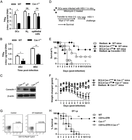
Cav-1-deficient mice are more resistant to HSV-1 infection.
To
investigate the potential role of Cav-1 in the host defense upon
infection, we initially infected wild-type (WT) C57BL/6 mice with
different doses of HSV-1 (50
l of 10
8, 10
9, and 10
9.47TCID
50
of
HSV-1) via the i.n. route, and the survival of the animals was
monitored. As shown in
Fig. 1A
, 50% of the mice died after
infec-tion with 10
9.47(3
⫻
10
9) TCID
50
of HSV-1, indicating that the
LD
50of HSV-1 was 3
⫻
10
9TCID
50in mice with the C57BL/6
background. To assess the role of Cav-1 in resistance to infection,
both WT and Cav-1
⫺/⫺mice were challenged with a lethal dose
(10 LD
50) of HSV-1, and the survival of the animals was
moni-tored. During the first 8 days after challenge, Cav-1
⫺/⫺mice had a
on August 17, 2020 by guest
http://cvi.asm.org/
higher survival rate than that of WT mice (75% in Cav-1
⫺/⫺mice
versus 0% in WT mice) (
Fig. 1B
), showing that Cav-1
⫺/⫺mice are
more resistant to HSV-1 infection.
Cav-1 deficiency correlates with reduced viral replication
and lung pathology.
Because virus replication and the consequent
tissue damage are thought to contribute to the mortality of mice
during viral infection, the influence of Cav-1 on both viral load
and histopathology was investigated. We challenged WT and
Cav-1
⫺/⫺mice with 10 LD
50
of HSV-1 via the i.n. route. On day 7
postinfection, we determined histology and viral replication in the
lungs, brain, heart, liver, spleen, and kidneys. HSV-1 did not cause
obvious tissue damage in most organs examined except the brain
and lungs (
Fig. 2A
). There was no difference between Cav-1
⫹/⫹and Cav-1
⫺/⫺mice in the degree of damage in brain sections,
indicating that less brain damage did not account for the greater
resistance of the Cav-1
⫺/⫺mice (
Fig. 2A
). However, the lungs
from Cav-1
⫺/⫺mice showed much less lung destruction than that
of the WT mice, which had substantial cell infiltration edema,
thickening of the alveolar wall, and alveolar septum capillary
con-gestion (
Fig. 2B
). Despite the greater cell infiltration observed in
the WT mouse lung sections, no major differences in total BALF
cell counts were detectable between the two groups (data not
shown). Histopathology scores were significantly lower in
Cav-1
⫺/⫺mice than those in WT mice (average score, 2.6 in Cav-1
⫺/⫺mice and 3.8 in WT mice;
Fig. 2C
). When viral load in the organs
was assessed by quantitative PCR (qPCR) at day 7 after HSV-1
infection, viral load was correlated with the histochemical results,
being barely detected in the liver, spleen, and kidneys (
Fig. 2D
).
The viral load was also remarkably less in Cav-1
⫺/⫺lung cells and
in BALF (10
1.67-fold lower), whereas no obvious difference in load
was seen in the brain at 7 dpi (
Fig. 2D
). Therefore, combined with
results shown in
Fig. 1
, Cav-1
⫺/⫺mice may have been more
resis-tant to HSV-1 infection because of enhanced elimination of virus
and milder damage in the lungs.
Cav-1 facilitates virus replication in the lungs.
Since Cav-1
may act as a gateway for the entry of viruses, such as SV40, we
tested whether the milder lung pathology in Cav-1-deficient mice
was a result of a lower entry of HSV-1. We analyzed viral load in
DNA derived from lung cells at 4 h postinfection. As illustrated in
Fig. 3A
, no significant difference in the viral load inside the lung
cells of Cav-1
⫺/⫺and WT mice was observed after i.n. infection
(
P
⫽
0.67).
On the other hand, a notable role of Cav-1 is its involvement in
virus replication (
29
). We therefore tested if Cav-1 influenced
HSV-1 replication in the lungs. To address this, mice were
chal-lenged with a lower dose of HSV-1 (1 LD
50). After 1, 3, and 5 days
of infection, the genomic DNA of lung cells and whole lung
pro-tein was collected, and we examined viral load by qPCR and
HSV-1 protein levels by Western blot. Increases in both viral load
and glycoprotein D expression in the lung cells of WT mice were
observed at 1 and 3 dpi compared to those in Cav-1
⫺/⫺mice (
Fig.
3B
and
C
). The increased viral load in Cav-1 KO mice was not
statistically significant from that of WT mice at day 3 p.i. Viruses
were also cleared in both groups by 5 dpi, probably because
C57BL/6 mice are resistant to low doses of viral infection. In
ad-dition, the viruses isolated from the challenged mice were
quanti-fied by measuring the TCID
50in Vero cells.
Figure 3D
shows that
Cav-1 expression drastically increased the viral load in the lungs
(5.33
⫾
0.44 log
10TCID
50in WT versus 3.27
⫾
0.501 log
10TCID
50in Cav-1
⫺/⫺) but not in the brain at 7 dpi. Histochemical analysis
of the expression of HSV-1 proteins showed a similar trend (
Fig.
3E
and
F
), further suggesting that Cav-1 facilitates virus
replica-tion. These results indicate that the greater resistance of Cav-1
⫺/⫺lungs to HSV-1 infection was associated with inhibition of virus
replication.
The effect of Cav-1 deficiency is dendritic cell dependent.
The
lung is one of the organs most affected by acute HSV-1 infection.
DCs, macrophages, and epithelial cells of the lung have been
re-ported to play critical roles in viral infection (
38
,
39
). Therefore,
we next examined these cellular populations, which might have
been responsible for the increased resistance of Cav-1
⫺/⫺mice.
Both WT and Cav-1
⫺/⫺mice were challenged with 10 LD
50of
HSV-1. The viral loads in isolated DCs, macrophages, and
epithe-lial cells were determined at 3 dpi. No difference in viral load was
observed in macrophages and epithelial cells between the
Cav-1
⫺/⫺and WT groups. However, DCs from Cav-1
⫺/⫺mice showed
a significantly lower viral load than that of the WT DCs (
Fig. 4A
).
To further assess this, CD11c
⫹DCs were isolated from WT and
Cav-1
⫺/⫺mice and infected at an MOI of 5 for 24 h and 48 h
in
vitro
. Viral load was substantially reduced in Cav-1
⫺/⫺DCs
com-pared to that in WT DCs (
Fig. 4B
), suggesting that DCs played a
role against HSV-1 infection, but the role was limited by the Cav-1
protein. In addition, there was little difference between WT and
Cav-1
⫺/⫺mice in the number of DCs recruited to the lungs (data
not shown), indicating Cav-1 does not influence DC migration to
the lungs. To further elucidate the function of Cav-1 expressed by
lung DCs during HSV-1 infection, a Cav-1 knockdown DC2.4 cell
line (Cav-1
kdDCs) and a Cav-1 overexpression DC cell line
(Cav-FIG 1Cav-1-deficient mice are relatively resistant to HSV-1 infection. (A) Groups of WT C57BL/6 mice (n⫽6) were challenged i.n. with HSV-1 at 1⫻
108, 1⫻109, or 3⫻109TCID
50, and the percent survival was observed until 30 dpi. (B) Groups of WT and Cav-1⫺
/⫺mice (n⫽8) were challenged with 10 LD50of HSV-1 (3⫻1010TCID50). Survival is represented according to the Mantel-Cox log rank test. The data are representative of three individual
experiments. **,P⬍0.01.
on August 17, 2020 by guest
http://cvi.asm.org/
1
overDCs) were constructed. The differential expression of Cav-1
in these cell lines was confirmed by Western blot (
Fig. 4C
). The
genetically modified DCs were infected with HSV-1 at an MOI of
5 for 2 h
in vitro
and then treated with mitomycin C at 50
g/ml
for 20 min at 37°C to prevent uncontrolled proliferation. A total of
2
⫻
10
6cells in 50
l of DMEM were adoptively transferred into
the lungs of WT or Cav-1
⫺/⫺recipients by i.t. injection. Twelve
hours later, mice were i.n. infected with 10 LD
50of HSV-1, and
FIG 2Cav-1 deficiency results in reduced viral replication and lung pathology. WT and Cav-1⫺/⫺mice were challenged with 10 LD
50of HSV-1. Next, the heart,
liver, spleen, kidneys, lungs, and brain were separated at 7 dpi.n⫽6. (A) Representative H&E-stained thin sections of organs (other than lung) from infected and uninfected mice. The bars indicate 50m or 100m. (B) Representative histopathology in the left lobe of the lung from infected and uninfected mice. Indicated are inflammatory cells infiltrating bronchial lumen (black arrows), edema and inflammatory cell infiltrations around the bronchi and vessels (white arrows), alveolar septum capillary congestion (open triangles), and inflammatory cells infiltrating alveolar space (black triangles). H&E stain was used. Scale bar indicates 100M. (C) Histological scores of the lung pathology in panel B. Severity of lung damage was assessed by the eye, scored, and analyzed. (D) Viral load in the infected organs. Total genomic DNA was extracted and the viral load assessed by qPCR. All data are representative of three independent experiments. UD, undetermined; *,P⬍0.05; **,P⬍0.01; ***,P⬍0.001.
on August 17, 2020 by guest
http://cvi.asm.org/
survival was monitored as outlined in
Fig. 4D
. The WT mice that
received Cav-1
kdDCs had a significantly increased survival of 33%
at 8 dpi, whereas WT mice that received Cav-1
overDCs were all
dead at 4 dpi (
Fig. 4E
). As HSV-1 infection might lead to body
weight loss and Cav-1
⫺/⫺mice are resistant to a lethal dose of
HSV-1 infection, weight loss in Cav-1
⫺/⫺mice was monitored
and the function of Cav-1
kdDCs evaluated during HSV-1
infec-tion. Infected Cav-1
⫺/⫺mice that received Cav-1
kdDCs also
showed growth and body weight gains similar to those of
nonin-fected Cav-1
⫺/⫺mice (
Fig. 4F
). In contrast, transfer of the
Cav-1
overDCs into infected Cav-1
⫺/⫺mice was accompanied by
sub-stantial weight loss (about 2.8 g decrease by day 11;
Fig. 4F
).
To further investigate the critical role of endogenous DCs
dur-ing HSV-1 infection, mice lackdur-ing both Cav-1 and DCs
(CD11c-DTR/Cav-1
⫺/⫺[double knockout {DKO} mice]) were generated;
the WT, CD11c-DTR, Cav-1
⫺/⫺, and DKO mice were challenged
with 10 LD
50of HSV-1, and then the CD11c
⫹DC population was
transiently depleted by DT injection. The survival of these mice
was monitored over 12 days. Although the depletion efficiency of
CD11c
⫹DCs is different in various organs, DT depletion shows a
systemic depletion effect (
40
). The efficiency of depletion of
CD11c
⫹DCs was assessed by flow cytometry at 24 h after
injec-tion. The CD11c
⫹DC population was successfully reduced to
0.014% by DT treatment (
Fig. 4G
). As shown in
Fig. 4H
, the
sur-vival rate was significantly impaired in DC-depleted DKO mice
(0% at 6 dpi) relative to Cav-1 single-deficient mice (75% at 6
dpi), and mortality rates returned to a level comparable to that of
infected CD11c-DTR mice. Our data demonstrate that DCs are
capable of, and essential for, immune defense against HSV-1
in-fection, but their antiviral function is downregulated by HSV-1 via
Cav-1.
Cav-1 suppresses NO production by DCs during HSV-1
in-fection.
Since DCs lacking Cav-1 might confer resistance to
HSV-1 infection, we next explored which cytokines or other
me-diators were involved. The levels of IL-6 and tumor necrosis factor
alpha (TNF-
␣
) in BALF were tested at days 0 and 4 postinfection.
However, HSV-1 stimulated similar production of IL-6 and
TNF-
␣
in both WT and Cav-1
⫺/⫺mice at 4 dpi (
Fig. 5A
). When
the mRNA levels of antiviral cytokines and signaling molecules
(IFN-

, IL-1

, IL-10, CD40, COX-2, PD-L1, ERK1, eNOS, and
SOCS-1) in HSV-1-infected lung cells were measured at 1 and 3
dpi, no significant changes were observed, except for iNOS (
Fig.
5B
and
C
). iNOS, as an antiviral factor, is potently induced in
virus-infected cells. We found that the mRNA level of iNOS was
significantly augmented in Cav-1
⫺/⫺lung cells but slowed in WT
lung cells at 1 dpi (
P
⫽
0.0015) and 3 dpi (
P
⫽
0.0168), as depicted
in
Fig. 5C
. Consistent with this, the iNOS mRNA level was also
more strongly increased
in vitro
in freshly purified Cav-1
⫺/⫺DCs
than in WT DCs at 12, 24, and 36 hpi (
Fig. 5D
). Furthermore, as
shown in
Fig. 5E
, Cav-1
kdDCs had a much greater abundance of
FIG 3Cav-1 facilitates virus replication in lung cells. (A) Viral load in WT and Cav-1⫺/⫺lung cells 4 h after infection with 1 LD50in vitro. The Pol gene content
in total extracted DNA was determined by qPCR analysis. (B and C) Viral load in infected mice. Cells were separated from WT and Cav-1⫺/⫺mice at 0, 1, 3, and 5 days after infection with 1 LD50of HSV-1. (B) Viral load in lung cells assessed by qPCR. (C) Amount of HSV-1 glycoprotein D in pooled whole-cell lysates of
isolated lung and brain cells. Immunoblots of lysates were stained with Abs specific for glycoprotein D or GAPDH as a reference. Total proteins of HSV-1-infected Vero cells were loaded as a control. KO refers to Cav-1⫺/⫺mice. (D to F) Results from mice that were challenged with 10 LD
50of HSV-1 and examined at 7 dpi.
(D) Viral load in lungs and brain quantified as TCID50in Vero cells according to the readout at day 4. (E and F) Representative immunohistochemical staining
of HSV-1 in lung (E) and brain (F) sections. The scale bars on the sections indicate 100M for lungs and 50M for brain. The data are representative of three independent experiments. Student’sttest was applied for differences between two groups. Two-way ANOVA was also applied. Nonsignificant (ns),P⬎0.05; *,
P⬍0.05; **,P⬍0.01; ***,P⬍0.001.
on August 17, 2020 by guest
http://cvi.asm.org/
FIG 4Cav-1⫺/⫺-enhanced survival is DC dependent. (A) Viral load in DCs, macrophages (M), and epithelial cells from HSV-1-infected WT and Cav-1⫺/⫺ mice. Mice were challenged with 10 LD50HSV-1, cells were collected from the lungs at 3 dpi, genomic DNA was extracted, and the viral load was assessed by
qPCR. (B) Viral load in DCs that were infectedin vitro. DCs were isolated from WT and Cav-1⫺/⫺mice with the CD11c MicroBeads kit, infected with HSV-1 at an MOI of 5, and the total DNA was extracted at 24 hpi and 48 hpi for qPCR. (C) Cav-1 protein expression levels in recombinant DC2.4 cell lines. Cav-1 knockdown (Cav-1kd) and Cav-1 overexpression (Cav-1over) DC2.4 cell lines were constructed. Thirty micrograms of whole-cell extract was subjected to
electrophoresis on an SDS-PAGE gel, transferred to a PVDF membrane, and the Cav-1 protein was assessed by Western blotting, with-actin as control. (D) Outline protocol of i.t. adoptive transfer of Cav-1kdDCs and Cav-1overDCs. (E and F) Effect of caveolin-1 on resistance of mice to challenge with 10 LD
50of
HSV-1. (E) Survival curves. Survival was enhanced in caveolin-1 KO mice and in mice receiving i.t. DC2.4 in which caveolin-1 expression was reduced (Cav-1kd).
Survival was decreased in mice receiving DC2.4 overexpressing caveolin-1 (Cav-1over). The reference mice received i.t. DMEM.n⫽6 mice per group. (F) Body
weight change curves. Weight was determined daily after a lethal dose of HSV-1 in Cav-1⫺/⫺mice that received recombinant DC2.4 lines i.t. The reference Cav-1⫺/⫺mice received medium only and were not infected. (G) Efficiency of DC depletion by DT in CD11c-DTR mice. DT was injected i.p. in 8-ng/g doses at 12-h intervals. Peripheral blood mononuclear cells (PBMC) were collected at 24 hpi and stained with CD11c-PercP-Cy5.5. Next, flow cytometry was performed to detect the percentage of DCs. (H) Resistance of Cav-1⫺/⫺mice after DC depletion. WT, CD11c-DTR, Cav-1⫺/⫺, and DKO mice were pretreated with DT and i.n. challenged with 10 LD50of HSV-1.n⫽6. DT injections were performed at 12-h intervals during infection. **, Cav-1⫺/⫺versus DKO. The results are
presented as the mean⫾SEM from three independent experiments. For survival curve analysis, a log rank (Mantel-Cox) test was performed. Nonsignificant (ns),
P⬎0.05; *,P⬍0.05; **,P⬍0.01.
on August 17, 2020 by guest
http://cvi.asm.org/
iNOS protein than did WT DCs at 24, 48, and 72 hpi
in vitro
, and
at 48 hpi, the release of NO from Cav-1
⫺/⫺DCs was 1.56-fold
greater than that from the WT cells (
Fig. 5F
). The dynamics of NO
release were associated with the change in expression of Cav-1.
The DCs with silenced Cav-1 exhibited a peak production of NO
at
⬃
30
M at 24 hpi, whereas the WT counterpart peak was at 16.7
M, and the peak for Cav-1-overexpressed DCs was at 8.6
M
(
Fig. 5G
), indicating that the degree of NO release was strongly
associated with the level of Cav-1 expression. To further confirm
the retardation of the expression of NO by Cav-1, the NO levels in
serum and BALF were analyzed. In agreement with the results
in
vitro
, there was significantly less NO at 0.5, 1, and 4 dpi in the
serum and in BALF of WT mice than that in the Cav-1
⫺/⫺coun-terparts (
Fig. 5H
). Furthermore, as Cav-1 regulated the
produc-tion of NO during HSV-1 infecproduc-tion, the penetraproduc-tion of virus entry
in Cav-1-deficient DCs needs to be evaluated. The results showed
the virus entry into DCs was not significantly changed in Cav-1
knockdown DCs at an MOI of 0.5, 1, and 5 (data not shown),
FIG 5Cav-1 suppresses NO production by DCs during HSV-1 infection. (A to C) Cytokine and NO production in lungs of infected WT and Cav-1⫺/⫺mice.
Mice were infected with 10 LD50HSV-1, and BALF was collected 0 to 4 dpi. (A) IL-6 and TNF-␣in BALF quantified with the cytometric bead assay with specific
antibody-conjugated beads. (B and C) Inflammatory cytokine, chemokine, and iNOS expression in BAL lung cells. Cells were collected at 0, 1, and 3 dpi, total RNA was extracted, and mRNA expression was assessed by qPCR. The fold induction relative to GAPDH is shown. (B) Expression of cytokines and chemokines. (C) Expression of iNOS. (D) Expression of iNOS in CD11c⫹DCs purified from WT and Cav-1⫺/⫺mice and then infected with HSV-1 at an MOI of 5. The fold changes of iNOS expressions were normalized to GAPDH. (E) iNOS protein increase in HSV-1-infected Cav-1kdDCs and not in WT DCs. Total cell protein was
extracted for Western blot analysis at 0, 24, 48, and 72 hpi. iNOS and GAPDH expression levels were detected with specific antibodies. (F) NO levels in supernatants of CD11c⫹DCs that had been purified from WT and Cav-1⫺/⫺mice and infected with HSV-1 at an MOI of 5. (G) NO levels in supernatants of WT (DC2.4), Cav-1kd, and Cav-1overDCs after infection with HSV-1 at an MOI of 5. (H) NO levels in serum and BALF of 10 LD
50infected WT and Cav-1⫺ /⫺mice. In each panel, the data shown are representative of three independent experiments. Nonsignificant (ns),P⬎0.05; *,P⬍0.05; **,P⬍0.01; ***,P⬍0.001.
on August 17, 2020 by guest
http://cvi.asm.org/
which indicated that Cav-1-mediated HSV-1 entry into DCs
re-sulted in the impairment of NOS2 activity by mechanisms that
were Cav-1 dependent. Collectively, these findings indicate that
Cav-1 suppresses NO production by DCs during HSV-1 infection,
which may be responsible for the greater susceptibility of WT mice
to HSV-1 infection.
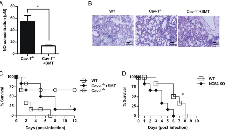
Inhibition of iNOS with SMT abolishes the resistance of
Cav-1
ⴚ/ⴚmice to HSV-1 challenge.
Since NO was reported to inhibit
HSV-1 infection (
17
), lower production of NO might account for
greater viral reproduction and exacerbated mortality in WT
com-pared to that in Cav-1
⫺/⫺mice. We found that daily i.p.
admin-istration of SMT (iNOS inhibitor) to Cav-1
⫺/⫺mice during the
course of HSV-1 infection resulted in a significantly smaller
amount of NO in serum at 48 hpi (
Fig. 6A
). In keeping with
pre-vious data, the lungs of SMT-treated Cav-1
⫺/⫺mice exhibited
pathological changes of greater severity than that in the
non-treated Cav-1
⫺/⫺mice (
Fig. 6B
). Consistent with the histological
data, this inhibition of iNOS with SMT nearly abolished the
Cav-1
⫺/⫺-mediated protection against HSV-1 infection (
P
⫽
0.03)
(
Fig. 6C
). Furthermore, mice lacking iNOS (NOS2 KO) were
sig-nificantly more susceptible to lethal HSV-1 infection than WT
(iNOS
⫹/⫹) mice (
P
⫽
0.019) (
Fig. 6D
). We conclude that NO is
involved in the resistance of Cav-1
⫺/⫺mice and alleviates the lung
pathology generated by HSV-1 infection.
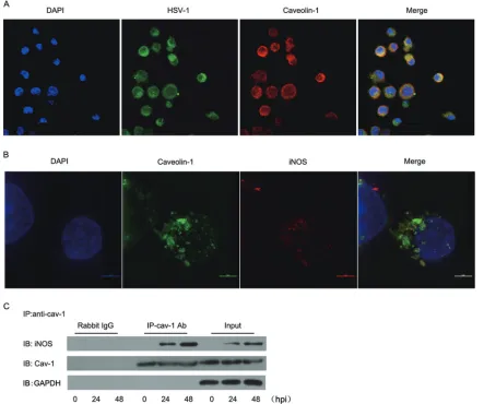
Colocalization of HSV-1 with Cav-1 and iNOS in caveolae.
Since Cav-1 modulated iNOS activity in HSV-1-infected DCs, we
investigated the subcellular localization of these proteins.
Confo-cal microscopy showed that 2 h after infection of DCs, HSV-1
colocalized with Cav-1 in caveolae, the cytoplasm, and
mem-branes (
Fig. 7A
). In addition, HSV-1-induced iNOS was
ob-served to colocalize with Cav-1 in caveolae at 24 hpi (
Fig. 7B
).
Furthermore, immunoprecipitation indicated that there might
be protein-protein interactions between Cav-1 and iNOS in the
caveolae and that this interaction increases as infection
pro-gresses (
Fig. 7C
).
DISCUSSION
Since Cav-1 modulates HSV-1 replication in infected cells, it
ap-pears to be an important component of cell defense. However, the
molecular cross talk that mediates the effect of Cav-1 on antiviral
activity is largely unknown. In this study, we uncovered a novel
mechanism exploited by HSV-1, using Cav-1 to disarm the
pro-duction by DCs of the antiviral factor NO. We showed that
defi-ciency of Cav-1 protected mice from fatal HSV-1 infection,
result-ing in much milder lung morphological changes and a lower level
of viral load. The resistance of Cav-1
⫺/⫺mice against virus
infec-tion was DC dependent. Mechanistic studies demonstrated that
the functions of Cav-1 include negatively regulating the
produc-tion of iNOS and NO in DCs. Depleproduc-tion of CD11c
⫹DCs in
Cav-1
⫺/⫺mice or iNOS inhibition by SMT significantly impaired the
production of NO and diminished the resistance conferred by the
Cav-1
⫺/⫺state. Furthermore, we observed that Cav-1 expression
colocalized with iNOS and sequestered together with virus in
caveolae in HSV-1-infected DCs. Therefore, this study identified a
previously unrecognized role of Cav-1 during HSV-1 infection in
the lungs. We demonstrated for the first time that Cav-1 facilitates
HSV-1 infection and leads to lung injury and mortality by
retar-dation of the NO response in the lung DCs.
Although caveolae are best known as an important regulatory
element for lipid metabolism, recent studies suggested that
cave-olae, and particularly the Cav-1 structural protein, can be involved
in the internalization of pathogens, signal transduction, and host
defenses (
19
,
26
). Indeed, previous studies have demonstrated
that Cav-1 facilitates H1N1 and HBV virus replication in cell
cul-FIG 6Decreased NO and increased mortality in SMT-treated Cav-1⫺/⫺mice. Mice were challenged by 10 LD
50of HSV-1 i.n. and given daily i.p. injections of
the iNOS inhibitor (50 mg/kg), as described in Materials and Methods. (A) Serum levels of NO in the SMT-treated and nontreated Cav-1⫺/⫺mice. (B) Histopathology in lung sections stained with H&E stain. The scale bar is 100m. (C) Percentage of survival. *,P⬍0.05 between SMT-treated and nontreated Cav-1⫺/⫺groups.n⫽6. (D) Survival of iNOS KO mice and WT mice after i.n. challenge with 10 LD
50of HSV-1. The mice were observed daily. The data are
representative of two independent experiments. *,P⬍0.05.
on August 17, 2020 by guest
http://cvi.asm.org/
ture (
29
,
30
), and Cav-1 was found to regulate cytokine expression
in macrophages via the p38/mitogen-activated protein kinase
(MAPK) pathway (
41
). However, the role of Cav-1 in herpesvirus
infection is largely unclear. To investigate this, we first established
an i.n. HSV-1 challenge system in C57BL/6 WT and Cav-1
⫺/⫺mice and found that the induction of significant mortality caused
by i.n. infection required a dose of 9.47 log
10TCID
50of HSV-1 in
WT mice (
Fig. 1A
), although only 8.47 log
10TCID
50was required
by intraperitoneal (i.p.) challenge (data not shown). WT animals
that were heavily challenged i.n. with 10 LD
50of HSV-1 all died,
while Cav-1
⫺/⫺mice exhibited 75% protection (
Fig. 1B
),
reveal-ing that Cav-1 played a suppressive role in host immunity durreveal-ing
HSV-1 infection via the i.n. route.
HSV-1 spreads to the brain, lungs, liver, spleen, and other
tis-sues after i.n. infection, causing encephalitis, pneumonia,
hepati-tis, and keratitis (
42
). Although it mainly infects the nervous
system and induces encephalitis in mice, HSV-1-induced
pneu-monia is common in immunocompromised patients, newborns,
healthy persons, and mice (
5
,
7
). Accordingly, when we used a
high titer of HSV-1 (10 LD
50) to infect mice via the i.n. route, this
resulted in severe disease in the lungs, with edema formation and
alveolar septum capillary congestion. In contrast, in Cav-1
⫺/⫺mice, there was remarkably less pneumonitis induced by the
HSV-1 infection, consistent with their increased survival (
Fig. 2B
and
C
). Previous studies noted that both imbalanced host
im-mune responses and viral pathogenic factors are critical for
virus-induced pneumonia (
4
,
43
). In agreement with evidence that
Cav-1 might facilitate virus replication (
29
), we observed that the
expression of Cav-1 significantly elevated HSV-1 titers in lung
cells and BALF. Interestingly, although the i.n. infection by HSV-1
also induced encephalitis in the brain, there were no significant
differences between the Cav-1
⫺/⫺and WT mice in either degree of
pathology (
Fig. 2A
) or viral load (
Fig. 3D
to
F
). The different effect
of Cav-1 on organs (brain versus lungs) might due to the
ex-tremely high dose of HSV-1 and the route of infection (intranasal
injection). The different expression of Cav-1 in epithelial cells of
FIG 7Cav-1 sequesters iNOS in caveolae in HSV-1-infected DCs. (A) Localization of HSV-1 in caveolae. DC2.4 cells were infected at an MOI of 50 and stained
at 2 hpi with goat anti-HSV-1 and rabbit anti-Cav-1 antibodies. Confocal microscopy was performed after secondary goat-anti-rabbit IgG-PE and bovine anti-goat-FITC antibody staining. (B) Localization of iNOS in caveolae. DC2.4 cells were infected at an MOI of 5 and stained 24 hpi with goat anti-iNOS antibody, followed by bovine anti-goat-PE. Bar, 5m. (C) Coimmunoprecipitation (Co-IP) of Cav-1 with iNOS. DCs were infected with HSV-1 at an MOI of 5 and lysed at different times with IP/wash lysis buffer. The lysate contents of iNOS, Cav-1, and GAPDH proteins were determined by Western blot analysis with specific Abs. Input, lysate before immunoprecipitation; IB, immunoblot; IP-Cav-1 Ab, immunoprecipitate; rabbit IgG, negative control. Similar results were obtained in two additional independent experiments.
on August 17, 2020 by guest
http://cvi.asm.org/
lung and brain cells may also contribute to the differences, and this
needs to be investigated further. We concluded that the lungs were
selectively susceptible after i.n. infection and that the pathology
and viral load were enhanced by Cav-1 availability. The HSV-1
infection-induced pneumonitis in WT C57BL/6 mice was
there-fore adopted as a model system to further define the role of Cav-1.
Although the mechanisms and types of cells mediating
resis-tance against HSV-1 infection in lungs are ill defined, published
studies suggest that certain cell subsets of the innate immune
sys-tem may be involved in controlling virus replication (
44
,
45
). DCs,
macrophages, and epithelial cells form the first line of host defense
in lungs against invading pathogens, like HSV-1, lymphocytic
choriomeningitis virus (LCMV), and influenza A (
43
,
46–48
). In
the case of RSV-induced pneumonia, adoptive transfer of DCs
contributed to a reduction of RSV and Sindbis virus titers, with
limited virus replication and airway hyperresponsiveness (
49
,
50
).
Previous studies of HSV-1 infection showed that an ablation of
CD11c
⫹DCs
in vivo
increased susceptibility to infection (
47
), but
the role that DCs play during HSV-1 infection required
clarifica-tion. Our results shown in
Fig. 4A
and
B
clearly show that the
absence of Cav-1 significantly suppressed infection in lung DCs
but not in lung macrophages or epithelial cells. Studies showed
iCD8
⫹DCs isolated from
Mycobacterium bovis
BCG-infected
mice were adoptively transferred to mice and both enhanced
bac-terial clearance and reduced pathological reactions following
chal-lenge. But the noninfected CD8 DC control and PBS sham
treat-ment exhibited no improvetreat-ment in bacterial load in the lungs
(
51
). In addition, DC pretreatment is able to initiate T-cell
im-mune responses against malaria and viruses in mice (
52
,
53
). In
the experiment outlined in the
Fig. 4
, DCs were preinfected with
HSV-1 before adoptive transfer. The importance of the DCs was
further supported by our adoptive transfer experiments:, as WT
mice that received the Cav-1
kdDCs showed better survival at 8 dpi
(33%) than that of mice that received Cav-1
overDC (0% survival at
4 dpi;
Fig. 4E
). Mitomycin C was applied to avoid unlimited
DC2.4 cell proliferation before the transfer (
54
). Previous studies
showed that mitomycin C treatment blocked cellular DNA
inte-gration but did not affect Visna virus and SV40 replication (
55
,
56
). In addition, the extent of T-cell stimulation by mitomycin
C-treated DCs is dose dependent, indicating that the function of
mitomycin C is diverse and needs to be studied in the future (
57
).
The depletion of DCs in Cav-1
⫺/⫺mice also abolished the
protec-tive effect that was conferred by the absence of Cav-1 (
Fig. 4H
).
Our findings indicated that HSV-1 exploits Cav-1 to disarm the
antiviral effects of DCs in lungs.
Cells in the innate immune system are equipped with antiviral
molecules for the clearance of invading pathogens.
Proinflamma-tory and inflammaProinflamma-tory cytokines are key regulators of the innate
cellular defense against viral infections and might be the
deter-mining factors against respiratory infections, including HSV-1,
H1N1, and respiratory syncytial virus (RSV) (
58–60
). NO is a
product of the cellular immune system that is well known as a
reactive free radical molecule that modulates cytokine expression,
and it is beneficial to the host defense against virus infections (
61
).
For example, cells with iNOS inhibited by 1-NG-monomethyl
ar-ginine (1-NMMA) failed to restrict Japanese encephalitis virus
(JEV) replication (
62
), and IFN-
␥
-induced iNOS and NO
produc-tion inhibited the replicaproduc-tion of vaccinia virus (VV) and HSV-1
(
17
,
63
). Our observations implicating NO in resistance to HIV-1
are consistent with this. We observed that the deficiency of Cav-1
markedly enhanced iNOS and NO production in BALF and in
DCs during HSV-1 infection (
Fig. 5
). In addition, lower levels of
NO were observed in infected DC-depleted mice (data not shown)
and in the serum from SMT-treated Cav-1
⫺/⫺mice (
Fig. 6A
) than
those in Cav-1
⫺/⫺mice and are associated with higher mortality.
Thus, consistent with a general antiviral role for iNOS, NO from
lung DCs appears to be the key to resistance to HSV-1 infection.
Furthermore, the connection between Cav-1 and iNOS has been
documented in cells responding to LPS stimulation and in a
tu-morigenesis model (
32
,
34
). Based on these observations, we
hy-pothesized that HSV-1 disarms DCs by using Cav-1 to reduce
iNOS production of NO.
Although IL-6 and TNF-
␣
have been reported to play critical
roles in immunity against HSV-1 (
64
), in this study, the deficiency
in Cav-1 resulted in only slightly upregulated the expression of
IL-6 and TNF-
␣
in BALF, and this was not statistically significant
(
Fig. 5A
). On the other hand, we did observe that Cav-1
upregu-lated transforming growth factor beta 1 (TGF-

1) expression in
total lung cells (data not shown). Because TGF-

1 is a suppressive
factor during immune responses, it may repress the expression of
iNOS or other antiviral molecules and thereby contribute to virus
replication (
65
). Whether Cav-1 regulation of TGF-

1, IL-6, or
TNF-
␣
is involved in resistance to HSV-1 infection in lungs needs
further study.
We propose that HSV-1-induced NO production was
sup-pressed through iNOS sequestration in caveolae. Our
observa-tions that Cav-1 both suppressed iNOS expression and colocalized
with iNOS and HSV-1 in infected DC cells (
Fig. 7B
and
C
) is
consistent with recent reports: in an LPS-induced sepsis model,
Cav-1 downregulated LPS-induced iNOS and NO production,
resulting in aggravated lung edema formation (
34
,
41
); in a
hu-man colon carcinoma model, Cav-1 was found to cofractionate
with iNOS and detain iNOS protein in caveolae, leading to iNOS
proteolysis (
32
). Since there is also evidence that SV40 uses
cave-olae as a gateway to mediate viral entry and avoid host antiviral
responses (
27
,
66
), it appears that HSV-1 may use such a gateway
to cause sequestration and the consequent inactivation of iNOS in
the caveolae of lung DCs.
In sum, in this study, we demonstrate that Cav-1 plays a key
role in susceptibility to HSV-1 infection, with higher viral load
and aggravated lung pathology occurring in its presence; a lack of
Cav-1 reversed this susceptibility and provided stronger
protec-tion against HSV-1 infecprotec-tion. Notably, NO and DCs were found to
be critically important to the host defense against HSV-1 infection
in Cav-1
⫺/⫺mice. Thus, this study provides a new insight into a
novel immunity evasion mechanism of HSV-1 and might indicate
a valuable approach to controlling herpesvirus infection.
ACKNOWLEDGMENTS
This work was supported in part by the National Science Foundation of China (31430027 and 30930068), MOST national 863 project of China (2012AA02A407), and the National Science and Technology Major Pro-gram of Infectious Diseases (2013ZX10002001) to Bin Wang.
We thank Jane Q. L. Yu, Zhonghuai He, and Xianghua Shi for their assistance in this work.
REFERENCES
1.Qiu X, Zhong M, Xiang Y, Qu C, Pei Y, Zhang Y, Yang C, Gasteiger J,
Xu J, Liu Z, Wang Y.2013. Self-organizing maps for the classification of
gallic acylate polyphenols as HSV-1 inhibitors. Med Chem10:388 – 401.
on August 17, 2020 by guest
http://cvi.asm.org/
Trouillet JL, Capron F, Agut H, Gibert C, Chastre J. 2007. Herpes simplex virus lung infection in patients undergoing prolonged mechanical ventilation. Am J Respir Crit Care Med175:935–942.http://dx.doi.org/10 .1164/rccm.200609-1322OC.
6.Costa C, Sidoti F, Saldan A, Sinesi F, Balloco C, Simeone S, Lorusso M,
Mantovani S, Merlino C, Solidoro P, Cavallo R.2012. Clinical impact of
HSV-1 detection in the lower respiratory tract from hospitalized adult patients. Clin Microbiol Infect18:E305–E307.http://dx.doi.org/10.1111/j .1469-0691.2012.03882.x.
7.Adler H, Beland JL, Del-Pan NC, Kobzik L, Brewer JP, Martin TR,
Rimm IJ.1997. Suppression of herpes simplex virus type 1
(HSV-1)-induced pneumonia in mice by inhibition of inducible nitric oxide syn-thase (iNOS, NOS2) J Exp Med185:1533–1540.
8.de Jong MA, de Witte L, Bolmstedt A, van Kooyk Y, Geijtenbeek TB.
2008. Dendritic cells mediate herpes simplex virus infection and transmis-sion through the C-type lectin DC-SIGN. J Gen Virol89:2398 –2409.http: //dx.doi.org/10.1099/vir.0.2008/003129-0.
9.Cook WJ, Kramer MF, Walker RM, Burwell TJ, Holman HA, Coen
DM, Knipe DM.2004. Persistent expression of chemokine and
chemo-kine receptor RNAs at primary and latent sites of herpes simplex virus 1 infection. Virol J1:5.http://dx.doi.org/10.1186/1743-422X-1-5.
10. Frank GM, Buela KA, Maker DM, Harvey SA, Hendricks RL.2012.
Early responding dendritic cells direct the local NK response to control herpes simplex virus 1 infection within the cornea. J Immunol188:1350 – 1359.http://dx.doi.org/10.4049/jimmunol.1101968.
11. Kassim SH, Rajasagi NK, Zhao X, Chervenak R, Jennings SR.2006.In
vivoablation of CD11c-positive dendritic cells increases susceptibility to herpes simplex virus type 1 infection and diminishes NK and T-cell re-sponses. J Virol80:3985–3993.http://dx.doi.org/10.1128/JVI.80.8.3985 -3993.2006.
12. Kohno S, Murata T, Sugiura A, Ito C, Iranshahi M, Hikita K, Kaneda
N.2011. Methyl galbanate, a novel inhibitor of nitric oxide production in mouse macrophage RAW264.7 cells. J Nat Med65:353–359.http://dx.doi .org/10.1007/s11418-010-0505-7.
13. Lu L, Bonham CA, Chambers FG, Watkins SC, Hoffman RA, Simmons
RL, Thomson AW.1996. Induction of nitric oxide synthase in mouse
dendritic cells by IFN-gamma, endotoxin, and interaction with allogeneic T cells: nitric oxide production is associated with dendritic cell apoptosis. J Immunol157:3577–3586.
14. Vallance P, Moncada S.1993. Role of endogenous nitric oxide in septic
shock. New Horiz1:77– 86.
15. Croen KD.1993. Evidence for antiviral effect of nitric oxide. Inhibition of herpes simplex virus type 1 replication. J Clin Invest91:2446 –2452.
16. Imanishi N, Andoh T, Sakai S, Satoh M, Katada Y, Ueda K, Terasawa
K, Ochiai H.2005. Induction of inducible nitric oxide (NO) synthase
mRNA and NO production in macrophages infected with influenza A/PR/8 virus and stimulated with its ether-split product. Microbiol Im-munol49:41– 48.http://dx.doi.org/10.1111/j.1348-0421.2005.tb03638.x.
17. Gamba G, Cavalieri H, Courreges MC, Massouh EJ, Benencia F.2004.
Early inhibition of nitric oxide production increases HSV-1 intranasal infection. J Med Virol73:313–322.http://dx.doi.org/10.1002/jmv.20093.
18. Tucker PC, Griffin DE, Choi S, Bui N, Wesselingh S.1996. Inhibition of
nitric oxide synthesis increases mortality in Sindbis virus encephalitis. J Virol70:3972–3977.
19. Stuart ES, Webley WC, Norkin LC.2003. Lipid rafts, caveolae,
caveo-lin-1, and entry by chlamydiae into host cells. Exp Cell Res287:67–78. http://dx.doi.org/10.1016/S0014-4827(03)00059-4.
20. Volonté D, Galbiati F, Pestell RG, Lisanti MP. 2001. Cellular stress
induces the tyrosine phosphorylation of caveolin-1 (Tyr(14)) via activa-tion of p38 mitogen-activated protein kinase and c-Src kinase. Evidence for caveolae, the actin cytoskeleton, and focal adhesions as mechanical sensors of osmotic stress. J Biol Chem276:8094 – 8103.
/10.4161/cc.9.11.11848.
23. Li J, Geng S, Xie X, Liu H, Zheng G, Sun X, Zhao G, Wan Y, Wu Y,
Chen X, Zhong Y, Wang B.2012. Caveolin-1-mediated negative
signal-ing plays a critical role in the induction of regulatory dendritic cells by DNA and protein coimmunization. J Immunol189:2852–2859.http://dx .doi.org/10.4049/jimmunol.1102828.
24. Huang WR, Wang YC, Chi PI, Wang L, Wang CY, Lin CH, Liu HJ.
2011. Cell entry of avian reovirus follows a caveolin-1-mediated and dy-namin-2-dependent endocytic pathway that requires activation of p38 mitogen-activated protein kinase (MAPK) and Src signaling pathways as well as microtubules and small GTPase Rab5 protein. J Biol Chem286: 30780 –30794.http://dx.doi.org/10.1074/jbc.M111.257154.
25. Van Gorp H, Van Breedam W, Delputte PL, Nauwynck HJ.2009. The
porcine reproductive and respiratory syndrome virus requires trafficking through CD163-positive early endosomes, but not late endosomes, for productive infection. Arch Virol 154:1939 –1943. http://dx.doi.org/10 .1007/s00705-009-0527-1.
26. Norkin LC, Kuksin D.2005. The caveolae-mediated sv40 entry pathway
bypasses the golgi complex en route to the endoplasmic reticulum. Virol J 2:38.http://dx.doi.org/10.1186/1743-422X-2-38.
27. Pelkmans L, Kartenbeck J, Helenius A.2001. Caveolar endocytosis of
simian virus 40 reveals a new two-step vesicular-transport pathway to the ER. Nat Cell Biol3:473– 483.http://dx.doi.org/10.1038/35074539.
28. Beer C, Andersen DS, Rojek A, Pedersen L.2005. Caveola-dependent
endocytic entry of amphotropic murine leukemia virus. J Virol79:10776 – 10787.http://dx.doi.org/10.1128/JVI.79.16.10776-10787.2005.
29. Sun L, Hemgård GV, Susanto SA, Wirth M.2010. Caveolin-1 influences
human influenza A virus (H1N1) multiplication in cell culture. Virol J 7:108.http://dx.doi.org/10.1186/1743-422X-7-108.
30. Macovei A, Radulescu C, Lazar C, Petrescu S, Durantel D, Dwek RA,
Zitzmann N, Nichita NB.2010. Hepatitis B virus requires intact
caveo-lin-1 function for productive infection in HepaRG cells. J Virol84:243– 253.http://dx.doi.org/10.1128/JVI.01207-09.
31. Chidlow JH, Jr, Sessa WC.2010. Caveolae, caveolins, and cavins:
com-plex control of cellular signalling and inflammation. Cardiovascular re-search86:219 –225.http://dx.doi.org/10.1093/cvr/cvq075.
32. Felley-Bosco E, Bender FC, Courjault-Gautier F, Bron C, Quest AF.
2000. Caveolin-1 down-regulates inducible nitric oxide synthase via the proteasome pathway in human colon carcinoma cells. Proc Natl Acad Sci U S A97:14334 –14339.http://dx.doi.org/10.1073/pnas.250406797.
33. Felley-Bosco E, Bender F, Quest AF.2002. Caveolin-1-mediated
post-transcriptional regulation of inducible nitric oxide synthase in human colon carcinoma cells. Biol Res35:169 –176.
34. Garrean S, Gao XP, Brovkovych V, Shimizu J, Zhao YY, Vogel SM,
Malik AB.2006. Caveolin-1 regulates NF-kappaB activation and lung
inflammatory response to sepsis induced by lipopolysaccharide. J Immu-nol177:4853– 4860.http://dx.doi.org/10.4049/jimmunol.177.7.4853.
35. Geng S, Zhong Y, Wang S, Liu H, Zou Q, Xie X, Li C, Yu Q, He Z,
Wang B.2012. Amiloride enhances antigen specific CTL by faciliting [sic]
HBV DNA vaccine entry into cells. PLoS One7:e33015.http://dx.doi.org /10.1371/journal.pone.0033015.
36. Jung S, Unutmaz D, Wong P, Sano G, De los Santos K, Sparwasser T,
Wu S, Vuthoori S, Ko K, Zavala F, Pamer EG, Littman DR, Lang RA. 2002.In vivodepletion of CD11c⫹dendritic cells abrogates priming of CD8⫹T cells by exogenous cell-associated antigens. Immunity17:211– 220.http://dx.doi.org/10.1016/S1074-7613(02)00365-5.
37. Wu B, Zou Q, Hu Y, Wang B. 2013. Interleukin-22 as a molecular
adjuvant facilitates IL-17-producing CD8⫹T-cell responses to HBV DNA vaccine in mice. Hum Vaccin Immunother9:2133–2141.
38. Guilliams M, Lambrecht BN, Hammad H.2013. Division of labor
be-tween lung dendritic cells and macrophages in the defense against