(PRRS) Modified Live Virus Vaccine Followed by Challenge with
PRRS Virus and Porcine Circovirus Type 2 (PCV2) Protects against
PRRS but Enhances PCV2 Replication and Pathogenesis Compared to
Results for Nonvaccinated Cochallenged Controls
Megan C. Niederwerder,aBhupinder Bawa,aNick V. L. Serão,bBenjamin R. Trible,aMaureen A. Kerrigan,aJoan K. Lunney,c Jack C. M. Dekkers,bRaymond R. R. Rowlanda
Department of Diagnostic Medicine/Pathobiology, College of Veterinary Medicine, Kansas State University, Manhattan, Kansas, USAa; Department of Animal Science, Iowa State University, Ames, Iowa, USAb; U.S. Department of Agriculture, Agricultural Research Service, Beltsville Agricultural Research Center, Beltsville, Maryland, USAc
Coinfections involving porcine reproductive and respiratory syndrome virus (PRRSV) and porcine circovirus type 2 (PCV2)
contribute to a group of disease syndromes known as porcine circovirus-associated disease (PCVAD). Presumably, PRRSV
infec-tion enhances PCV2 replicainfec-tion as a result of modulainfec-tion of host immunity. The purpose of this study was to evaluate PCV2
rep-lication and pathogenesis in pigs vaccinated with a PRRS modified live virus (MLV) vaccine and subsequently challenged with a
combination of PRRSV and PCV2. During the early postchallenge period, the number of pigs with PRRSV-associated clinical
signs was decreased, and average daily gain (ADG) was increased, in the vaccinated group, demonstrating the protective effect of
PRRS vaccination. However, during the later postchallenge period, more pigs in the vaccinated group showed increased PCV2
viremia, decreased ADG, increased PCVAD clinical signs, and increased mortality. In this disease model, the early benefits of
PRRSV vaccination were outweighed by the later amplification of PCVAD.
P
orcine circovirus type 2 (PCV2), a single-stranded DNA virus
in the family
Circoviridae
, contributes to a group of
syn-dromes collectively termed porcine circovirus-associated disease
(PCVAD) (
1
). Two important clinical syndromes associated with
PCVAD are PCV2-associated pneumonia and postweaning
mul-tisystemic wasting syndrome (PMWS) (
1
,
2
). Management of
PCV2 through the use of inactivated and subunit vaccines has led
to the effective control of PCVAD in North America and Europe.
However, the emergence of new PCV2 strains and the lack of
PCV2 vaccination programs in other countries create an
uncer-tain future for continued disease control.
Porcine reproductive and respiratory syndrome virus (PRRSV) is
a single-stranded RNA virus in the family
Arteriviridae
(
3
,
4
). For
the past 20 years, PRRSV has remained the most costly disease
affecting swine production worldwide (
5
). PRRSV infection
con-tributes to a number of immunological outcomes that increase the
susceptibility of the host to secondary infections by primary and
secondary pathogens (
6–8
). PRRSV is frequently isolated along
with PCV2 (
9
) and is one of the major cofactors linked with
in-creasing PCV2 replication and pathogenesis (
10–12
). Previous
work by us and others has shown that a principal contribution of
PRRSV is to increase PCV2 viremia (
13
). Increased PCV2
replica-tion is likely the result of immune stimulareplica-tion that results in more
PCV2-permissive cells combined with PRRSV-induced
immuno-modulation. The complex etiology of PCVAD, including the role
of PRRSV infection, has yet to be fully understood. In an extensive
body of work, we identified the aberrant recognition of a
nonneu-tralizing decoy epitope on the PCV2 capsid protein (CP) as a
con-tributing factor in PCVAD immunopathogenesis. Natural PCV2
infection of a population produces a mixture of pigs that
recog-nize the decoy and neutralizing epitopes, which may explain why
only a subpopulation of infected pigs goes on to develop PCVAD
(
13–15
).
In this study, we took advantage of a host genetics study to
evaluate clinical and virological outcomes after experimental
chal-lenge with PCV2 and PRRSV in pigs with or without prior
vacci-nation with a commercial PRRS modified live virus (MLV). The
results demonstrate the protective properties of vaccination;
how-ever, the short-term benefit is outweighed by the longer-term
im-pact of MLV on PCVAD.
MATERIALS AND METHODS
Animals and housing.Experiments involving animals and viruses were performed in accordance with theGuide for the Care and Use of
Agricul-tural Animals in Research and Teachingby the Federation of Animal
Sci-ence Societies (FASS) (16) and with the USDA Animal Welfare Act and Animal Welfare Regulations and were approved by the Kansas State Uni-versity Institutional Animal Care and Use Committees and Institutional
Received30 July 2015 Returned for modification24 August 2015 Accepted1 October 2015
Accepted manuscript posted online7 October 2015
CitationNiederwerder MC, Bawa B, Serão NVL, Trible BR, Kerrigan MA, Lunney JK, Dekkers JCM, Rowland RRR. 2015. Vaccination with a porcine reproductive and respiratory syndrome (PRRS) modified live virus vaccine followed by challenge with PRRS virus and porcine circovirus type 2 (PCV2) protects against PRRS but enhances PCV2 replication and pathogenesis compared to results for nonvaccinated cochallenged controls. Clin Vaccine Immunol 22:1244 –1254.
doi:10.1128/CVI.00434-15. Editor:D. W. Pascual
Address correspondence to Megan C. Niederwerder, mniederwerder@vet.k-state.edu.
Copyright © 2015, American Society for Microbiology. All Rights Reserved.
on August 17, 2020 by guest
http://cvi.asm.org/
Biosafety Committees. The study was conducted as part of the evaluation of a previously described genomic marker, WUR (17). The population used in this study was composed of pigs with two genotypes: 50% had WUR genotype AA, and 50% had WUR genotype AB or BB. The AB and BB genotypes were predicted to have beneficial effects on the response to PRRSV infection. Both the vaccine group and the nonvaccine group were balanced according to WUR genotype; therefore, WUR was not a factor in the comparison of the outcomes of the vaccine and nonvaccine groups. Three-week-old barrows (n⫽226; average age, 19.4⫾1.8 days) were obtained from a high-health commercial source negative for PRRSV. While the pigs were derived from a sow herd previously vaccinated with a PCV2 capsid subunit vaccine, the piglets were not vaccinated for PCV2 and were obtained after weaning without regard to maternal antibody levels. All pigs were housed in two environmentally controlled rooms at the Kansas State University Large Animal Research Center and were maintained under biosafety level 2 (BSL-2) conditions. Rooms were chemically disinfected, cleaned with a high-heat pressure washer, and gas decontaminated with vaporized hydrogen peroxide prior to use. Both rooms were empty for at least 19 days prior to the start of the study. Pigs were housed in 20 pens, each with an area of 144 ft2, with 11 to 12 pigs per
pen. Pigs were given access to food and waterad libitum.
Experimental design.A total of 226 pigs were randomly allocated to two identical rooms by use of a random number assignment protocol and were housed in groups of 11 to 12 pigs per pen. After acclimation for 4 days, 115 pigs in one room were vaccinated with a 2-ml dose of a com-mercial PRRS MLV vaccine (Ingelvac PRRS MLV; Boehringer Ingelheim Animal Health; GenBank accession no.AF159149) administered intra-muscularly according to the vaccine label instructions. At 28 days post-vaccination (dpv), all pigs in both rooms were challenged with a combi-nation of PRRSV and PCV2b. Individual body weights were determined on days⫺3, 0, 7, 14, 21, 28, 35, 42, 49, 56, 63, and 70 postvaccination. Blood samples were collected from all pigs at 0, 4, 7, 11, 14, 21, 28, 35, and 42 days postinfection (dpi). Blood was also collected from the vaccinated group at 0, 4, 7, 11, 14, and 21 dpv. At 11 dpi, 10 vaccinated and 10 nonvaccinated pigs were randomly selected for humane euthanasia, and complete necropsies were performed. Between days 32 and 42 postinfec-tion, 11 pigs showing clinical signs of PCVAD and 7 pigs not showing such signs were humanely euthanized, and complete necropsies were per-formed. These pigs were selected on the basis of clinical disease without regard to vaccine status.
Challenge inoculum.The PRRSV and PCV2b isolates used to prepare the inoculum were originally derived from the lymph node of a pig with severe PMWS, as described previously (13,14). PRRSV (isolate KS62; GenBank accession no. KM035803) was isolated by propagation on MARC-145 cells. The PRRSV component of the challenge inoculum, KS62, shared 88.06% identity with the MLV (GenBank accession no. AF159149) at the peptide sequence level of GP5. Since wild-type PCV2b (GenBank accession no.JQ692110) does not propagate to high levels in cell culture, we took advantage of the heat stability of PCV2 to make a virus preparation from a lymph node suspension enriched for PCV2. The suspension was heat treated at 55°C for 30 min to remove PRRSV, bacte-ria, and other heat-labile agents. The treated homogenate was recombined with the isolated PRRSV in order to infect cesarean-derived, colostrum-deprived (CD/CD) pigs. A combination lung/lymph node homogenate was prepared from the CD/CD pigs, and PRRSV and PCV2 were isolated from the homogenate by the methods described above. Analysis of the heat-treated preparation for common agents showed that the preparation was negative for most heat-stable agents, such as parvovirus, but still pos-itive for torque teno sus virus (TTSuV) and porcine oncovirus (PCOV), which are ubiquitous.
The titers of PRRSV were determined on MARC-145 cells. Briefly, the virus was serially diluted 1:10 in minimal essential medium (MEM; Corn-ing) supplemented with 7% fetal bovine serum (FBS; Sigma-Aldrich), penicillin-streptomycin (Pen Strep; 80 U/ml and 80g/ml, respectively; Gibco), 3 g/ml amphotericin B (Fungizone) (Gibco), and 25 mM
HEPES (Life Technologies). The dilutions were then added in quadrupli-cate to confluent MARC-145 cells in a 96-well tissue culture plate (BD Falcon). Following a 4-day incubation at 37°C under 5% CO2, wells were
examined for PRRSV-induced cytopathic effects, and the 50% tissue cul-ture infectious dose (TCID50) per milliliter was calculated using the
method of Reed and Muench (18).
The quantity of PCV2 was determined by titration on swine testicle (ST) cells. Briefly, serial 10-fold dilutions of the PCV2 challenge stock were plated in quadruplicate into rapidly dividing ST cells in a 96-well tissue culture plate (BD Falcon). Dilutions were prepared in Eagle’s min-imal essential medium (EMEM; Sigma-Aldrich) supplemented with 7% FBS (Sigma-Aldrich) and 50g/ml of gentamicin (Lonza). Following a 3-day incubation at 37°C under 5% CO2, the cells were fixed,
permeabil-ized with 80% acetone, and then stained with fluorescein isothiocyanate (FITC)-labeled porcine anti-PCV (Veterinary Medical Research and De-velopment, Inc.). Infected cells were visualized using an inverted fluores-cence microscope, and the TCID50per milliliter was calculated using the
method of Reed and Muench (18).
The challenge viruses were recombined to yield a 2-ml dose consisting of 103.6TCID
50PCV2 and 10 5TCID
50PRRSV in MEM. The 2-ml dose
was split, with 1 ml administered intranasally and the remaining 1 ml administered intramuscularly.
Clinical evaluation.Pigs were evaluated daily for the presence of clin-ical signs associated with PCVAD, including dyspnea, aural cyanosis, coughing, nasal discharge, open-mouth breathing, poor body condition, muscle wasting, pallor or jaundice, lameness, joint effusion, depression, and lethargy. Each pig was visually examined by a veterinarian or veteri-nary assistant each day during the study period. Appropriate treatments were initiated for pigs that presented with moderate to severe clinical disease. Examples of clinical presentations where treatment was adminis-tered included (i) difficult respiration, (ii) mucoid nasal discharge, (iii) lameness with associated joint effusion, (iv) pallor or jaundice associated with muscle wasting, and (v) lethargy or depression, with a rectal temper-ature ofⱖ104°F. For clinically affected pigs, antibiotic therapy was ad-ministered, including ceftiofur hydrochloride for respiratory or systemic disease, oxytetracycline for infectious arthritis, and enrofloxacin for cases unresponsive to the previous two antibiotics. All pigs with overt clinical disease and rectal temperatures ofⱖ104°F were administered flunixin meglumine, a nonsteroidal anti-inflammatory drug (NSAID). Pigs with intractable fevers of⬎4 days’ duration were given a 2-day washout period and were then administered oral meloxicam. All treatments were admin-istered as directed by a veterinarian. Clinical signs and systemic treat-ments unrelated to PRRSV or PCVAD (e.g., lacerations, dermatitis, hoof wounds, congenital hernias) were documented but were not included in the data analysis related to clinical outcomes. Animals were humanely euthanized with pentobarbital sodium. Pigs that died or were humanely euthanized due to circumstances unrelated to the effects of coinfection were excluded from the mortality analysis. Average daily gain (ADG) was calculated as the change in weight divided by the number of days and was reported in kilograms per day.
Gross pathology and histopathology.Lungs were removedin toto
immediately after euthanasia. Gross lung lesions were scored using two techniques. First, the percentage of the lung affected by pneumonia was estimated for both the dorsal and ventral aspects of each lung lobe during gross necropsy. The results were reported as the percentage of the whole lung affected by pneumonia (ranging from 0 to 100%) (19). Second, the dorsal and ventral aspects of the whole lung were photographed (with an Olympus Stylus 7010 camera), and digital images were evaluated after gross necropsy using a photo scoring system. Gross anatomical photo scores were determined on a scale of 0 to 4, as follows: 0, no macroscopic lesions; 1, pneumonia affecting⬍25% of gross lung; 2, pneumonia affect-ing 25 to 50% of gross lung; 3, pneumonia affectaffect-ing 50 to 75% of gross lung; 4, pneumonia affecting⬎75% of gross lung. The evaluator was blinded to the sources of the lung pictures.
For histopathology, tissues collected from the lung, tracheobronchial
on August 17, 2020 by guest
http://cvi.asm.org/
lymph node, and inguinal lymph node were immediately placed in 10% neutral buffered formalin and were allowed to fix for at least 7 days. Fixed tissues were processed in an automated tissue processor and were embed-ded in paraffin. Slide-mounted tissue sections were stained with hematox-ylin and eosin (H&E) and were evaluated by a blinded board-certified pathologist. Microscopic lung lesions were estimated based on the follow-ing scorfollow-ing system: 0, no significant microscopic lesions; 1, mild intersti-tial pneumonia with⬍50% lung lobe involvement; 2, mild to moderate multifocal interstitial pneumonia with 50 to 75% lung lobe involvement; 3, moderate to severe multifocal interstitial pneumonia with 50 to 75% lung lobe involvement; 4, severe diffuse interstitial pneumonia with ⬎75% lung lobe involvement. The final score assigned to each pig was an average from two separate evaluations by the same pathologist, who re-mained blinded to the source of the lung tissue.
PCV2 immunohistochemical (IHC) staining.PCV2 antigen staining in paraffin-embedded thin sections of tissue was performed by personnel in the Kansas State Veterinary Diagnostic Laboratory. Briefly, deparaf-finized slide-mounted thin sections were first treated with proteinase K (1.2 mg/ml diluted in Bond enzyme diluent with 0.35% ProClin 950) for 10 min at room temperature (Bond enzyme pretreatment kit; Leica Bio-systems). A rabbit anti-PCV2 antibody (Iowa State University) was di-luted 1:500 in Bond primary antibody diluent (Leica Biosystems) and was applied to the tissue section for 15 min at room temperature. Bound antibody was detected by incubation with 25g/ml Poly-AP anti-rabbit IgG (Leica Biosystems) in antibody diluent for 25 min at room tempera-ture. The complex was visualized using Fast Red chromogen (Bond Poly-mer Refine Red Detection kit; Leica Biosystems) and was counterstained with hematoxylin.
Measurement of PRRSV and PCV2 viremia.Viral DNA and RNA were extracted simultaneously from 50l of serum by using Ambion’s MagMAX-96 viral isolation kit (Applied Biosystems) in accordance with the manufacturer’s instructions. PRRS viral RNA was quantified by using EZ-PRRSV MPX 4.0 real-time reverse transcription-PCR (RT-PCR) tar-get-specific reagents (Tetracore) according to the manufacturer’s instruc-tions. For consistency, each plate contained Tetracore quantification stan-dards and control sets for use with EZ-PRRSV MPX 4.0 RT-PCR reagents. All PCRs were carried out on a CFX96 Touch real-time PCR detection system (Bio-Rad) in a 96-well format using the recommended cycling parameters. PCV2 DNA was quantified using SsoAdvanced Universal SYBR green supermix (Bio-Rad). The forward and reverse PCR primers were 5=-AATGCAGAGGCGTGATTGGA-3=and 5=-CCAGTATGTGGT TTCCGGGT-3=, respectively. The primers were used at a final concentra-tion of 300M. Nuclease-free water was used to bring the master mix volume to 18l per reaction. The addition of 2l of template nucleic acid brought the final reaction volume for each sample to 20l. Standard curves and positive and negative controls were included on each plate. Plasmid DNA was used for the PCV2 standard curve and positive-control template. DNA inserted into the plasmid was obtained from a field strain of PCV2 (PCV2b 321/393). Plasmid DNA was isolated by using the PureYield Plasmid Miniprep system (Promega) according to the manu-facturer’s instructions. The DNA for the standard curve was quantified using a NanoDrop 8000 spectrophotometer. The standard curve was pro-duced by diluting the purified plasmid DNA 1:1,000 in nuclease-free wa-ter, followed by five serial 1:10 dilutions in nuclease-free water. The final standard curve contained 6 points ranging from 107to 102copies of
tem-plate DNA, which produced threshold crossing values between 15 and 33 cycles. Standard curves were run in duplicate with nuclease-free water as a negative control. The PCV2 PCR was carried out on a CFX96 Touch real-time PCR detection system using the following settings: activation at 98°C for 2 min, followed by 40 cycles of denaturation at 98°C for 5 s and annealing/extension at 60°C for 10 s. The melting curve was performed between 65 and 95°C using 0.5°C increments. The PCR assay results were reported as log10PRRSV RNA starting quantity (copy number) per 50-l
reaction volume or log10PCV2 DNA starting quantity per 20-l reaction volume.
Microsphere immunoassay for detection of anti-PCV2 antibodies. PCV2b capsid protein (CP) polypeptide fragments CP(43–233) and CP(160 –233) were cloned and expressed in theEscherichia colivector pHUE as described previously (14). For protein expression, bacteria were grown in Luria-Bertani (LB) broth plus ampicillin (0.01 mg/ml) and were incubated at 37°C with shaking. When the optical density at 600 nm (OD600) reached 0.4 to 0.6, protein expression was induced with isopropyl -D-1-thiogalactopyranoside (IPTG; final concentration, 1 mM), and bacteria were harvested 4 h later. Protein was purified by using the USB PrepEase histidine-tagged protein purification kit (Affymetrix) under nondenaturing conditions, according to the manufacturer’s directions. Purity was assessed by SDS-PAGE, and total protein was measured using the Bio-Rad protein assay.
Proteins were coupled to carboxylated Luminex MagPlex polystyrene microspheres according to the manufacturer’s directions. For the assay, approximately 2,500 antigen-coated beads, suspended in 50 l phos-phate-buffered saline with 10% goat serum (PBS-GS), were placed in each well of a 96-well round-bottom polystyrene plate (Costar). Sera were di-luted 1:400 in PBS-GS, and 50l was added to each well. The plate was wrapped in foil and was incubated for 30 min at room temperature with gentle shaking. The plate was placed on a magnet, and beads were washed three times with 190l of PBS-GS. For the detection of IgG, 50l of a Biotin-SP-conjugated, affinity-purified goat swine secondary anti-body (IgG; Jackson ImmunoResearch) was diluted to 2g/ml in PBS-GS, and 100l was added to each well. The plate was incubated at room temperature for 30 min and was washed three times, followed by the addition of 50l of streptavidin-conjugated phycoerythrin (SAPE) (2 g/ml in PBS-GS). After 30 min, the plate was washed, and the micro-spheres were resuspended in 100l of PBS-GS. The microspheres were analyzed using a Magpix instrument (Luminex) and Luminex xPONENT software, version 4.2. A minimum of 100 microspheres were used for the calculation of mean fluorescence intensity (MFI). The sample-to-positive (S/P) ratio was calculated as (MFI of sample⫺MFI of negative control)/ (MFI of standard positive control⫺MFI of negative control).
Statistical analyses.A logistic mixed model was used to evaluate the effect of vaccination on the binary traits of blue ear, veterinary treatment for PCVAD, and mortality. The model included the design effects of vac-cination, WUR, and the interaction between vaccination and WUR, with initial body weight as a covariate. Random effects included sire, dam, and pen. Odds ratios for the effect of vaccination were estimated as the vacci-nated group over the nonvaccivacci-nated group (treatment and mortality) and the nonvaccinated group over the vaccinated group (blue ear). Odds ra-tios included Wald’s confidence intervals (CI).
Data on the percentage of lung lobe involvement were analyzed as a normal quantitative response variable, whereas gross anatomical photo scores and microscopic lung lesion scores were analyzed as ordinal cate-gorical variables in a mixed multinomial regression. The model included the fixed effects of PRRS vaccination, WUR, their interaction, and clinical signs (presence/absence). Sire and dam were included as random effects. All analyses were performed using the GLIMMIX procedure of SAS, ver-sion 9.3 (Statistical Analysis System Institute, Inc.). Comparisons of aver-age daily gain, viremia, and antibody response between groups were per-formed using GraphPad Prism software, version 5.00, using the unpaired
ttest.
RESULTS
PRRSV viremia is decreased after PRRS vaccination, but PCV2
viremia is increased.
PRRS MLV replication in the vaccine group
prior to challenge was assessed by RT-PCR on serum samples
collected at 11 dpv. Of the 84 pig sera tested, 78 (93%) had
detect-able levels of the vaccine virus, confirming that the pigs supported
active MLV replication (
Fig. 1
). The mean level of viremia at 11
dpv was 2.7
⫾
1.7 log10
templates per PCR. Prior to challenge, the
nonvaccine group was negative for PRRSV nucleic acid in serum
(data not shown).
on August 17, 2020 by guest
http://cvi.asm.org/
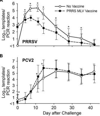
The results for PRRSV and PCV2 viremia after challenge are
shown in
Fig. 2
. PRRSV infection in the nonvaccine group
fol-lowed the typical course of viremia, peaking between 7 and 11 dpi,
followed by decay and the eventual disappearance of virus from
the blood by 42 dpi. In the vaccine group, 90% of pigs (102/113)
had detectable levels of virus nucleic acid in serum at 28 days after
vaccination or at the time of challenge. The PRRSV viremia in the
vaccine group peaked at about 7 days after challenge and then
decayed. Except for the day of challenge and day 42, the mean
PRRSV level was significantly lower on all days in the vaccine
group (
Fig. 2A
). Peak viremia for the vaccine group at days 7 and
11 was reduced by
⬎
1 log unit from that for nonvaccinated pigs.
The results demonstrated that vaccination was effective in
reduc-ing PRRS viremia in a heterologous challenge model.
Mean PCV2 viremia levels for the vaccine and nonvaccine
groups are presented in
Fig. 2B
. In the nonvaccine group, mean
PCV2 viremia peaked at about 21 days after challenge and
re-mained elevated for the remainder of the study. In contrast, mean
PCV2 viremia for the vaccine group peaked at 14 dpi, when the
mean virus level was approximately 1.5 log units greater for the
vaccine group than for the nonvaccine group (
P
⬍
0.0001). Thus,
PCV2 viremia in the vaccine group peaked much earlier.
PCV2 immunization of dams provides temporary protection
of piglets from PCV2 infection. However, by 35 days after
wean-ing, passive immunity decays to the point that pigs become
sus-ceptible to PCV2, a virus that is normally present in the
environ-ment. At the time of challenge, low but detectable levels of PCV2
nucleic acid were present in 7 of 115 vaccinated pigs (6%) and 25
of 111 nonvaccinated pigs (22%). Mean PCV2 viremia levels prior
to challenge were 2.3 and 2.9 log
10templates per PCR for
PCR-positive vaccinated and nonvaccinated pigs, respectively. The
in-creased proportion of nonvaccinated pigs with evidence of PCV2
exposure prior to challenge may account for the differences in the
outcomes between the two groups. However, exclusion of these 32
pigs did not alter the conclusions of the study.
PRRSV vaccination results in reduced clinical signs and
pa-thology during the first 21 days after coinfection.
Prior to virus
challenge, no clinical signs were apparent in either the vaccinated
or the nonvaccinated group. After infection, two clinical
syn-dromes emerged. The first was a PRRSV-associated syndrome,
aural cyanosis, commonly known as “blue ear” (
20
,
21
), which was
easily identified in pigs by the presence of red, cyanotic, or blue
discoloration of the ear tissue. Even though blue ear is not
patho-gnomonic for PRRS, it often coincides with acute infection. A
representative example of a pig with blue ear is shown in
Fig. 3
. No
blue ear was observed among either the vaccinated or the
nonvac-cinated pigs prior to challenge. However, during the postchallenge
period, 64 of all 226 pigs (28.3%) were documented as having blue
ear on one or more days. As shown in
Fig. 4A
, the percentage of
pigs with blue ear peaked between 8 and 17 dpi, which
corre-sponded to the peak in PRRS viremia (compare
Fig. 2A
). Overall,
19% of vaccinated pigs (22/115) and 38% of nonvaccinated pigs
(42/111) were documented with blue ear. A nonvaccinated pig
was 3.02 times (95% CI, 1.7, 5.9) more likely to develop blue ear
than a vaccinated pig (
P
⫽
0.001). The total numbers of days with
blue ear were 65 and 201 for the vaccine and nonvaccine
groups, respectively. The reduction in the number of pigs with
blue ear in the vaccine group is consistent with a beneficial
effect of the PRRS MLV.
The primary clinical sign associated with acute PRRSV
infec-tion is respiratory disease resulting from interstitial pneumonia.
Lungs were removed from 20 euthanized pigs (10 pigs from each
group), which were randomly selected at 11 dpi.
Figure 5
shows a
summary of the gross and microscopic lung scores. Mean scores
for the percentage of gross lung lobe involvement and microscopic
scoring for interstitial pneumonia were higher in the nonvaccine
group; however, the differences were not statistically significant
(
P
⫽
0.62 for the percentage of lung involvement and 0.30 for the
histopathology score). Together, the results showed that
vaccina-tion with the PRRS MLV had an overall protective effect by
reduc-ing PRRSV viremia and decreasreduc-ing PRRS-associated clinical signs.
FIG 1Distribution of viremia at 11 days after vaccination with PRRS MLV. Shown are PRRSV RT-PCR results for 84 pigs in the vaccine group.
FIG 2PRRSV and PCV2 viremia in vaccinated and nonvaccinated pigs. Val-ues are means⫾1 standard deviation. Asterisks indicate statistically significant differences between groups (P⬍0.015 by Student’sttest).
FIG 3Aural cyanosis, or blue ear. (A) Photograph representative of the ear discoloration associated with aural cyanosis during PRRSV infection. The photograph was taken at 11 days after virus challenge. (B) A normal ear is shown for comparison.
on August 17, 2020 by guest
http://cvi.asm.org/
PRRS vaccination results in increased clinical signs and
pa-thology at 22 to 42 days after coinfection.
Beyond the acute
pe-riod of infection, a second clinical syndrome appeared, which first
became apparent by an increase in the number of pigs receiving
systemic veterinary treatment due to clinical signs associated with
PCVAD, such as tachypnea, dyspnea, pyrexia, loss of condition,
muscle wasting, mucoid nasal discharge, lethargy, and pallor or
jaundice (
Fig. 4B
). Lesions typical of PCVAD were found by gross
anatomical and microscopic examinations of lungs and lymph
nodes from pigs that died or were euthanized. Representative
pic-tures and photomicrographs showing the lesions associated with
clinically affected pigs are presented in
Fig. 6
. Lungs showed
mul-tifocal to diffuse interstitial pneumonia with mottling of lung
tis-sue, hemorrhage, and consolidation (
Fig. 6A
). At the microscopic
level, multifocal to diffuse interstitial pneumonia with
lymphohis-tiocytic infiltration into the alveolar septa and peribronchiolar
areas was easily visible (
Fig. 6B
). The lymph nodes of affected pigs
showed depletion of lymphocytes (
Fig. 6C
). Positive staining for
PCV2 antigen was observed in the lymph nodes and lungs of
af-fected pigs (
Fig. 6D
). Analysis of gross and microscopic lesions
combined with the accumulation of PCV2 antigen in target organs
confirmed the presence of PCVAD.
The number of pigs undergoing treatment as a result of
PCVAD-associated clinical signs peaked between 22 and 35 dpi
(
Fig. 4B
). During this time, 39 pigs received at least 1 day of
vet-erinary treatment, including 12 nonvaccinated pigs (12/101
[12%]) and 27 pigs in the vaccine group (27/105 [26%]). A
vacci-nated pig was 2.67 times (95% CI, 1.23, 5.80) more likely to receive
veterinary treatment during peak PCVAD than a nonvaccinated
pig (
P
⫽
0.01). Large amounts of PCV2 in serum were associated
with the 39 pigs that went on to develop PCVAD. At 14 dpi,
sig-nificantly higher levels of circulating PCV2 were present in the 39
PCVAD-affected pigs (mean, 5.8 log10
templates/PCR) than in the
163 pigs without clinical signs (mean, 4.8 log10
templates/PCR)
(
P
⫽
0.004). The different treatments administered to the 39 pigs
with clinical signs included a single antibiotic and an NSAID
(16/39 [41%]), multiple antibiotics and an NSAID (7/39 [18%]), a
single antibiotic (6/39 [15%]), multiple antibiotics (3/39 [8%]),
and an NSAID alone (3/39 [8%]). Four of the 39 pigs (10%) were
humanely euthanized after the initial treatment due to the severity
of the clinical presentations. The decline in the percentage of pigs
with PCVAD clinical signs was largely the result of increased
mor-tality or the euthanization of pigs that were moribund or
nonre-sponsive to treatment (compare
Fig. 4B
and
C
). Over the entire
study period, 49 pigs received at least 1 day of systemic veterinary
treatment: 16% in the nonvaccine group (18/111) and 27% in the
vaccine group (31/115). A vaccinated pig was 1.79 times (90% CI,
0.99, 3.25) more likely to receive veterinary treatment than a
non-vaccinated pig (
P
⫽
0.11) over the entire study period.
Macroscopic and microscopic changes in organs and tissues
were evaluated between 32 and 42 dpi for 11 clinically affected
pigs, which were humanely euthanized as a result of failure to
respond to treatment. For comparison, 7 pigs without clinical
signs were also necropsied. As summarized in
Table 1
, all 11 pigs
with clinical signs showed some form of macroscopic lung
in-volvement, as determined by the photographic score. The
macro-scopic scores for the group without clinical signs were significantly
lower (
P
⫽
0.04). A similar trend for clinically affected versus
nonaffected pigs appeared at the microscopic level; however, the
difference was not statistically significant (
P
⫽
0.16). Mild to
se-vere lymphoid depletion was observed in 8 of the 11 pigs with
clinical signs (73%), compared to only 3 of the 7 (43%) pigs
with-out clinical signs. Even though the pigs withwith-out clinical signs
ap-peared normal, almost all showed some form of pathology related
to PCVAD, such as mild to moderate pneumonia and/or mild
lymphoid depletion.
Effect of PRRS MLV vaccination on mortality.
As shown in
Fig. 4C
, of the 101 nonvaccinated pigs, 9 died, resulting in an
overall survival rate of 91.1%. Of the 105 vaccinated pigs, 14 died,
for an overall survival rate of 86.7% (
Fig. 4C
). A vaccinated pig was
1.7 times (95% CI, 0.89, 3.72) more likely to die during the overall
study period than a nonvaccinated pig (
P
⫽
0.35). Increased
mor-tality became apparent after 20 days and was associated with the
appearance of PCVAD. Of the 39 pigs that developed clinical signs
FIG 4Clinical outcomes for the vaccinated and nonvaccinated groups after a dual challenge with PRRSV and PCV2. Panel A shows the percentage of pigs with aural cyanosis, a PRRSV-associated syndrome. Clinical signs were as-sessed as described in Materials and Methods.
FIG 5Assessment of lung pathology at 11 days after PRRSV/PCV2 challenge. Ten vaccinated pigs (filled bars) and 10 nonvaccinated pigs (shaded bars) were randomly removed from the study at 11 days and were assessed for pneumo-nia. Results (means⫾standard deviations) are presented as the percentage of gross lung involvement and the histopathology score. The differences between the vaccine and nonvaccine groups were not significant (P⬎0.05).
on August 17, 2020 by guest
http://cvi.asm.org/
of PCVAD, 21 died prior to the end of the study (14 vaccinated
and 7 nonvaccinated), resulting in a mortality rate of
approxi-mately 54% in pigs exhibiting clinical signs. Between 22 and 35
dpi, a vaccinated pig was 2.1 times (90% CI, 1.03, 5.87) more likely
to die than a nonvaccinated pig (
P
⫽
0.09). Even though mortality
was higher in the vaccine group, the differences between the
vac-cine and nonvacvac-cine groups were not significantly different.
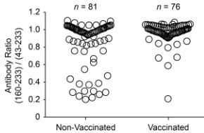
Vaccination increases the appearance of antibodies against a
PCV2 decoy epitope.
In previous work, we identified the presence
of antibodies against a decoy epitope in the capsid protein of
PCV2, CP(160 –180), correlated with PCVAD (
13–15
). The
pres-ence of an anti-CP(160 –180) response is associated with the
ab-sence of PCV2 neutralizing activity in serum. Pigs vaccinated for
PCV2 and protected from disease produce little anti-CP(160 –
180) activity and preferentially recognize a larger epitope, CP(43–
233). Pigs that are naturally infected with PCV2 show a mixture of
the two antibody responses. As a means to standardize results
across plates, the anti-PCV2 antibody responses for the vaccine
and nonvaccine groups were presented as CP(160 –233)/CP(43–
233) ratios. The higher the ratio, the more the immune response is
skewed toward the recognition of the decoy epitope. The results,
presented in
Fig. 7
, showed a significant (
P
⫽
0.0006) difference
between the CP(160 –233)/CP(43–233) mean ratios: 0.85 for
non-FIG 6Gross and microscopic lesions associated with PCVAD. The images shown are representative of the lesions of PCVAD-affected pigs necropsied between 32 and 42 days after combined PRRSV and PCV2 challenge. (A) Set of lungs from a challenged pig with pneumonia, mottling, and consolidation. A normal set of lungs from an age-matched pig is shown for comparison. (B) H&E-stained lung from a challenged pig showing moderate to severe multifocal interstitial pneumonia with lymphohistiocytic infiltration of alveolar septa. A normal lung is shown for comparison. (C) Lymphoid depletion in a lymph node from a challenged pig. A normal lymph node with prominent germinal centers (GC) is shown for comparison. (D) Immunohistochemical staining showing the accumulation of PCV2 antigen in a lung and a lymph node from a challenged pig.
TABLE 1Lung and lymph node lesions of pigs with clinical signs associated with PCVAD and apparently healthy pigs between 32 and 42 dpia
Score or severity
No. (%) of pigs:
With clinical signs
Apparently healthy
Macroscopic lung lesion scoreb
0 0 (0) 1 (14)
1 4 (36) 4 (57)
2 2 (18) 1 (14)
3 1 (9) 1 (14)
4 4 (36) 0 (0)
Microscopic lung lesion scorec
0 0 (0) 0 (0)
1 1 (9) 1 (14)
2 3 (27) 4 (57)
3 7 (64) 2 (29)
4 0 (0) 0 (0)
Level of lymphoid depletiond
None 3 (27) 4 (57)
Mild 4 (36) 3 (43)
Moderate 2 (18) 0 (0)
Severe 2 (18) 0 (0)
a
A total of 18 pigs, of which 11 showed clinical signs associated with PCVAD and 7 were apparently healthy, were necropsied.
b
Determined by evaluation of ventral and dorsal photographs of lungs. Scores were assigned as follows: 0, no macroscopic lesions; 1, pneumonia affecting⬍25% of gross lung; 2, pneumonia affecting 25 to 50% of gross lung; 3, pneumonia affecting 50 to 75% of gross lung; 4, pneumonia affecting⬎75% of gross lung. The data for pigs with clinical signs were statistically different (P⫽0.0426) from those for apparently healthy pigs, based on ordinal logistic regression.
c
Determined by evaluation of tissue sections stained with hematoxylin and eosin. Scores were assigned as follows: 0, no significant microscopic lesions; 1, mild interstitial pneumonia with⬍50% lung lobe involvement; 2, mild to moderate multifocal interstitial pneumonia with 50 to 75% lung lobe involvement; 3, moderate to severe multifocal interstitial pneumonia with 50 to 75% lung lobe involvement; 4, severe diffuse interstitial pneumonia with⬎75% lung lobe involvement.
d
Determined by evaluation of lymph node and tonsil tissue sections stained with hematoxylin and eosin. Characterization as mild, moderate, or severe indicates a small, intermediate, or large extent of lymphocyte depletion, respectively, with replacement by histiocytes.
FIG 7PCV2 CP(160 –233)/CP(43–233) ratio at 42 days after coinfection. Sera were analyzed for anti-PCV2 CP antibodies. The serum antibody ratio was calculated as the MFI for reactivity with the CP(160 –233) decoy epitope di-vided by the MFI for the CP(43–233) conformational antigen. The difference between the means for the PRRSV-vaccinated and nonvaccinated groups was significant (P⫽0.0006 by Student’sttest).
on August 17, 2020 by guest
http://cvi.asm.org/
vaccinated pigs versus 0.97 for vaccinated pigs. Exclusion of the 32
pigs that showed the presence of PCV2 nucleic acid at the time of
challenge changed the ratios but did not affect the conclusion;
CP(160 –233)/CP(43–233) mean ratios were 0.92 for
nonvacci-nated pigs (
n
⫽
61) and 0.98 for vaccinated pigs (
n
⫽
72) (
P
⫽
0.03).
As discussed above, previous work showed that pigs with low
CP(160 –233)/CP(43–233) ratios are protected from disease.
Therefore, nonvaccinated pigs with ratios of
⬍
0.5 (
n
⫽
13) were
compared to nonvaccinated pigs with PCV2 antibody ratios of
⬎
0.5 (
n
⫽
68). Pigs with antibody ratios of
⬍
0.5 had significantly
lower levels of circulating PCV2 in the serum at 21 (
P
⫽
0.0001),
28 (
P
⫽
0.0008), 35 (
P
⫽
0.007), and 42 (
P
⫽
0.03) dpi than pigs
with higher ratios. The results confirm earlier findings describing
the nonprotective effect of anti-CP(160 –233) antibodies (
13
).
Effect of PRRS MLV vaccination on ADG.
Over the entire
70-day study period, the mean average daily gain (ADG) for the
vaccine group (
n
⫽
91) was 0.65
⫾
0.11 kg, compared to 0.68
⫾
0.10 kg for the nonvaccine group (
n
⫽
92). The means differed
significantly between the two groups (
P
⫽
0.029). Decreased mean
ADG was also observed in the vaccine group during the 42-day
post–virus challenge period. However, the difference between the
vaccine group (ADG, 0.82
⫾
0.14 kg) (
n
⫽
91) and the nonvaccine
group (ADG, 0.86
⫾
0.14 kg) (
n
⫽
92) was not statistically
signif-icant (
P
⫽
0.061). In addition, ADG differences between the two
groups during the 70-day study period were no longer significant
after the exclusion of the 32 pigs with PCV2 detected prior to
challenge.
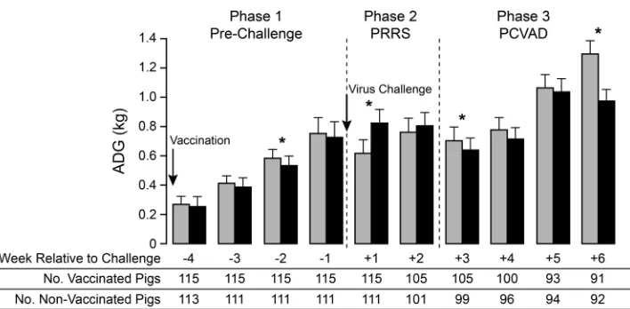
Therefore, a more-detailed analysis was conducted by
calculat-ing ADG on a weekly basis (
Fig. 8
). The results showed that ADG
differences between the nonvaccine and vaccine groups could be
divided into three distinct phases. In the first phase, covering the
prechallenge period, mean ADG after vaccination was reduced,
with a significant difference between the vaccinated and
nonvac-cinated groups appearing at 3 weeks after vaccination. The second
phase covered the period of acute PRRSV infection. At 1 week
after virus challenge, mean ADG was significantly increased for
the vaccine group. ADG remained higher for the vaccine group in
the second week postchallenge, but the difference was not
signif-icant. The improved ADG for the vaccinated pigs likely resulted
from the positive effect of vaccination in reducing
PRRS-associ-ated clinical signs and virus load (
Fig. 2A
and
4A
). The third phase
covered the period from the onset of PCVAD, beginning at about
3 weeks after virus challenge, to the end of the study. During this
phase, ADG was lower in the vaccinated group at every time point,
with significantly lower mean ADG values on week 6 after virus
challenge. A significant decrease in ADG was also initially detected
for vaccinated pigs on week 3 postchallenge; however, this
differ-ence was no longer significant after the 32 PCV2-positive pigs had
been excluded (
P
⫽
0.08). The lower mean ADG values are
con-sistent with the effects of PCVAD, which include poor growth
performance and muscle wasting.
The negative effect of vaccination on ADG could have been the
result of increased numbers of pigs with clinically apparent
PCVAD. Therefore, a separate analysis was performed after the 26
pigs with clinical signs that survived the length of the study had
been excluded from the vaccine and nonvaccine groups. The
re-moval of pigs with clinical signs increased the ADGs of the two
groups to 0.69
⫾
0.09 kg for nonvaccinated pigs without clinical
signs (
n
⫽
83) and 0.66
⫾
0.10 kg for vaccinated pigs without
clinical signs (
n
⫽
74) (
P
⫽
0.047). However, even in the absence
of overt clinical signs, PRRSV vaccination had a negative effect on
weight gain.
DISCUSSION
Enhanced PCV2 infection leading to PCVAD is typically
associ-ated with immune stimulation (
22
,
23
). PCV2 replication is
lo-cated in the nuclei of permissive cells and is dependent on cellular
enzymes expressed during the S phase of the cell cycle (
24
). It is
presumed that actively dividing lymphocytes, in response to an
immune stimulus, provide a cellular environment ideal for
sup-porting PCV2 replication. PRRSV, porcine parvovirus, and
My-coplasma hyopneumoniae
are common copathogens linked with
lymphoproliferation and increased PCV2 pathogenesis (
10–13
,
25–31
). Examples of noninfectious immunostimulators include
immunization with keyhole limpet hemocyanin in incomplete
Freund’s adjuvant (
22
,
32
) and inactivated vaccines, such as
My-coplasma hyopneumoniae
(
32–34
) and
Actinobacillus
pleuropneu-FIG 8Weekly average daily gain (ADG) before and after virus challenge. Mean ADG values for the nonvaccinated (shaded bars) and vaccinated (filled bars) groups were calculated on a weekly basis. The data show means and standard deviations. The numbers of pigs used in the analysis are given below the bar graph. Asterisks indicate statistically significant differences in ADG between groups (P⬍0.03 by Student’sttest).
on August 17, 2020 by guest
http://cvi.asm.org/
moniae
(
33
,
34
). The results of this study showed that the PRRS
MLV initially had a beneficial effect in reducing PRRS-associated
clinical signs and PRRS viremia; however, PRRSV-vaccinated pigs
showed increased PCV2 replication, reduced average daily gain,
and increased clinical signs associated with PCVAD.
Previous experimental studies documenting interactions
be-tween the PRRS MLV and PCV2 infection have yielded conflicting
results. Allan et al. (
35
) found that colostrum-deprived,
specific-pathogen-free (SPF) pigs infected with PCV2 at the age of 5 weeks
and administered the PRRS MLV 1 week later had greater
amounts of PCV2 antigen in tissues and more-severe histologic
lesions, characteristic of PMWS, than pigs infected with PCV2
alone. However, the pigs failed to exhibit clinical signs or gross
lesions typical of PCVAD (
35
). In contrast, Opriessnig et al. (
36
)
evaluated the effects of PCV2 infection on the efficacy of the PRRS
MLV in groups of 10 early-weaned SPF pigs. Pigs were inoculated
with PCV2 at the age of 6 weeks, vaccinated with the PRRS MLV 2
weeks later, and then challenged with PRRSV at the age of 12
weeks. The group with PCV2 and the MLV exhibited lower ADG
and more-severe lung lesions after PRRSV challenge than the
group that was vaccinated and received a PRRSV challenge
with-out PCV2. Because PCV2 was not detected in affected lungs by
IHC, the authors attributed the lesions to PRRSV infection and
further concluded that the effect of PCV2 was to reduce the
effi-cacy of the PRRS MLV (
36
). Park et al. (
37
) investigated the
po-tential for the PRRS MLV to reduce PRRSV-associated
amplifica-tion of PCV2 pathogenesis after coinfecamplifica-tion with PRRSV and
PCV2. Groups of 8 conventional pigs were subjected to a variety of
treatments involving different combinations of the PRRS MLV,
wild-type PRRSV, a PCV2 vaccine, and PCV2. Pigs were
vacci-nated with the PRRS MLV, a PCV2 subunit vaccine, or both and
were then challenged, 4 weeks later, with PRRSV, PCV2, or both.
The group that received the PRRS MLV followed by coinfection
showed no differences in PCV2 viremia, PCV2-associated
pathol-ogy, or the number of PCV2-positive cells in lymph nodes and
lungs from the coinfected group that was not vaccinated with the
PRRS MLV (
37
). In contrast to our study, in which vaccination
enhanced PCVAD, these authors found that coinfected pigs, with
or without previous PRRS MLV vaccination, had similar PCV2
replication and pathogenesis.
Field studies have also yielded conflicting results. A survey
con-ducted on 70 pig farms in the Netherlands included questions
regarding PRRS MLV use on farms with and without PMWS or
PCVAD. The results showed that the PRRS MLV is a significant
risk factor for PMWS outbreaks (
38
). In contrast, an analysis of
the effect of the PRRS MLV on farms affected by PCVAD in the
United States found that farms incorporating the PRRS MLV have
significantly lower levels of PCV2 viremia than nonvaccinating
farms in the age group in which peak wasting disease occurred
(
39
). In that study, quantitative PCR (qPCR) was used to measure
PCV2 in serum samples collected at different time points from 6
herds vaccinated with the PRRS MLV and 12 nonvaccinated
herds. These results suggest that the PRRS MLV can reduce PCV2
viremia (
39
).
The failure of experimental studies to find a consistent link
between the PRRS MLV and PCVAD was likely due in part to the
length of the observation period. For example, the studies
de-scribed above were terminated on day 25 after infection with
PCV2 (
35
) and 21 days after challenge with PRRSV and PCV2
(
37
). In the current study, peak PCVAD occurred between 22 and
35 days after challenge with PRRSV and PCV2. Another
impor-tant difference is the sizes of the experimental groups. Typically,
the prevalence of clinically ill pigs on farms affected by PCVAD is
only 2 to 25% (
40–42
). In small groups of pigs, animals with
clin-ical disease may not be apparent or present. The current study
utilized more than 200 pigs, mimicking the environment found in
the field and providing the depth of data required for the
obser-vation and quantification of low-percentage outcomes. PCVAD
morbidity rates were 11.9% and 25.7% in nonvaccinated and
vac-cinated pigs, respectively. Therefore, PCVAD should be assessed
by comparing mortality rates, clinical disease presentation,
viremia, and weight gain in relatively large groups evaluated for
several weeks after infection.
Because large groups of pigs were required to ensure clinical
disease expression, some control groups, including pigs
chal-lenged with PCV2 or PRRSV alone, were not incorporated into
the study design. Therefore, conclusions are based on the findings
for vaccinated and nonvaccinated cochallenged pigs. Caution
should be exercised in generalizing results to single infections (i.e.,
the effect of the PRRS MLV on PCV2-challenged pigs).
PCV2 is ubiquitous in swine populations, and elimination of
the virus from the environment is extremely difficult. As
demon-strated in this study, 32 of 226 pigs had detectable PCV2 in serum
at the time of challenge. Although these pigs were in the minority
(14%), and group titers were relatively low (2.3 and 2.9 log
10tem-plates per PCR for vaccinated and nonvaccinated pigs,
respec-tively), the presence of PCV2 prior to challenge had to be
consid-ered a possible factor in postchallenge response. This was
especially true due to the difference between the proportions of
PCV2-positive pigs in the two groups, despite randomized
alloca-tion of pigs and balanced genotypes. This difference was likely due
to the failure of randomization to distribute PCV2-positive pigs
equally across the two groups. Therefore, analyses were also
com-pleted after the 32 initially PCV2 positive pigs were excluded, and
all conclusions of the study were confirmed. Regardless, this
high-lights the difficulty of eliminating PCV2 from the environment
and also suggests that pigs should be balanced according to PCV2
status prior to challenge.
Significantly increased levels of PCV2 viremia in PRRS
MLV-treated pigs were observed at 11 and 14 days after challenge, but
not at later time points (
Fig. 2B
). This effect of the PRRS MLV on
PCV2 infection is similar to that seen in a previous study by us,
which showed a significant increase in the level of PCV2 viremia at
23 days after coinfection with PRRSV and PCV2 (
13
). In the
cur-rent study, it is noteworthy that PCV2 viremia levels did not differ
significantly between the vaccine and nonvaccine groups during
peak PCVAD. However, the 39 clinically ill pigs did maintain
sig-nificantly higher levels (
P
⬍
0.02) of circulating PCV2 during
these later time points (21 and 35 dpi) (data not shown). The
increased incidence of PCVAD in vaccinated pigs between 22 and
35 dpi may also be the result of greater levels of localized PCV2 in
tissues. Further studies are needed to assess differences in the
quantity and tissue distribution of PCV2 between vaccinated and
nonvaccinated pigs.
Average daily gain (ADG) is used in swine production as an
objective measure of overall health and performance. The
nega-tive effect of the PRRS MLV on growth performance is well
doc-umented. For example, Opriessnig et al. (
43
) reported that pigs
vaccinated with the PRRS MLV at the age of 2 weeks exhibited
significantly lower ADG than nonvaccinated pigs. Pretzer et al.
on August 17, 2020 by guest
http://cvi.asm.org/
(
44
) found that PRRS MLV-vaccinated weaned pigs had lower
ADG between 0 and 14 dpv than nonvaccinated pigs. While this
effect on ADG was no longer apparent between 21 and 42 dpv, the
vaccinated pigs maintained lighter weights overall (
44
). We
con-firmed the negative effect of the PRRS MLV on ADG during the
28-day period prior to coinfection; significantly reduced ADG was
observed during the third week after vaccination (
Fig. 8
). The
benefit of PRRSV vaccination was documented during the first 2
weeks after PCV2 and PRRSV challenge, when the MLV had a
positive effect on ADG. Although vaccinated pigs in this study had
increased ADG in the first 2 weeks postchallenge, this effect was
quickly outweighed when ADG was decreased in the presence of
PCVAD. Vaccination decreased ADG in both clinically affected
pigs and pigs without clinical signs during the study period,
dem-onstrating that poor growth performance may be a subclinical
manifestation of PCVAD in apparently healthy pigs.
At least three mechanisms may be involved in the
enhance-ment of PCVAD following PRRSV vaccination. First, the PRRS
MLV may function to stimulate the immune system and increase
the number of PCV2-permissive cells. As with wild-type viruses,
lymphocytes undergo mitosis in response to vaccination with the
PRRS MLV, thereby increasing the population of cells with the
ability to support PCV2 replication. In addition, the vaccine likely
stimulates PRRSV-specific lymphocyte populations that are
re-stimulated after challenge with a wild-type PRRSV. Second, like
wild-type PRRS viruses, the PRRS MLV may suppress innate
im-munity, thereby blocking anti-PCV2 responses. For example,
PRRSV nonstructural proteins, such as nsp1 and nsp2, block the
induction of interferon and response of cells to interferon (
5
).
Viral proteins such as nsp1
␣
and nsp1

antagonize the type I
interferon response by degrading key components needed for
in-terferon gene expression and inhibiting inin-terferon signaling
path-ways (
45
,
46
). Finally, the third mechanism is based on the
possi-bility that the PRRS MLV may skew the immune response toward
the production of nonneutralizing PCV2-specific antibodies.
PCV2 has circulated in the swine population for at least 25
years. In 2005, the emergence of PCV2b in North America was
attributed to outbreaks of PCVAD (
47–49
). Since then, the disease
has been effectively managed through the use of PCV2 vaccines
(
50–52
). Therefore, the negative effect of the MLV on PCV2
in-fection may not be relevant. However, there remain several
coun-tries in which PRRS MLV vaccination is in wide use, but in the
absence of a comprehensive PCVAD vaccination program.
Fur-ther, there is the potential for new and emerging PCV2 strains to
escape current vaccine protection. Emerging PCV2 mutant strains
have been documented in China (
53
) and more recently have been
associated with PCVAD outbreaks in vaccinated herds in the
United States and Korea (
54
,
55
). Overall, this study supports the
notion that maintaining a successful PCV2 control program and
assessing the risk of virulent PRRSV exposure is critical to
weigh-ing the benefits of the PRRS MLV.
ACKNOWLEDGMENT
This work was supported by USDA NIFA award 2013-68004-20362.
REFERENCES
1.Baekbo P, Kristensen CS, Larsen LE.2012. Porcine circovirus diseases: a review of PMWS. Transbound Emerg Dis59:60 – 67.http://dx.doi.org/10 .1111/j.1865-1682.2011.01288.x.
2.Ramamoorthy S, Meng XJ.2009. Porcine circoviruses: a minuscule yet
mammoth paradox. Anim Health Res Rev10:1–20.http://dx.doi.org/10 .1017/S1466252308001461.
3.Benfield DA, Nelson E, Collins JE, Harris L, Goyal SM, Robison D, Christianson WT, Morrison RB, Gorcyca D, Chladek D.1992. Charac-terization of swine infertility and respiratory syndrome (SIRS) virus (iso-late ATCC VR-2332). J Vet Diagn Invest4:127–133.http://dx.doi.org/10 .1177/104063879200400202.
4.Conzelmann KK, Visser N, Van Woensel P, Thiel HJ.1993. Molecular characterization of porcine reproductive and respiratory syndrome virus, a member of the arterivirus group. Virology193:329 –339.http://dx.doi .org/10.1006/viro.1993.1129.
5.Chand RJ, Trible BR, Rowland RR.2012. Pathogenesis of porcine re-productive and respiratory syndrome virus. Curr Opin Virol2:256 –263. http://dx.doi.org/10.1016/j.coviro.2012.02.002.
6.Gómez-Laguna J, Salguero FJ, Pallares FJ, Carrasco L.2013. Immuno-pathogenesis of porcine reproductive and respiratory syndrome in the respiratory tract of pigs. Vet J195:148 –155.http://dx.doi.org/10.1016/j .tvjl.2012.11.012.
7.Opriessnig T, Gimenez-Lirola LG, Halbur PG. 2011. Polymicrobial respiratory disease in pigs. Anim Health Res Rev12:133–148.http://dx.doi .org/10.1017/S1466252311000120.
8.Renukaradhya GJ, Alekseev K, Jung K, Fang Y, Saif LJ.2010. Porcine reproductive and respiratory syndrome virus-induced immunosuppres-sion exacerbates the inflammatory response to porcine respiratory coro-navirus in pigs. Viral Immunol 23:457– 466.http://dx.doi.org/10.1089 /vim.2010.0051.
9.Pallarés FJ, Halbur PG, Opriessnig T, Sorden SD, Villar D, Janke BH, Yaeger MJ, Larson DJ, Schwartz KJ, Yoon KJ, Hoffman LJ. 2002. Porcine circovirus type 2 (PCV-2) coinfections in US field cases of postweaning multisystemic wasting syndrome (PMWS). J Vet Diagn In-vest14:515–519.http://dx.doi.org/10.1177/104063870201400614. 10. Allan GM, McNeilly F, Ellis J, Krakowka S, Meehan B, McNair I,
Walker I, Kennedy S.2000. Experimental infection of colostrum de-prived piglets with porcine circovirus 2 (PCV2) and porcine reproductive and respiratory syndrome virus (PRRSV) potentiates PCV2 replication. Arch Virol145:2421–2429.http://dx.doi.org/10.1007/s007050070031. 11. Harms PA, Sorden SD, Halbur PG, Bolin SR, Lager KM, Morozov I,
Paul PS.2001. Experimental reproduction of severe disease in CD/CD pigs concurrently infected with type 2 porcine circovirus and porcine re-productive and respiratory syndrome virus. Vet Pathol38:528 –539.http: //dx.doi.org/10.1354/vp.38-5-528.
12. Rovira A, Balasch M, Segales J, Garcia L, Plana-Duran J, Rosell C, Ellerbrok H, Mankertz A, Domingo M.2002. Experimental inoculation of conventional pigs with porcine reproductive and respiratory syndrome virus and porcine circovirus 2. J Virol76:3232–3239.http://dx.doi.org/10 .1128/JVI.76.7.3232-3239.2002.
13. Trible BR, Ramirez A, Suddith A, Fuller A, Kerrigan M, Hesse R, Nietfeld J, Guo B, Thacker E, Rowland RR.2012. Antibody responses following vaccination versus infection in a porcine circovirus-type 2 (PCV2) disease model show distinct differences in virus neutralization and epitope recognition. Vaccine 30:4079 – 4085. http://dx.doi.org/10 .1016/j.vaccine.2012.04.022.
14. Trible BR, Kerrigan M, Crossland N, Potter M, Faaberg K, Hesse R, Rowland RR.2011. Antibody recognition of porcine circovirus type 2 capsid protein epitopes after vaccination, infection, and disease. Clin Vac-cine Immunol18:749 –757.http://dx.doi.org/10.1128/CVI.00418-10. 15. Trible BR, Suddith AW, Kerrigan MA, Cino-Ozuna AG, Hesse RA,
Rowland RR.2012. Recognition of the different structural forms of the capsid protein determines the outcome following infection with porcine circovirus type 2. J Virol86:13508 –13514.http://dx.doi.org/10.1128/JVI .01763-12.
16. Federation of Animal Science Societies.2010. Guide for the care and use of agricultural animals in research and teaching. Federation of Animal Science Societies, Champaign, IL.
17. Boddicker N, Waide EH, Rowland RR, Lunney JK, Garrick DJ, Reecy JM, Dekkers JC.2012. Evidence for a major QTL associated with host response to porcine reproductive and respiratory syndrome virus chal-lenge. J Anim Sci90:1733–1746.http://dx.doi.org/10.2527/jas.2011-4464. 18. Reed LJ, Muench H.1938. A simple method of estimating fifty per cent
endpoints. Am J Hyg27:493– 497.
19. Halbur PG, Paul PS, Frey ML, Landgraf J, Eernisse K, Meng XJ, Lum MA, Andrews JJ, Rathje JA.1995. Comparison of the pathogenicity of two US porcine reproductive and respiratory syndrome virus isolates with