OncoTargets and Therapy 2019:12 2615–2621
OncoTargets and Therapy
This article was published in the following Dove Medical Press journal: OncoTargets and Therapy
Dove
press
submit your manuscript | www.dovepress.com 2615
O r i g i n a l r e s e a r c h
open access to scientific and medical research
Open Access Full Text Article
mirna-99b-5p targets FZD8 to inhibit non-small
cell lung cancer proliferation, migration and
invasion
rui liu1,2
Yajuan chen1
Tao shou2
Jing hu2
chen Qing1
1Yunnan Key laboratory of
Pharmacology for natural Products, Kunming Medical University, Kunming 650031, People’s republic of china; 2Department of Oncology,
The First People’s hospital of Yunnan Province, Kunming 650032, Yunnan, People’s republic of china
Background: miRNAs were found to play crucial roles in regulating cellular behaviors. The
aim of this study was to investigate the biological function of miRNA-99b-5p (miR-99b-5p) in non-small cell lung cancer (NSCLC).
Materials and methods: miR-99b-5p expression level in NSCLC cell lines was detected by quantitative real-time PCR (qRT-PCR). Cell proliferation, migration and invasion were exam-ined by cell counting kit-8 (CCK-8) assay, wound-healing assay and Transwell invasion assay, respectively. Dual-luciferase activity reporter assay and Western blot assay were conducted to validate the target of miR-99b-5p.
Results: The expression of miR-99b-5p was decreased in NSCLC cell lines compared with normal cell line. Overexpression of miR-99b-5p inhibits cell proliferation, migration and inva-sion in vitro. FZD8 was validated as a direct target of miR-99b-5p. Overexpresinva-sion of FZD8 partially abolished the effects of miR-99b-5p mimic on NSCLC cell behaviors.
Conclusion: Collectively, our results demonstrated that miR-99b-5p inhibits NSCLC cell pro-liferation, migration and invasion through targeting FZD8. This newly identified miR-99b-5p/ FZD8 axis provided novel insights into the mechanisms underlying NSCLC progression.
Keywords: miR-99b-5p, FZD8, non-small cell lung cancer, migration, invasion
Introduction
Lung cancer is one of the most common causes of cancer-related death worldwide.1
There are two major subgroups of lung cancer: non-small cell lung cancer (NSCLC) and small cell lung cancer (SCLC).2 NSCLC alone represents 85% of all lung cancer cases.3
Five-year overall survival of NSCLC patients remains undesirable even though there are significant improvements in treatment measures.4 Investigation of biomarkers closely
associated with NSCLC tumorigenesis will contribute to control NSCLC progression. miRNAs are noncoding RNAs that were capable to regulate gene expres-sion through 3′-UTR binding and thus affect cell behaviors, disease initiation and progression.5 miRNAs were reported to have dual roles in the tumorigenesis of human
cancers: either tumor-suppressive miRNA or oncogenic miRNA.6 Oncogenic miRNAs
refer to miRNAs frequently upregulated, while tumor-suppressive miRNAs represent miRNAs often downregulated in human cancers.6
Mounting studies have been carried out to investigate the abnormal expression of miRNA in NSCLC tissues and cell lines.7–10 Guan et al7 reviewed previously published
literatures concerning miRNA expression profile in NSCLC tissues and identified 184 differentially expressed miRNAs. Wang et al8 investigated miRNA profiles in
seven NSCLC datasets and identified 11 key miRNAs in NSCLC carcinogenesis. correspondence: chen Qing
Yunnan Key laboratory of Pharmacology for natural Products, Kunming Medical University, no. 1168, West chunrong road, Yuhua street, chenggong District, Kunming 650500, Yunnan, People’s republic of china
Tel +86 871 6533 2413 email qingchen_yn@163.com
Journal name: OncoTargets and Therapy Article Designation: Original Research Year: 2019
Volume: 12
Running head verso: Liu et al Running head recto: Liu et al DOI: 199196
OncoTargets and Therapy downloaded from https://www.dovepress.com/ by 118.70.13.36 on 25-Aug-2020
Dovepress
liu et al
These identified miRNAs might be profitable to diagnosis or prognostic prediction of NSCLC.8 Besides that, Chen
et al9 found that miR-148a expression was downregulated
in NSCLC tissues compared with normal lung tissues. They also found that low miR-148a expression was correlated with advanced tumor grade, lymph node metastasis and a higher risk of tumor-related death.9 Also, miR-148a
over-expression was shown to inhibit NSCLC cell migration and invasion through targeting Wnt family member 1.9 Wang
et al10 investigated that miR-142-5p overexpression decreased
phosphatidylinositol-4,5-bisphosphate 3-kinase and catalytic subunit alpha expression to inhibit NSCLC proliferation in vitro and in vivo. These results indicated the importance of miRNAs in NSCLC carcinogenesis.
FZD8 expression was revealed to be significantly overexpressed in primary lung cancer tissue samples and cell lines compared with their adjacent normal tissues and normal cell line.11 Importantly, knockdown of FZD8
expression in NSCLC cells inhibited proliferation in vitro and suppressed tumor growth in vivo.11 Another study
con-ducted by Jin et al12 showed that FZD8 was a direct target
of miR-375 in SCLC cell line. These results indicated the importance of FZD8 in lung cancer. miRNA-99b-5p (miR-99b-5p) was reported to function as a tumor suppressor in multiple human cancers including gastric cancer, clear-cell renal cell carcinoma and colorectal cancer.13–15 However,
the biological roles of miR-99b-5p in NSCLC remain unclear. In this study, we analyzed miR-99b-5p expression in NSCLC cell lines and normal cell line. Furthermore, we analyzed the effects of miR-99b-5p expression on NSCLC cell proliferation, migration and invasion. Moreover, the effects of miR-99b-5p on the overall survival of NSCLC patients were analyzed at Kaplan–Meier plotter website. Luciferase activity reporter assay and Western blot assay were conducted to validate whether FZD8 was a functional target of miR-99b-5p.
Materials and methods
cell culture and cell transfection
NSCLC cell lines (A549 and NCI-H292), SCLC cell line (NCI-H69), human bronchial epithelial cells (16-HBE) and human embryonic kidney-derived cell line (HEK293T) were pur-chased from the American Type Culture Collection (ATCC, Manassas, VA, USA). These cell lines were maintained at DMEM (Thermo Fisher Scientific, Waltham, MA, USA) supplemented with 10% FBS (Thermo Fisher Scientific), streptomycin (100 mg/mL) and penicillin (100 U/mL) in a 37°C humidified incubator containing 5% of CO2.
Cells were transfected with 100 nM of miR-99b-5p mimic or negative control (NC-miRNA) obtained from GenePharma (Shanghai, People’s Republic of China) and 4 μg FZD8 overexpression construct (pcDNA3.1-FZD8) or pcDNA3.1 (GenScript, Nanjing, People’s Republic of China) using Lipofectamine 2000 reagent (Thermo Fisher Scientific) as per the manufacturer’s instructions.
cell proliferation assay
Cell counting kit-8 (CCK-8) assay was performed to measure the rate of cellular proliferation. Cells were seeded at the 96-well plate at a density of 3,000 cells/well and incubated for 0, 24, 48 or 72 h. Then, CCK-8 reagent was added to each well and incubated at 37°C for additional 3 h, after which the optical density was measured at 450 nm using MRX II microplate reader (Dynex Technologies, Chantilly, VA, USA).
Wound-healing assay
Cells were seeded in six-well plate and incubated until approximately 100% confluence. Then, a wound at cell surface was created using pipette tip, after which cells were washed to remove cell debris. Cell images were captured at 0 or 48 h after wound creation to assess cellular migra-tion rate.
Transwell invasion assay
Cellular invasion ability was analyzed using 24-well Tran-swell chamber with a Matrigel (BD Biosciences, San Jose, CA, USA) pre-coated 8 μm corning filter (Corning Inc., New York, NY, USA). 1×10⁴ cells in serum-free DMEM
were plated to the upper chamber of Transwell plate. The lower chamber was filled with DMEM supplemented with 10% FBS. Forty-eight hours after incubation, noninvasive cells were removed using cotton, while the invaded cells were fixed with 4% paraformaldehyde and stained with 0.5% crystal violet. Invasive cell numbers were counted from five randomly selected fields using microscope.
Quantitative real-time Pcr (qrT-Pcr)
Total RNA was extracted from cultured cells using Trizol reagent (Beyotime, Haimen, People’s Republic of China) fol-lowing the manufacturer’s instructions. cDNA libraries were synthesized using Reverse Transcription Kit (Takara, Dalian, People’s Republic of China). qRT-PCR was conducted at ABI 7500 PCR system (Bio-Rad Laboratories Inc., Hercules, CA, USA) using SYBR Premix Ex Taq (Takara). miR-99b-5p expression level was analyzed using comparative
OncoTargets and Therapy downloaded from https://www.dovepress.com/ by 118.70.13.36 on 25-Aug-2020
Dovepress liu et al
threshold cycle method with U6 snRNA as internal control. The primers used were as follows: miR-99b-5p: forward: 5′-CACCCGTAGAACCGACCTT-3′, reverse: 5′-GTC GTATCCAGTGCGTGTCGTGGAGTCGGCAATTG CACTGGATACGACCGCAAGG-3′; U6 snRNA: forward: 5′-GCTTCGGCAGCACATATACTAAAAT-3′, reverse: 5′-CGCTTCACGAATTTGCGTGTCAT-3′.
Western blot
RIPA lysis buffer containing protease inhibitors and phospha-tase inhibitors (Beyotime) was employed to extract proteins from cultured cells. Equal amounts of protein lysates were separated using 10% SDS-PAGE and transferred to polyvi-nylidene fluoride membranes (Beyotime). Membranes were blocked with 5% skim milk, then incubated with primary antibodies (anti-FZD8: ab155093; anti-GAPDH: ab181602; both from Abcam, Cambridge, MA, USA) for overnight at 4°C. After washing with TBST, the membranes were incu-bated with secondary antibody (ab6721; Abcam) in room temperature at 1 h. Bands were visualized using BeyoECL kit (Beyotime) and ImageJ 1.42 software (NIH, Bethesda, MD, USA).
Bioinformatical analysis and luciferase
activity reporter assay
TargetScan (http://www.targetscan.org) was used to predict the potential targets of miR-99b-5p. Wild-type (wt) 3′-UTR of FZD8 was inserted into pGL3 vector (Promega Corpora-tion, Fitchburg, WI, USA) and named as FZD8-wt. FZD8-mt was constructed based on FZD8-wt using site-directed
mutagenesis kit (Takara). Cells were co-transfected with FZD8-wt or FZD8-mt and miR-99b-5p mimic or NC-miRNA using Lipofectamine 2000. Forty-eight hours after transfection, relative luciferase activity was measured using a dual-luciferase reporter assay kit (Promega Corporation).
Kaplan–Meier survival analysis
Kaplan–Meier plotter (www.kmplot.com) was used to inves-tigate the effect of miR-99b-5p expression on overall survival of NSCLC patients.16 Cutoff value was auto selected in the
algorithm. Log-rank test was used to calculate the difference between low or high expression groups.
statistical analyses
Data were presented as mean ± SD. Differences between groups or among three or above groups were evaluated using the Student’s t-test or ANOVA and Tukey’s post hoc test, respectively. GraphPad Prism 5 (GraphPad Software, Inc., La Jolla, CA, USA) was used for all data analyses. P,0.05 was regarded as statistical significance.
Results
mir-99b-5p expression was reduced in
nsclc
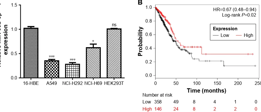
We found that miR-99b-5p expression was significantly reduced in NSCLC cell lines investigated compared with 16-HBE or HEK293T cell line (P,0.001; Figure 1A). Meanwhile, we showed that miR-99b-5p expression was also lower in SCLC cell line than in 16-HBE or HEK293T cell line (P,0.05; Figure 1A). In addition, we analyzed
Figure 1 Expression and clinical significance of miR-99b-5p expression in NSCLC. (A) expression level of mir-99b-5p in nsclc cell lines, sclc cell line and normal cell line. (B) Kaplan–Meier curve of cancer patients with high or low mir-99b-5p expression. *P,0.05, ***P,0.001.
Abbreviations: miR-99b-5p, miRNA-99b-5p; ns, not significant; NSCLC, non-small cell lung cancer; SCLC, small cell lung cancer.
$
+%( $ 1&,+
1&,+ +(.7 QV
3UREDELOLW\
5HODWLYHPL5ES
H[SUHVVLRQ
7LPHPRQWKV
/RZ
1XPEHUDWULVN
+LJK
+5 ± /RJUDQN3
%
([SUHVVLRQ
/RZ +LJK
OncoTargets and Therapy downloaded from https://www.dovepress.com/ by 118.70.13.36 on 25-Aug-2020
Dovepress
liu et al
the effect of miR-99b-5p expression on overall survival of NSCLC patients and found that low miR-99b-5p expression was correlated with poor overall survival (Figure 1B). These results indicated that miR-99b-5p plays a crucial role in the carcinogenesis of NSCLC.
Overexpression of mir-99b-5p inhibits
nsclc cell proliferation, migration and
invasion
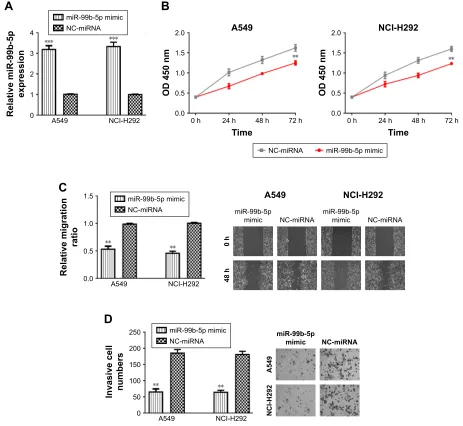
We further investigated the effects of miR-99b-5p on cell proliferation, migration and invasion by introducing the synthetic miRNAs into NSCLC cell lines. We found that miR-99b-5p mimic transfection significantly enhanced the levels of miR-99b-5p in NSCLC cell lines (Figure 2A).
CCK-8 assay revealed that cellular proliferation rate of NSCLC cell lines was significantly reduced by miR-99b-5p mimic compared with NC-miRNA (Figure 2B). Similarly, wound-healing assay demonstrated that cell migration ability was inhibited by miR-99b-5p mimic (Figure 2C). We also found that NSCLC cell invasion ability was remarkably sup-pressed by miR-99b-5p mimic compared with NC-miRNA (Figure 2D). These results indicated that miR-99b-5p over-expression repressed NSCLC cell proliferation, migration and invasion.
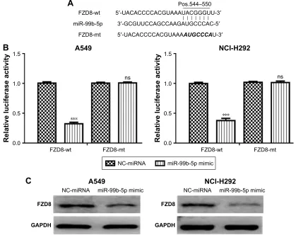
FZD8 was a direct target of mir-99b-5p
TargetScan analysis results showed that miR-99b-5p could bind to the 3′-UTR of FZD8 (Figure 3A). Luciferase activity
$
K
2'QP
K K
7LPH 1&,+
K
PL5ES PLPLF
K
K
1&PL51$
$ 1&,+
PL5ES
PLPLF 1&PL51$
5HODWLYHPLJUDWLRQ
UDWLR
$ 1&,+
PL5ESPLPLF 1&PL51$
PL5ES
PLPLF 1&PL51$
$
1&,+
,QYDVLYHFHOO QXPEHUV
$ 1&,+
PL5ESPLPLF 1&PL51$
5HODWLYHPL5ES
H[SUHVVLRQ
$ 1&,+
PL5ESPLPLF 1&PL51$
$
%
&
'
1&PL51$ PL5ESPLPLF K
2'QP
K K
7LPH
K
Figure 2 Overexpression of mir-99b-5p inhibits nsclc cell proliferation, migration and invasion. (A) mir-99b-5p expression, (B) cell proliferation, (C) cell migration and (D) cell invasion in cells transfected with mir-99b-5p mimic or nc-mirna. **P,0.01, ***P,0.001.
Abbreviations: mir-99b-5p, mirna-99b-5p; nc-mirna, negative control mirna; nsclc, non-small cell lung cancer.
OncoTargets and Therapy downloaded from https://www.dovepress.com/ by 118.70.13.36 on 25-Aug-2020
Dovepress liu et al
reporter assay validated that relative luciferase activity in cells transfected with FZD8-wt was repressed by miR-99b-5p mimic (Figure 3B). Furthermore, we found that FZD8 protein expression in NSCLC cells was inhibited by miR-99b-5p mimic (Figure 3C). Thus, these results implied that FZD8 was a direct target of miR-99b-5p.
FZD8 was involved in mir-99b-5p
inhibitory effects on nsclc cell
behaviors
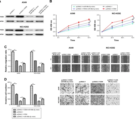
Based on the abovementioned findings, we hypothesized that miR-99b-5p regulates NSCLC cell behaviors through target-ing FZD8. Western blot confirmed the overexpression status of FZD8 after pcDNA3.1-FZD8 transfection (Figure 4A). As shown in Figure 4B–D, the overexpression of FZD8 significantly increased cell proliferation, migration and invasion. Moreover, the inhibitory effects of miR-99b-5p on NSCLC cell behaviors could partially reversed by FZD8 overexpression (Figure 4B–D).
Discussion
Recent studies have investigated that biomarkers are associ-ated with cancer progression with the aim to develop novel treatment strategies for cancer.17–19 Previously, miR-99b-5p
was reported to display a crucial role in human cancers to regulate various cellular behaviors.13–15 It was found that
miR-99b-5p functions as a tumor suppressor in gastric cancer through targeting insulin-like growth factor 1 receptor.13
Furthermore, miR-99b-5p was found to suppress colorectal cancer metastasis to liver through targeting mTOR.15 However,
the role of miR-99b-5p in NSCLC remains unclear till now. We found that miR-99b-5p expression was significantly reduced in NSCLC cell lines compared with normal cell line. Moreover, we found that low miR-99b-5p expression was a predictor of poor overall survival in NSCLC patients. These results suggested the tumor-suppressive role of miR-99b-5p in NSCLC, which is in consistent with the previous studies.13–15
In addition, we confirmed that the elevated expression of miR-99b-5p could inhibit NSCLC cell proliferation, migration
3RV± ′8$&$&&&&$&*8$$$8$&***88′
′*&*88&&$*&&$$*$8*&&&$&′
′8$&$&&&&$&*8$$$$8*&&&$8′
)='ZW PL5ES
)='PW
$
5HODWLYHOXFLIHUDVHDFWLYLW\
)='ZW
$
)='PW QV
5HODWLYHOXFLIHUDVHDFWLYLW\
)='ZW
1&,+
)='PW QV
1&PL51$ PL5ESPLPLF
%
1&PL51$
)='
*$3'+
PL5ESPLPLF
$
1&PL51$
)='
*$3'+
PL5ESPLPLF
1&,+
&
Figure 3 FZD8 was a direct target of mir-99b-5p. (A) The predicted mir-99b-5p binding site within the 3′-UTr of FZD8 and a mutated version are shown. (B) luciferase reporter assay illustrating direct binding of mir-99b-5p to the FZD8-wt, but not FZD8-mt. (C) The protein expression of FZD8 was regulated by mir-99b-5p in nsclc cells. ***P,0.001.
Abbreviations: miR-99b-5p, miRNA-99b-5p; mt, mutant; NC-miRNA, negative control miRNA; ns, not significant; NSCLC, non-small cell lung cancer; wt, wild-type.
OncoTargets and Therapy downloaded from https://www.dovepress.com/ by 118.70.13.36 on 25-Aug-2020
Dovepress
liu et al
and invasion in vitro. These results collectively indicated that miR-99b-5p plays a crucial role in NSCLC carcinogenesis.
FZD8, a member of FZD receptor family, is reported to be a crucial receptor for the Wnt/β-catenin signaling pathway.20
FZD8 was also reported to be a target of p53 to promote prostate cancer metastasis to bone through the Wnt/β-catenin signaling pathway.21 In addition, FZD8 expression was found
to be directly regulated by miRNAs in human cancers.22,23
For example, miR-520b was found to suppress spinal osteo-sarcoma cell proliferation, migration and invasion via down-regulation of FZD8.22 In addition, miR-100 could suppress
breast cancer cell migration and invasion by targeting FZD-8 and inhibiting Wnt/β-catenin signaling pathway.23
Here, by TargetScan analysis, we identified that FZD8 might be a target of miR-99b-5p. Luciferase activity reporter assay and Western blot assay validated this prediction. Moreover, rescue experiments showed that the overexpression of FZD8 could partially reversed the inhibitory effects of miR-99b-5p on NSCLC cell behaviors.
Conclusion
miR-99b-5p plays a crucial role in regulating NSCLC prolif-eration, migration and invasion through suppressing FZD8. These results indicated that miR-99b-5p functions as a tumor suppressor in NSCLC and might be developed as therapeutic target for NSCLC in the near future.
C
A549 NCI-H2922.0 **
* *
**
1.5
Relative migration ratio
1.0
0.5
0.0
A549 NCI-H292
0
h
48
h
pcDNA3.1 pcDNA3.1-FZD8
pcDNA3.1+miR-99b-5p mimic miR-99b-5p mimicpcDNA3.1-FZD8+pcDNA3.1+miR-99b-5p mimic pcDNA3.1-FZD8 pcDNA3.1 miR-99b-5p mimicpcDNA3.1-FZD8+
pcDNA3.1 pcDNA3.1-FZD8 pcDNA3.1+miR-99b-5p mimic pcDNA3.1-FZD8+
miR-99b-5p mimic
A54
9
NCI-H29
2
D
400 *** *
**
300
Invasive cell numbers
200
100
0
A549 NCI-H292
pcDNA3.1-FZD8+miR-99b-5p mimic pcDNA3.1
pcDNA3.1-FZD8 pcDNA3.1+miR-99b-5p mimic
A
B
2.5
**
*
2.0
1.5
OD 450 nm
1.0
0.5
0.0
0 h 24 h
Time A549
48 h 72 h 2.0
**
*
1.5
OD 450 nm
1.0
0.5
0.0
0 h 24 h
Time NCI-H292
48 h 72 h
FZD8
A549
pcDNA3. 1
pcDNA3.1-FZD 8
pcDNA3.1+ miR-99b-5p mimi
c
pcDNA3.1-FZD8+ miR-99b-5p mimi
c
GAPDH
FZD8
GAPDH
NCI-H292
pcDNA3.1+miR-99b-5p mimic pcDNA3.1-FZD8+miR-99b-5p mimic
pcDNA3.1 pcDNA3.1-FZD8
Figure 4 Overexpression of FZD8 reversed the inhibitory effects of mir-99b-5p on nsclc cell behaviors. (A) FZD8 expression, (B) cell proliferation, (C) cell migration and (D) cell invasion in cells transfected with pcDna3.1-FZD8, pcDna3.1, pcDna3.1-FZD8 and mir-99b-5p mimic or pcDna3.1 and mir-99b-5p. *P,0.05, **P,0.01. Abbreviations: miR-99b-5p, miRNA-99b-5p; ns, not significant; NSCLC, non-small cell lung cancer.
OncoTargets and Therapy downloaded from https://www.dovepress.com/ by 118.70.13.36 on 25-Aug-2020
OncoTargets and Therapy
Publish your work in this journal
Submit your manuscript here: http://www.dovepress.com/oncotargets-and-therapy-journal OncoTargets and Therapy is an international, peer-reviewed, open access journal focusing on the pathological basis of all cancers, potential targets for therapy and treatment protocols employed to improve the management of cancer patients. The journal also focuses on the impact of management programs and new therapeutic agents and protocols on
patient perspectives such as quality of life, adherence and satisfaction. The manuscript management system is completely online and includes a very quick and fair peer-review system, which is all easy to use. Visit http://www.dovepress.com/testimonials.php to read real quotes from published authors.
Dovepress
Dove
press
liu et al
Acknowledgment
This work was supported by Yunnan Health Science and Technology Project (2018NS0276).
Author contributions
All authors contributed to data analysis, drafting and revising the article, gave final approval of the version to be published, and agree to be accountable for all aspects of the work.
Disclosure
The authors report no conflicts of interest in this work.
References
1. Bray F, Ferlay J, Soerjomataram I, Siegel RL, Torre LA, Jemal A. Global cancer statistics 2018: GLOBOCAN estimates of incidence and mortality worldwide for 36 cancers in 185 countries. CA Cancer J Clin. 2018;68(6):394–424. doi:10.3322/caac.21492
2. Cai Y, Dong ZY, Wang JY. MiR-520b inhibited metastasis and pro-liferation of non-small cell lung cancer by targeting CHAF1A. Eur Rev Med Pharmacol Sci. 2018;22(22):7742–7749. doi:10.26355/ eurrev_201811_16396
3. Song YJ, Tan J, Gao XH, Wang LX. Integrated analysis reveals key genes with prognostic value in lung adenocarcinoma. Cancer Manag Res. 2018;10:6097–6108. doi:10.2147/CMAR.S168636
4. Inamura K, Ishikawa Y. MicroRNA in lung cancer: novel biomarkers and potential tools for treatment. J Clin Med. 2016;5(3):36. doi:10.3390/ jcm5030036
5. Inamura K. Diagnostic and therapeutic potential of microRNAs in lung cancer. Cancers (Basel). 2017;9(12):E49. doi:10.3390/cancers9050049 6. Zhang B, Pan X, Cobb GP, Anderson TA. microRNAs as oncogenes
and tumor suppressors. Dev Biol. 2007;302(1):1–2. doi:10.1016/j. ydbio.2006.08.028
7. Guan P, Yin Z, Li X, Wu W, Zhou B. Meta-analysis of human lung cancer microRNA expression profiling studies comparing cancer tissues with normal tissues. J Exp Clin Cancer Res. 2012;31(1):54. doi:10.1186/1756-9966-31-95
8. Wang K, Chen M, Wu W. Analysis of microRNA (miRNA) expres-sion profiles reveals 11 key biomarkers associated with non-small cell lung cancer. World J Surg Oncol. 2017;15:175. doi:10.1186/ s12957-017-1244-y
9. Chen Y, Min L, Ren C, et al. miRNA-148a serves as a prognostic factor and suppresses migration and invasion through Wnt1 in non-small cell lung cancer. PLoS One. 2017;12(2):e0171751. doi:10.1371/journal. pone.0171751
10. Wang Z, Liu Z, Fang X, Yang H. MiR-142-5p suppresses tumorigen-esis by targeting PIK3CA in non-small cell lung cancer. Cell Physiol Biochem. 2017;43(6):2505–2515. doi:10.1159/000484459
11. Wang HQ, Xu ML, Ma J, Zhang Y, Xie CH. Frizzled-8 as a putative therapeutic target in human lung cancer. Biochem Biophys Res Commun. 2012;417(1):62–66.
12. Jin Y, Liu Y, Zhang J, et al. The expression of miR-375 is associ-ated with carcinogenesis in three subtypes of lung cancer. PLoS One. 2015;10(12):e0144187. doi:10.1371/journal.pone.0144187
13. Wang Z, Zhao Z, Yang Y, et al. MiR-99b-5p and miR-203a-3p function as tumor suppressors by targeting IGF-1R in gastric cancer. Sci Rep. 2018;8(1):10119. doi:10.1038/s41598-018-27583-y
14. Lukamowicz-Rajska M, Mittmann C, Prummer M, et al. MiR-99b-5p expression and response to tyrosine kinase inhibitor treatment in clear cell renal cell carcinoma patients. Oncotarget. 2016;7(48):78433–78447. doi:10.18632/oncotarget.12618
15. Li W, Chang J, Wang S, et al. miRNA-99b-5p suppresses liver metas-tasis of colorectal cancer by down-regulating mTOR. Oncotarget. 2015;6(27):24448–24462. doi:10.18632/oncotarget.4423
16. Lanczky A, Nagy A, Bottai G, et al. miRpower: a web-tool to validate survival-associated miRNAs utilizing expression data from 2,178 breast cancer patients. Breast Cancer Res Treat. 2016;160(3):439–446. doi:10.1007/s10549-016-4013-7
17. Zhao X, Tang D, Yang T, Wang C. Facile preparation of biocompatible nanostructured lipid carrier with ultra-small size as a tumor-penetration delivery system. Colloids Surf B Biointerfaces. 2018;170:355–363. doi:10.1016/j.colsurfb.2018.06.017
18. Wang C, Chen S, Wang Y, et al. Lipase-triggered water-responsive “Pandora’s Box” for cancer therapy: toward induced neighboring effect and enhanced drug penetration. Adv Mater. 2018;30(14):e1706407. doi:10.1002/adma.201706407
19. Tang D, Zhao X, Zhang L, Wang Z, Wang C. Identification of hub genes to regulate breast cancer metastasis to brain by bioinformatics analyses. J Cell Biochem. Epub 2018 Dec 03. doi:10.1002/jcb.28228 20. Murillo-Garzón V, Gorroño-Etxebarria I, Åkerfelt M, et al. Frizzled-8
integrates Wnt-11 and transforming growth factor-β signaling in
prostate cancer. Nat Commun. 2018;9:1747. doi:10.1038/s41467-018- 04042-w
21. Li Q, Ye L, Zhang X, et al. FZD8, a target of p53, promotes bone
metas-tasis in prostate cancer by activating canonical Wnt/β-catenin signaling.
Cancer Lett. 2017;402:166–176. doi:10.1016/j.canlet.2017.05.029 22. Wang J, Pang W, Zuo Z, Zhang W, He W. MicroRNA-520b suppresses
proliferation, migration, and invasion of spinal osteosarcoma cells via downregulation of Frizzled-8. Oncol Res. 2017;25(8):1297–1304. doi:10.3727/096504017X14873430389189
23. Jiang Q, He M, Guan S, et al. MicroRNA-100 suppresses the migration and invasion of breast cancer cells by targeting FZD-8 and inhibiting
Wnt/β-catenin signaling pathway. Tumour Biol. 2016;37(4):5001–5011.
doi:10.1007/s13277-015-4342-x
OncoTargets and Therapy downloaded from https://www.dovepress.com/ by 118.70.13.36 on 25-Aug-2020
![(3aS,8aS,9R,9aS) 9 Hydroxy 2,3,3a,7,8,8a,9,9a octahydrofuro[3,2 f]indolizin 6(4H) one](data:image/gif;base64,R0lGODlhAQABAIAAAP///wAAACH5BAEAAAAALAAAAAABAAEAAAICRAEAOw==)