1
MicroRNA cross-involvement in Autism Spectrum Disorders and Atopic Dermatitis: a literature reviewAuthors: Alessandro Tonacci1, Gianluca Bagnato2, Gianluca Pandolfo3, Lucia Billeci1, Francesco Sansone1, Raffaele Conte1, Sebastiano Gangemi2
1Clinical Physiology Institute – National Research Council of Italy (IFC-CNR), Via Moruzzi 1, 56124 Pisa, Italy; 2School and Division of Allergy and Clinical Immunology, Department of Clinical and Experimental Medicine, University Hospital “G. Martino”, Via Consolare Valeria SNC, 98125 Messina, Italy; 3Department of Biomedical and
Dental Sciences and Morphofunctional Imaging, University of Messina, Via Consolare Valeria 1, 98125 Messina, Italy
Corresponding author: Dr. Alessandro Tonacci, Clinical Physiology Institute – National Research Council of Italy
(IFC-CNR), Pisa, Italy. Tel.: +39.050.3152175, Fax: +39 050 315 2166. e-mail: [email protected]
2
AbstractAutism Spectrum Disorders (ASD) are neurodevelopmental disturbances affecting social skills, whose incidence
worldwide is dramatically increasing. Together with the rise of ASD prevalence, several immune conditions are
following the same trend, including Atopic Dermatitis (AD), with a possible clinical relationship with ASD. To date,
their pathogenesis is still unknown, but several studies highlighted the relevance of gene-environment interactions to the
onset of both disorders. Among potential contributing factors, microRNAs (miRNAs), small molecules capable of
controlling gene expression and targeting mRNA transcripts, might represent one of the major circulating link,
unraveling the connections between neurodevelopmental and immune conditions.
We conducted a systematic literature review, under the PRISMA guidelines, trying to define the panel of common
miRNAs involved in both ASD and AD. The review retrieved articles published until December 13, 2018, in PubMed,
ScienceDirect, PsycARTICLES and Google Scholar.
We found a handful works dealing with miRNAs in ASD and AD, with the most overlapping dysregulated miRNAs
being miR-146 and miR-155.
Two possible compounds are abnormally regulated in both ASD and AD subjects, possibly cross-contributing to the
interactions between the two disorders, setting the basis to investigate more precisely the possible link between ASD
and AD from another, not just clinical, perspective.
3
1. IntroductionAutism spectrum disorders (ASD) are a heterogeneous group of neurodevelopmental disorders characterized by
impairments in social interaction and communication and restricted or stereotyped interests and behaviors [1], typically
occurring before the fourth year of life [2]. Their prevalence has dramatically increased in last decades, from 4/10,000
in 2008 [3] to 1/68 cases in children living in the United States nowadays [4], with a gender (males vs. females) ratio of
4:1. It still remains unsolved whether this exponential increment may be due to a better knowledge on this topic and to
an improved awareness along with broadening of the diagnostic criteria or, rather, may reflect a true increase in the
incidence of ASD.
Nonetheless, ASD are the most heritable neuropsychiatric disorders, with genetic contributions accounting for more
than 50% of ASD risk [5-7], and higher risk seen in siblings of autistic children [8]. Although epidemiological studies
provide information on the genetic contribution to ASD, less is known about the putative genes involved and/or the
frequency of specific polymorphisms and variants (single-nucleotide or copy number variants).
Whole-genome and candidate‑gene analyses have shown a complex genetic background of ASD, characterized by high
individual differences and variability, with many ASD-risk genes involved in synaptic plasticity and gene products
modifying synaptic number and strength. In addition to inherited variants, individuals with ASD often carry de novo
genetic variants, defined as variants not present in the parental genome and found for the first time in the proband [9].
Such mutated variants affect biological pathways involved in synaptic plasticity and connectivity at different levels. It
has been proposed that both individual’s genetic background and de novo and rare mutations converge in disrupting
synaptic homeostasis [10].
The multigenic condition of ASD seems also be dependent on gene-environment interactions; epigenetic mechanisms
involving DNA methylation, transcriptional regulations, and post-translational changes in histone proteins, are all
relevant to neurodevelopmental processes that can be affected in-utero by maternal lifestyle factors [11]. Furthermore,
chemicals and/or heavy metals exposure, appears to strongly contribute to ASD development [12-14].
Consistently, recent studies report the clinical association between ASD and atopic disorders, such as asthma or atopic
dermatitis (AD) [15-18], strengthening the link between neurodevelopmental disorders and immune diseases.
In fact, in recent times, a clinical association between the two conditions was hypothesized [16,17], and the
investigation of possible common genetic basis is critical for the current scientific knowledge.
As evidenced by Billeci and colleagues, AD, defined as a chronic inflammatory disease, puts at higher risk of
developing one of more of the other atopic conditions, therefore it is considered as the beginning of the so-called
4
This condition, also determined by a close gene-environment interaction [19], appears to be correlated with a number ofmental health conditions including, according to recent literature, ASD [16].
Among the compounds which could possibly explain this link, up to now hypothesized from a clinical point of view,
microRNAs (miRNAs) were recently seen to play a role in several molecular and cellular mechanisms, including
neurodevelopment, brain plasticity and immunity [20,21].
miRNAs might participate in pathological process both in neurological conditions, including autism, and in atopic
disorders, including AD [22]. The overlapping microRNAs in ASD and AD could therefore allow to explore the role of
genetics in the hypothetical common pathophysiological pathways of these two conditions.
1.1 General insight into microRNAs
miRNAs are very short (18-25 nucleotides), single-stranded non-coding RNAs, able to control gene expression and to
target mRNA transcripts, possibly bringing on their translational degradation or their repression, with particular degrees
of complementarity [23]. The targeting of mRNA transcripts by miRNA occurs as one miRNA is able to target a
number of mRNA transcripts, conversely a single mRNA transcript can be targeted by many miRNAs.
Actually, miRNAs have rapidly induced a great interest in humans, being potential biomarkers for diagnostic and
prognostic aims [24.25], and being the number of classified miRNA increasing, till over 2,500 potential molecules in
the Homo Sapiens genetic makeup [23]. Despite being small molecules, not capable of encoding proteins, miRNAs hold
important structural, regulatory and catalytic functions.
miRNA genes are located in the introns of protein-coding genes or in independent non-coding DNA loci [26], whereas
nearly half of the total miRNAs are pooled on chromosomes with a common promoter [27].
The biogenesis of miRNA is extremely complex, consisting of several phases, including: i) in the nucleus, the
transcription of miRNA genes into primary miRNA transcripts by RNA polymerase II; ii) the freeing of pre-miRNA
hairpin, through the trim of the primary miRNA transcripts by the RNAse III Drosha endonuclease; iii) the active
exportation of the pre-miRNA hairpin out of the nucleus in a process involving the nucleocytoplasmic shuttler
Exportin-5; iv) the final maturation, in the cytoplasm, processed by Dicer RNase III endonuclease, splitting the
pre-miRNA into a single-stranded mature pre-miRNA [28]; v) the binding of the mature pre-miRNA to proteins of the Ago family,
and vi) the assembly of the RISC complexes together in order to employ its physiological functions.
The mature miRNA, once incorporated into the RISC, induces posttranscriptional gene silencing by binding RISC to be
partially complementary to the target mRNA found mainly within the 3’-untranslated region (UTR) [29,30].
Flaws in miRNA expression deeply affect several pathways related to cell regulation, including apoptosis, stress
5
mRNAs, while around 60% of human protein coding-genes are represented by conserved targets of miRNAs, thus anumber of mRNA targets are regulated by miRNAs [34].
1.2 miRNAs linked to brain function
MiRNAs approximately regulate two-thirds of human mRNAs [34] and are, as much as for the 70%, expressed in the
central nervous system (CNS), including the brain and spinal cord [35,36]. Their changes during childhood are different
depending on the affected brain region [37].
In particular, miRNAs are abundant in neurons and glia, often placed at the synaptic level, able to regulate the structure
of the dendritic spine, as happens with miR-134, that reduces, by targeting spine growth-promoting kinase Limk1, spine
growth [38].
Indeed, dendritic spines are bulges on a dendritic tree of a neuron, composing the post-synaptic termination of a
synapse, reflecting – through their structure – the degree of brain maturation and, somehow, brain plasticity.
Their density is abnormal in several conditions, including schizophrenia (dendritic spines loss) and ASD, the latter
featuring an increase in the quantity of spines in specific brain areas [39-42].
Several other miRNAs are associated with dendritic spine structure, including miR-125b [43], miR-132 [44], miR-137
[45] and miR-138 [46].
Overall, several miRNAs affect brain functions and development, neuronal plasticity, maturation and differentiation
[20,47]. Dysregulation of miRNA expression is particularly frequent within several neurological disorders, including
ASD, therefore the association between some common miRNA families and ASD, despite still largely unknown, is
nowadays less unclear than in the past.
1.3 miRNAs and skin disorders
Recently, many evidences were published about the role of miRNAs in several cellular processes, including immune
response, DNA repair, apoptosis, proliferation and differentiation [48], but also in morphogenesis, differentiation,
wound healing, psoriasis, and AD [49-51]. Specifically, also AD pathogenesis is associated with a complex
gene-environment interaction, as well as with an alteration of the skin barrier function, and a deregulation of the immune
system [52]. Several miRNAs, including miR-146a, miR-155, miR-203 and miR-483–5p, are also differentially
expressed in AD and in other immunologic and inflammatory disorders.
6
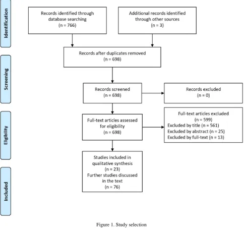
A literature review of the articles published between January 1, 2005 and December 13, 2018, was conducted in PubMed,ScienceDirect, PsycARTICLES and Google Scholar following the PRISMA guidelines.
Studies about ASD
The search strategy for this part was as follows: (("micrornas"[MeSH Terms] OR "micrornas"[All Fields] OR
"microrna"[All Fields]) AND (("autistic disorder"[MeSH Terms] OR ("autistic"[All Fields] AND "disorder"[All Fields])
OR "autistic disorder"[All Fields] OR "autism"[All Fields]) OR ("autism spectrum disorder"[MeSH Terms] OR
("autism"[All Fields] AND "spectrum"[All Fields] AND "disorder"[All Fields]) OR "autism spectrum disorder"[All
Fields]))).
Studies about AD
In this part, the search strategy was as follows: ((“microRNAs” [MeSH Terms]) AND (“skin” [MeSH Terms] OR
“dermatitis” [MeSH Terms] OR “urticaria” [MeSH Terms] OR “eczema” [MeSH Terms] OR “hypersensitivity” [MeSH
Terms])).
Overall, the search was limited to articles describing studies conducted on humans published in peer-reviewed journals.
After having discarded duplicates, the obtained results were sorted by relevance and the most significant works related to
ASD and miRNAs and to AD and miRNAs were selected. We will first present the results from the literature review and
then discuss the possible associations between miRNAs in ASD and AD according to such findings.
3. Results
7
Figure 1. Study selection3.1 Studies about ASD
According to the literature review, a number of miRNAs were found to be associated to ASD (Table 1). Specifically,
the most overlapping dysregulated miRNAs appeared to be let-7, miR-23, miR-106 and miR-146.
Study N
(case/control)
Design Findings
Up-regulated miRNA Down-regulated
miRNA
Abu-Elneel
et al. (2008)
[53]
26 (13/13) Measure of the expression
level of 466 human miRNAs
from postmortem cerebellar
miR-106a, miR-106b,
miR-140, miR-146b,
miR-181d, miR-193b,
7, 15a,
miR-15b, miR-21, miR-23a,
miR-8
tissue by multiplex real-timePCR. 377 miRNAs detected
and used for further analysis
miR-320a, miR-381,
miR-432, miR-539,
miR-550, miR-652
95, miR-128, miR-129,
miR-132, miR-148b, miR-212, miR-431, miR-484, miR-598 Sarachana et al. (2010) [54]
14 (5/9) Lymphoblasts derived from
peripheral lymphocytes were
obtained. miRNA expression
profiling performed by
high-throughput miRNA microarray
analysis. Differentially
expressed miRNAs confirmed
by qRT-PCR analysis, putative
target genes of two of the
confirmed miRNA validated
by knockdown and
overexpression of the
respective miRNAs miR-16-2, miR-106b, miR-132, miR-133b, miR-136, miR-139, miR-148b, miR-153, miR-182, miR-189, miR-190, miR-199b, miR-211, miR-219, miR-326, miR-367, miR-455, miR-495, miR-518a, miR-520b miR-23a, miR-23b,
25, 29b,
miR-30e, miR-93, miR-103,
miR-107, miR-185,
miR-186, miR-191,
miR-194, miR-195,
miR-205, miR-342,
miR-346,
376a-AS, 451,
miR-519c, miR-524
Talebizadeh
et al. (2008)
[55]
12 (6/6) 6 ASD (3 males, aged 5, 12
and 14 years, and 3 females,
aged 6, 11 and 13 years), 6
age- and gender-matched TD
controls. Lymphoblastoid cell
lines, quantitative PCR
miR-23a, miR-23b,
miR-132, miR-146a,
miR-146b, miR-663
92, 320,
miR-363
Mundalil
Vasu et al.
(2014) [56]
110 (55/55) 55 ASD (48 males, 6 females,
aged 11.29±5.45 years), 55 TD
controls (41 males, 14
females, aged 11.3±2.37
years). RNA extracted from
serum, mature miRNAs
selectively converted into
miR-19b-3p,
27a-3p, 101-27a-3p,
9
cDNA. The expression of 125mature miRNAs was
compared between pooled
control and ASD samples. The
differential expression of 14
miRNAs further validated by
SYBR Green quantitative PCR
of individual samples. Target
genes and pathways of
miRNAs predicted by DIANA
mirPath software
Popov et al.
(2012) [57]
55 (30/25) 30 ASD (24 males, 6 females,
aged 3-20), 25 TD controls (20
males, 5 females, aged 3-20
years). Whole blood
collection, analysis of gene
expression changes applying
LC expression profiling
service, using pooled whole
blood-derived total RNA
samples
miR-486-3p
Seno et al.
(2011) [58]
42 (20/22) 20 severe ASD (13 males and
7 females), 22 unaffected
siblings (19 males and 3
females). Lymphoblastoid cell
lines, RNA was extracted and
assayed using Illumina gene
and miRNA expression arrays.
Control quality in BeadStudio
(Illumina)
miR-10a, miR-30a,
miR-181a, miR-181b,
miR-181c,
199b-5p, 338-3p,
miR-486-3p, miR-486-5p,
miR-500, miR-502-3p,
miR-548
miR-199a-5p,
10
Mor et al.(2015) [59]
24 (12/12) Brain tissue samples taken
from postmortem Brodmann’s
area 10
miR-7-5p, miR-19a-3p,
miR-19b-3p,
21-3p, 21-5p,
miR-142-3p, miR-142-5p,
miR-144-3p,
146a-5p, 155-146a-5p,
miR-219-5p, miR-338-5p,
miR-379-5p, miR-451a,
miR-494, miR-3168
miR-34a-5p,
92b-3p, 211-5p,
miR-3960
Ander et al.
(2015) [60]
18 (10/8) Brain tissue samples taken
from postmortem Brodmann’s
areas 22, 41, 42
664-3p,
miR-4709-3p, miR-4753-5p
1, 297,
miR-4742-3p
Wu et al.
(2016) [61]
56 (28/28) Tissue samples taken from
postmortem cerebellar cortex,
Brodmann area 9
miR-10a-5p,
18b-5p, 20b-18b-5p,
miR-21-3p, miR-23a-3p,
miR-107,
129-2-3p, 130b-5p,
miR-148a-3p, miR-155-5p,
218-2-3p,
miR-221-3p, miR-223-3p,
miR-335-3p,
363-3p, 424-363-3p,
miR-424-5p, miR-425-3p,
449b-5p,
miR-450b-5p, miR-484,
miR-629-5p,
651-5p, 708-651-5p,
miR-766-3p, miR-874-3p,
miR-887-3p, miR-940,
1277-3p,
miR-miR-204-3p,
491-5p, 619-491-5p,
11
3938, miR-2277-5p,let-7g-3p
Huang et al.
(2015) [62]
40 (20/20) Peripheral blood sample taken,
microarray (5 ASD/5 controls)
and quantitative Real-Time
PCR (15 ASD/15 controls)
miR-34b-3p,
34c-3p, 483-5p,
494, 564,
miR-642a-3p, miR-574-5p, miR-575, miR-921, miR-1246, miR-1249, miR-1273c, miR-4270, miR-4299, miR-4436a, miR-4443, miR-4516, miR-4669, miR-4721, 4728-5p,
4788, 5739,
miR-6086, miR-6125
miR-15a-5p,
15b-5p, 16-15b-5p,
miR-19b-3p, miR-20a-5p,
miR-92a-3p,
103a-3p, 195-5p,
miR-451a, miR-574-3p,
miR-940, miR-1228-3p,
3613-3p,
miR-3935, miR-4436b-5p,
4665-5p,
miR-4700-3p, 7a-5p,
let-7d-5p, let-7f-5p
Hicks et al.
(2016) [63]
45 (24/21) Salivary samples miR-7-5p, miR-28-5p,
miR-127-3p,
140-3p, 191-5p,
miR-218-5p, miR-335-3p,
628-5p,
miR-2467-5p, miR-3529-3p
miR-23a-3p,
27a-3p, 30e-5p,
miR-32-5p
Nguyen et
al. (2016)
[64]
14 (8/6) Samples taken from olfactory
mucosal stem cells and skin
fibroblasts or Peripheral Blood
Mononuclear Cells. Measured
through microarray and
quantitative Real-Time PCR
validation
miR-146a miR-221, miR-654-5p,
12
Kichukovaet al. (2017)
[65]
60 (30/30) Blood samples. Quantitative
Real-Time PCR validation
18b-3p,
miR-106b-5p, miR-142-3p,
miR-210-5p,
365a-3p, 374b-5p,
miR-619-5p, miR-664a-3p,
3620-3p,
miR-4489, miR-8052
hsa-let-7i-3p, miR
15a5p, miR 20b3p, miR
-29c-5p, miR -96-5p,
miR 1455p, miR
-183-5p, miR -193b-3p,
miR -197-5p,
miR-199a-5p, miR -301a-3p,
miR 3283p, miR
-424-5p, miR -486-3p,
miR 487b3p, miR
-500a-5p, miR -504-5p,
miR 5765p, miR
-587-3p, miR-589-3p,
miR 664b3p, miR
-671-3p, miR -3064-5p,
miR 3135a, miR
-3674, miR -3687,
miR-3909, miR -6799-3p,
miR -6849-3p
Jyonouchi et
al. (2017)
[66]
96 (69/27) Peripheral blood monocytes
samples. miRNA expression
determined by
high-throughput sequencing
1,
7a-2, 7a-3,
let-7f-1, let-7f-2,
hsa-let-7g, hsa-let-7i,
17, 26a-2,
30b, 30c-1,
30c-2, 98,
106b, 130a,
148a, 148b,
150, 186,
301a, 374b,
miR-hsa-let-7b, miR-15a,
miR-15b, miR-16-1,
miR-16-2, miR-18a,
miR-19a, miR-19b-1,
miR-19b-2, miR-20a,
21, 27a,
27b, 29a,
miR-29b-1, miR-29b-2,
miR-29c, miR-30e,
miR-93, miR-101-1,
miR-103a-13
494, 1248,miR-3607, miR-3609
1, 103a-2,
107, 126,
142, 145,
146a, 151a,
miR-181a-1, miR-181a-2,
miR-199b, miR-221,
miR-222, miR-320a,
miR-376c, miR-409,
miR-423, miR-484,
miR-625, miR-4433b,
5701-1,
miR-5701-2
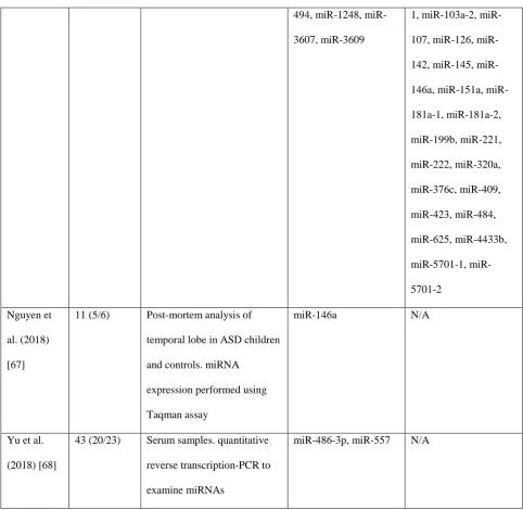
Nguyen et
al. (2018)
[67]
11 (5/6) Post-mortem analysis of
temporal lobe in ASD children
and controls. miRNA
expression performed using
Taqman assay
miR-146a N/A
Yu et al.
(2018) [68]
43 (20/23) Serum samples. quantitative
reverse transcription-PCR to
examine miRNAs
miR-486-3p, miR-557 N/A
Table 1. MicroRNAs directly involved in autism
3.2 Studies about AD
A few works studied microRNAs involved in AD (Table 2). Here, the main dysregulated miRNAs are 146,
miR-155, and miR-203.
Summarizing, the association between ASD and AD revealed a common unbalance for miR-146 and miR-155.
Study N (case/control) Design Findings
14
Sonkolyet al.
(2010)
[49]
47 (18/29) Skin
samples
miR-155
Lv et al.
(2014)
[69]
58 (30/28) Serum and
urine samples miR-203, miR-483-5p (serum) miR-203 (urine) Ralfkiaer et al. (2014) [70]
75 (20/55)* Skin
samples
miR-149, miR-Plus-C1070,
miR-205, miR-141, miR-23b,
miR-221, miR-27b, miR-203,
miR-7b, miR-19b, miR-27a,
455-3p, 200a,
miR-211, miR-23a, miR-214
181a, 342-5p,
miR-766, miR-7i, miR-186,
miR-342-3p, miR-664, miR-425, miR-9,
331-3p, 146b-5p,
10a, 663, 937,
miR-361-3p, miR-605, miR-146a,
miR-940, miR-150, miR-1913,
miR-155, miR-302c
Rebane
et al.
(2014)
[71]
18 (9/9) Skin
samples
miR-146a
Ma et al.
(2015)
[72]
64 (33/31) Skin
samples miR-155 Ding et al. (2016) [73]
22 (14/8) Skin
samples
148b, 152,
miR-324
Yang et
al.
(2017)
[74]
37 (37/0) Skin
samples
15
Table 2. MicroRNAs in atopic dermatitis (*control cohort represented by patients with early-stage mycosis fungoides(MF1))
4. Discussion
4.1 The overlap between atopy and autism
Recent data strongly support the clinical association between atopy and ASD [15,16]. It has been widely demonstrated
that allergic diseases, especially food allergies, are more frequent among ASD children [75,76].
Notably, a large observational study, comparing 14,812 atopic subjects with 6,944 non-atopic subjects, with no lifetime
atopic disease, highlighted a strong association between atopy and the risk of developing ASD [77]. Furthermore, also
autoimmune disorders, including psoriasis (2-fold risk) are frequently identified in ASD [78].
Beside the robust clinical evidence for the association between atopy and ASD, an intriguing neuroinflammatory
hypothesis has been advanced for ASD, involving the disruption of the brain blood barrier induced by inflammatory
molecules, brain mast cells activation and mast cells-microglia interactions [79].
In addition, specific environmental factors, including infectious pathogens, food allergens, toxins, toxic metals (e.g.,
aluminum, lead, mercury) may negatively act on neurodevelopment through the alteration of the immune response
[80-82].
However, the hypothesis that the pro-inflammatory cascade induced by AD could lead, in the presence of genetic
susceptibility, to ASD is supported by the clinical association and by a shared pattern of cellular damage, with
epigenetic changes as a common pathogenic mechanism.
4.2 Role for overlapping miRNAs in ASD and AD
The main literature finding concerning miRNAs in AD and ASD is represented by miR-146a. miR-146a, upregulated in
various neurodevelopmental disorders [64], was reported to be highly expressed throughout the cortex, hippocampus,
and amygdala, key structures for higher cognitive functioning [83]. Furthermore, it was demonstrated that reproducing
abnormal miR-146a expression in mouse primary cell cultures leads to impaired neuronal dendritic arborization -
producing shriveled dendritic trees with branching points at more proximal levels compared to controls, proving the
defective neural connectivity typical of ASD - and to increased astrocyte glutamate uptake capacities [64], in turn
modifying fast synaptic transmission at the CNS level. miR-146a, expressed in the developing brain, is enclosed within
neurons, with poor expression in the glial lineage in adult mice. However, it generally inhibits the expression of
16
In addition, the neuron excitation at cortex is probably affected by miR-146a deregulation through the involvement ofpotassium two pore domain channel subfamily K member 2 (KCNK2), having a key role in neural excitability and
migration at the cortex level of the developing mice, a critical issue in ASD.
Furthermore, miR-146a expression contributes to neuroinflammation in the brain of ASD subjects, having a role in
immune system regulation.
Its function in the regulation of inflammatory processes could partially fill the gap between ASD (and
neurodevelopmental disorders in general) and AD (and atopic conditions in extenso).
It is evident indeed that an increased miR-146a expression is present in the lesional skin of AD patients [84], as it
inhibits nuclear factor κ B (NF-κB)-mediated proinflammatory cytokines and chemokines, bringing to alleviation of the
inflammation directly linked to AD and similar conditions [71].
In AD, during skin inflammation, miR-146a is increased in keratinocytes, controlling chronic inflammatory processes
triggered by IFN-γ and the activation of NF-κB. Indeed, the relevance of miR-146a in inflammatory skin disorders is
confirmed by evidence from psoriasis research [85]. Furthermore, the expression of miR-146a is strongly dependent on
NF-κB, and the miRNA has been shown to suppress the NF-κB signaling pathway through a direct targeting of a
number of compounds, including IL-1 receptor–associated kinase 1 (IRAK1), TNF receptor–associated factor 6 [86],
v-rel avian reticuloendotheliosis viral oncogene homolog B (RELB) [87], and CARD10 [88].
Moreover, it was discovered that mice with a deficiency of miR-146a develop a late autoimmunity caused by an
impaired activation of NF-κB in T cells and signal transducer and STAT1 activator in regulatory T cells [89].
Of note, previous work demonstrated that both an enhanced opioidergic activity and reduced vitamin D levels could
represent shared features of AD [90] and ASD [91], and possibly miR-146a and miR-155 could interact with the genetic
milieu in subjects with these disorders. Indeed, both miR-146a and miR-155 have been tested in models of LPS
tolerance and miR-146 was able to amplify the severity of morphine-mediated hyper-inflammation [92].
Finally, the regulatory role of miR-146a also occurs at lung alveolar epithelial cells, where the release of IL-8 and
CCL5 occurs independently from IL-1β signaling.
Concerning miR-155, its role in ASD is not yet known, whereas in AD it appears to modulate T helper type 17 (Th17)
cells differentiation and function [72] and to directly target the suppressor of cytokine signalling-1 (SOCS1) gene,
taking part in a negative feedback loop to attenuate cytokine signaling [93]. Interestingly, miR-155 is also linked to
inflammation and immunity, thanks to its potent upregulation in immune cell lineages, including lymphocytes,
fibroblasts, macrophages, mast cells, and dendritic cells, in turn implicated in the pathogenesis of chronic skin
inflammation [94-99]. Briefly, miR-155 seems to be also involved in the regulation of T-cell responses through a
17
increased expression, in peripheral CD4 T cells of AD patients, was correlated to disease severity, supporting its role inAD pathogenesis [72].
5. Conclusions
Beyond the clinical evidence [16,17], a possible, yet speculative, role for genetics (miRNAs in particular) can be
hypothesized to justify the clinical association between AD and ASD.
Yet, both miR-146a and 155 appear to be involved in this common pathogenetic pathway, despite being the role of the
latter still poorly known. However, several other aspects differentiating these diseases remain elusive, including the
identification of putative environmental injuries and the complex role of vitamin D in immune and neurologic disorders.
Therefore, further studies focusing on the association between vitamin D and opiod receptors in skin and neurologic
disorders should investigate the role of target genes for common dysregulated miRNAs, in order to discover specific
overlapping features of these conditions.
It remains evident that an inflammatory component is active in both diseases and the actual data support future
applications for miR-146a both as a biomarker and as a target for therapy.
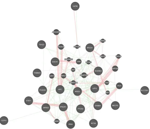
Interestingly, it can be speculated that a deregulation of miR-146a occurs earlier, during embryonic development, thus
participating in the development of ASD. Apart from the well-described effect on NF-κB activity and the associated
inflammatory pathways strictly linking miR-146a with AD, the deregulation of miR-146a and miR-155 could influence
a wide range of their validated targets (Figure 2), essential for brain development and function. However, it remains
complex to determine the major source (skin vs brain) of miR-146a and/or miR-155 and whether they are potentiating
each other or having more organ- or disease-specific effects. These aspects warrant future larger longitudinal studies.
Finally, a better understanding of the link ASD/AD might be useful to investigate whether a specific miRNA could act
as a biomarker for the risk of developing ASD for patients with AD and potentially represent a target for ASD
19
Acknowledgment: The authors wish to thank the library management service of the National Research Council ofItaly, Pisa Headquarter, for assistance in article retrieval.
Author contribution: Literature search, A.T., G.B., S.G.; Conceptualization, G.B., G.P., S.G.; Bias assessment, A.T.,
L.B., F.S., R.C.; Study selection, A.T., G.B., G.P., L.B., S.G.; Manuscript drafting, A.T., G.B., G.P., F.S., R.C., S.G.;
Critical revision and approval of the manuscript, All Authors.
Conflict of Interest: Author A.T., Author G.B., Author G.P., Author L.B., Author F.S., Author R.C. and Author S.G.
20
References1. American Psychiatric Association. Diagnostic and statistical manual of mental disorders, 5th ed. Washington, DC,
American Psychiatric Association, 2013.
2. Barger, B.D.; Campbell, J.M.; McDonough, J.D. Prevalence and onset of regression within autism spectrum
disorders: a meta-analytic review. J Autism Dev Disord. 2015, 43(4), 817-828.
3. CDC. Prevalence of autism spectrum disorders–autism and developmental disabilities monitoring network, 14 sites,
United States, 2008. MMWR Surveillance Summaries 2012, 61, 1–19.
4. Centers for Disease Control and Prevention (CDC), United States Department of Health and Human Services, 2014.
http://www.cdc.gov/ncbddd/autism/states/comm_report_autism_2014.pdf. Accessed December 10, 2018.
5. Gaugler, T.; Klei, L.; Sanders, S.J.; Bodea, C.A.; Goldberg, A.P.; Lee, A.B., et al. Most genetic risk for autism
resides with common variation. Nat Genet. 2014, 46(8), 881–885.
6. Gupta, A.R.; State, M.W. Recent advances in the genetics of autism. Biol Psychiatry. 2007, 61, 429–437.
7. Beaudet, A.L. Autism: highly heritable but not inherited. Nat Med. 2007, 13, 534–536.
8. Ozonoff, S.; Young, G.S.; Carter, A.; Messinger, D.; Yirmiya, N.; Zwaigenbaum, L., et al. Recurrence risk for autism
spectrum disorders: a Baby Siblings Research Consortium study. Pediatrics. 2011, 128(3), e488-495.
9. Vorstman, J.A.S.; Parr, J.R.; Moreno-De-Luca, D.; Anney, R.J.L.; Nurnberger, J.I. Jr.; Hallmayer, J.F. Autism
genetics: opportunities and challenges for clinical translation. Nat Rev Genet. 2017, 18, 362-376.
10. Bourgeron, T. From the genetic architecture to synaptic plasticity in autism spectrum disorder. Nat Rev Neurosci.
2015, 16, 551-563.
11. Hertz-Picciotto, I.; Schmidt, R.J.; Krakowiak, P. Understanding environmental contributions to autism: Causal
concepts and the state of science. Autism Res. 2018, 11(4), 554-586.
12. Meek, S.E.; Lemery-Chalfant, K.; Jahromi, L.B., Valiente, C. A review of gene-environment correlations and their
implications for autism: a conceptual model. Psychol Rev. 2013, 120(3), 497-521.
13. Gorini, F.; Muratori, F.; Morales, M.A. The Role of Heavy Metal Pollution in Neurobehavioral Disorders: a Focus
on Autism. Review J Autism Dev Disord. 2014, 1(4), 354-372.
14. Kalkbrenner, A.E.; Schmidt, R.J.; Penlesky, A.C. Environmental chemical exposures and autism spectrum
disorders: a review of the epidemiological evidence. Curr Probl Pediatr Adolesc Health Care. 2014, 44(10), 277-318.
15. Tonacci, A.; Billeci, L.; Ruta, L.; Tartarisco, G.; Pioggia, G.; Gangemi, S. A systematic review of the association
between allergic asthma and autism. Minerva Pediatr. 2017, 69(6), 538-550.
16. Billeci, L.; Tonacci, A.; Tartarisco, G.; Ruta, L.; Pioggia, G.; Gangemi, S. Association Between Atopic Dermatitis
21
17. Billeci, L.; Tonacci, A.; Tartarisco, G.; Ruta, L.; Pioggia, G.; Gangemi, S. Reply to Fluegge: Association BetweenAtopic Dermatitis and Autism Spectrum Disorders: A Systematic Review. Am J Clin Dermatol. 2016, 17(2), 189-190.
18. Heffler, E.; Allegra, A.; Pioggia, G.; Picardi, G.; Musolino, C.; Gangemi, S. MicroRnas Profiling in Asthma:
Potential Biomarkers and Therapeutic Targets. Am J Respir Cell Mol Biol. 2017, 57(6), 642-650.
19. Simpson, E.L. Atopic dermatitis: a review of topical treatment options. Curr Med Res Opin. 2010, 26(3), 633–640.
20. Tonelli, D.D.P.; Pulvers, J.N.; Haffner, C.; Murchison, E.P.; Hannon, G.J.; Huttner, W.B. miRNAs are essential for
survival and differentiation of newborn neurons but not for expansion of neural progenitors during early neurogenesis in
the mouse embryonic neocortex. Development. 2008, 135(23), 3911–3921.
21. Dai, R.; Ahmed, S.A. MicroRNA, a new paradigm for understanding immunoregulation, inflammation, and
autoimmune diseases. Transl Res. 2011, 157(4), 163-179.
22. Mannucci, C.; Casciaro, M.; Minciullo, P.L.; Calapai, G.; Navarra, M.; Gangemi, S. Involvement of microRNAs in
skin disorders: A literature review. Allergy Asthma Proc. 2017, 38(1), 9-15.
23. Kozomara, A.; Griffiths-Jones, S. MiRBase: integrating microRNA annotation and deep-sequencing data. Nucleic
Acids Res. 2011, 39(1), D152–157.
24. Radojicic, J.; Zaravinos, A.; Vrekoussis, T.; Kafousi, M.; Spandidos, D.A.; Stathopoulos, E.N. MicroRNA
expression analysis in triple-negative (ER, PR and Her2/neu) breast cancer. Cell Cycle. 2011, 10(3), 507–517.
25. Zaravinos, A.; Radojicic, J.; Lambrou, G.I.; Volanis, D.; Delakas, D.; Stathopoulos, E.N., et al. Expression of
miRNAs involved in angiogenesis, tumor cell proliferation, tumor suppressor inhibition, epithelial-mesenchymal
transition and activation of metastasis in bladder cancer. J Urol. 2012, 188(2), 615–623.
26. Krol, J.; Loedige, I.; Filipowicz, W. The widespread regulation of microRNA biogenesis, function and decay. Nat
Rev Genet. 2010, 11(9), 597–610.
27. Kim, V.N.; Han, J.; Siomi, M.C. Biogenesis of small RNAs in animals. Nat Rev Mol Cell Biol. 2009, 10(2), 126–
139.
28. Bartel, D.P. MicroRNAs: genomics, biogenesis, mechanism, and function. Cell. 2004, 116(2), 281–297.
29. Jansson, M.D.; Lund, A.H. MicroRNA and cancer. Mol Oncol. 2012, 6(6), 590–610.
30. Sabina, S.; Vecoli, C.; Borghini, A.; Guarino, R.; Andreassi, M.G. Analysis of miRNAs Targeting 3'UTR of
H2AFX Gene: a General in Silico Approach. Microrna. 2015, 4(1), 41-49.
31. Furdui, C. Ionizing radiation: mechanisms and therapeutics. Antioxid Redox Signal. 2014, 21(2), 218–220.
32. Li, X.Y.; Luo, Q.F.; Wei, C.K.; Li, D.F.; Li, J.; Fang, L. MiRNA-107 inhibits proliferation and migration by
22
33. Wojtas, B.; Ferraz, C.; Stokowy, T.; Hauptmann, S.; Lange, D.; Dralle, H., et al. Differential miRNA expressiondefines migration and reduced apoptosis in follicular thyroid carcinomas. Mol Cell Endocrinol. 2014, 388(1-2), 1–9.
34. Friedman, R.C.; Farh, K.K.H.; Burge, C.B.; Bartel, D.P. Most mammalian mRNAs are conserved targets of
microRNAs. Genome Res. 2009, 19(1), 92–105.
35. Adlakha, Y.K.; Saini, N. Brain miRNAs and insights into biological functions and therapeutic potential of brain
enriched miRNA-128. Mol Cancer. 2014, 13, 33.
36. Liu, N.K.; Xu, X.M. MicroRNA in central nervous system trauma and degenerative disorders. Physiol Genomics.
2011, 43(10), 571–580.
37. Ziats, M.N.; Rennert, O.M. Identification of differentially expressed microRNAs across the developing human
brain. Mol Psychiatry. 2014, 19(7), 848–852.
38. Schratt, G.M.; Tuebing, F.; Nigh, E.A.; Kane, C.G.; Sabatini, M.E.; Kiebler, M., et al. A brain-specific microRNA
regulates dendritic spine development. Nature. 2006, 439, 283–289.
39. Hutsler, J.J.; Zhang, H. Increased dendritic spine densities on cortical projection neurons in autism spectrum
disorders. Brain Res. 2000, 1309, 83–94.
40. Glantz, L.A.; Lewis, D.A. Decreased dendritic spine density on prefrontal cortical pyramidal neurons in
schizophrenia. Arch Gen Psychiatry. 2000, 57, 65–73.
41. Kolomeets, N.S.; Orlovskaya, D.D.; Rachmanova, V.I.; Uranova, N.A. Ultrastructural alterations in hippocampal
mossy fiber synapses in Schizophrenia: a postmortem morphometric study. Synapse. 2005, 57, 47–55.
42. Sweet, R.A.; Henteleff, R.A.; Zhang, W.; Sampson, A.R.; Lewis, D.A. Reduced dendritic spine density in auditory
cortex of subjects with schizophrenia. Neuropsychopharmacology. 2009, 34, 374–389.
43. Edbauer, D.; Neilson, J.R.; Foster, K.A.; Wang, C.F.; Seeburg, D.P.; Batterton, M.N., et al. Regulation of synaptic
structure and function by FMRP-associated microRNAs miR-125b and miR-132. Neuron. 2010, 65, 373–384.
44. Impey, S.; Davare, M.; Lasiek, A.;Fortin, D.; Ando, H.; Varlamova, O., et al. An activity-induced microRNA
controls dendritic spine formation by regulating Rac1-PAK signaling. Mol Cell Neurosci. 2010, 43, 146–156.
45. Smrt, R.D.; Szulwach, K.E.; Pfeiffer, R.L.; Li, X.; Guo, W.; Pathania, M., et al. MicroRNA miR-137 regulates
neuronal maturation by targeting ubiquitin ligase mind bomb-1. Stem Cells. 2010, 28, 1060–1070.
46. Siegel, G.; Obernosterer, G.; Fiore, R.; Oehmen, M.; Bicker, S.; Christensen, M., et al. A functional screen
implicates microRNA-138-dependent regulation of the depalmitoylation enzyme APT1 in dendritic spine
morphogenesis. Nat Cell Biol. 2009, 11, 705–716.
47. Davis, T.H.; Cuellar, T.L.; Koch, S.M.; Barker, A.J.; Harfe, B.D.; McManus, M.T., et al. Conditional loss of Dicer
23
48. Fiorucci, G.; Chiantore, M.V.; Mangino, G.; Percario, Z.A.; Affabris, E.; Romeo, G. Cancer regulator microRNA:Potential relevance in diagnosis, prognosis and treatment of cancer. Curr Med Chem. 2012, 19, 461–474.
49. Sonkoly, E.; Janson, P.; Majuri, M.L.; Savinko, T.; Fyhrquist, N.; Eidsmo, L., et al. MiR-155 is overexpressed in
patients with atopic dermatitis and modulates T-cell proliferative responses by targeting cytotoxic T
lymphocyte-associated antigen 4. J Allergy Clin Immunol. 2010, 126, 581–589.
50. Wei, T.; Orfanidis, K.; Xu, N.; Janson, P.; Ståhle, M.; Pivarcsi, A., et al. The expression of microRNA-203 during
human skin morphogenesis. Exp Dermatol. 2010, 19, 854–856.
51. Hawkes, J.E.; Nguyen, G.H.; Fujita, M.; Florell, S.R.; Callis Duffin, K.; Krueger, G.G., et al. microRNAs in
psoriasis. J Invest Dermatol. 2016, 136, 365–371.
52. Nutten, S. Atopic dermatitis: Global epidemiology and risk factors. Ann Nutr Metab. 2015, 66(1), 8–16.
53. Abu-Elneel, K.; Liu, T.; Gazzaniga, F.S.; Nishimura, Y.; Wall, D.P.; Geschwind, D.H., et al. Heterogeneous
dysregulation of microRNAs across the autism spectrum. Neurogenetics. 2008, 9, 153–161.
54. Sarachana, T.; Zhou, R.; Chen, G.; Manji, H.K.; Hu, V.W. Investigation of post-transcriptional gene regulatory
networks associated with autism spectrum disorders by microRNA expression profiling of lymphoblastoid cell lines.
Genome Med. 2010, 2, 23.
55. Talebizadeh, Z.; Butler, M.G.; Theodoro, M.F. Feasibility and relevance of examining lymphoblastoid cell lines to
study role of microRNAs in autism. Autism Res. 2008, 1, 240–250.
56. Mundalil Vasu, M.; Anitha, A.; Thanseem, I.; Suzuki, K.; Yamada, K.; Takahashi, T., et al. Serum microRNA
profiles in children with autism. Mol Autism. 2014, 5, 40.
57. Popov, N.T.; Madjirova, N.P.; Minkov, I.N.; Vachev, T.I. Micro RNA HSA-486-3P Gene Expression Profiling in
the Whole Blood of Patients with Autism. Biotechnol Biotechnol Equip. 2012, 26(6), 3385-3388.
58. Weber, F.; Teresi, R.E.; Broelsch, C.E.; Frilling, A.; Eng, C. A limited set of human microRNA is deregulated in
follicular thyroid carcinoma. J Clin Endocrinol Metab. 2006, 91, 3584-3591.
59. Mor, M.; Nardone, S.; Sams, D.S.; Elliott, E. Hypomethylation of miR-142 promoter and upregulation of
microRNAs that target the oxytocin receptor gene in the autism prefrontal cortex. Mol Autism. 2015, 6, 46.
60. Ander, B.P.; Barger, N.; Stamova, B.; Sharp, F.R.; Schumann, C.M. Atypical miRNA expression in temporal cortex
associated with dysregulation of immune, cell cycle, and other pathways in autism spectrum disorders. Mol Autism.
2015, 6, 37.
61. Wu, Y.E.; Parikshak, N.N.; Belgard, T.G.; Geschwind, D.H. Genome-wide, integrative analysis implicates
24
62. Huang, F.; Long, Z.; Chen, Z.; Li, J.; Hu, Z.; Qiu, R., et al. Investigation of gene regulatory networks associatedwith autism spectrum disorder based on MiRNA expression in China. PLoS One. 2015, 10(6), e0129052.
63. Hicks, S.D.; Ignacio, C.; Gentile, K.; Middleton, F.A. Salivary miRNA profiles identify children with autism
spectrum disorder, correlate with adaptive behavior, and implicate ASD candidate genes involved in neurodevelopment.
BMC Pediatr. 2016, 16, 52.
64. Nguyen, L.S.; Lepleux, M.; Makhlouf, M.; Martin, C.; Fregeac, J.; Siquier-Pernet, K., et al. Profiling olfactory stem
cells from living patients identifies miRNAs relevant for autism pathophysiology. Mol Autism. 2016, 7(1), 1.
65. Kichukova, T.M.; Popov, N.T.; Ivanov, I.S.; Vachev, T.I. Profiling of Circulating Serum MicroRNAs in Children
with Autism Spectrum Disorder using Stem-loop qRT-PCR Assay. Folia Med (Plovdiv). 2017, 59(1), 43-52.
66. Jyonouchi, H.; Geng, L.; Streck, D.L.; Dermody, J.J.; Toruner, G.A. MicroRNA expression changes in association
with changes in interleukin-1ß/interleukin10 ratios produced by monocytes in autism spectrum disorders: their
association with neuropsychiatric symptoms and comorbid conditions (observational study). J Neuroinflammation.
2017, 14(1), 229.
67. Nguyen, L.S.; Fregeac, J.; Bole-Feysot, C.; Cagnard, N.; Iyer, A.; Anink, J., et al. Role of miR-146a in neural stem
cell differentiation and neural lineage determination: relevance for neurodevelopmental disorders. Mol Autism. 2018, 9,
38.
68. Yu, D.; Jiao, X.; Cao, T.; Huang, F. Serum miRNA expression profiling reveals miR-486-3p may play a significant
role in the development of autism by targeting ARID1B. Neuroreport. 2018, 29(17), 1431-1436.
69. Lv, Y.; Qi, R.; Xu, J.; Di, Z.; Zheng, H.; Huo, W., et al. Profiling of serum and urinary microRNAs in children with
atopic dermatitis. PLoS One. 2014, 9(12), e115448.
70. Ralfkiaer, U.; Lindahl, L.M.; Litman, T.; Gjerdrum, L.M.; Ahler, C.B.; Gniadecki, R., et al. MicroRNA expression
in early mycosis fungoides is distinctly different from atopic dermatitis and advanced cutaneous T-cell lymphoma.
Anticancer Res. 2014, 34, 7207–7217.
71. Rebane, A.; Runnel, T.; Aab, A.; Maslovskaja, J.; Rückert, B.; Zimmermann, M., et al. MicroRNA-146a alleviates
chronic skin inflammation in atopic dermatitis through suppression of innate immune responses in keratinocytes. J
Allergy Clin Immunol. 2014, 134, 836–847.
72. Ma, L.; Xue, H.B.; Wang, F.; Shu, C.M.; Zhang, J.H. MicroRNA-155 may be involved in the pathogenesis of atopic
dermatitis by modulating the differentiation and function of T helper type 17 (Th17) cells. Clin Exp Immunol. 2015,
181(1), 142-149.
73. Ding, Y.; Shao, X.; Li, X.; Zhai, Y.; Zhang, Y.; Wang, S., et al. Identification of candidate genes in atopic
25
74. Yang, Z.; Zeng, B.; Wang, C.; Wang, H.; Huang, P.; Pan, Y. MicroRNA-124 alleviates chronic skin inflammationin atopic eczema via suppressing innate immune responses in keratinocytes. Cell Immunol. 2017, 319, 53-60.
75. Jyonouchi H. Autism spectrum disorders and allergy: observation from a pediatric allergy/immunology clinic. Exp
Rev Clin Immunol. 2010, 6(3), 397-411.
76. Bakkaloglu, B.; Anlar, B.; Anlar, F.Y.; Oktem, F.; Pehlivantürk, B.; Unal, F., et al. Atopic features in early
childhood autism. Eur J Paediatr Neurol. 2008, 12, 476–479.
77. Chen, M.H.; Su, T.P.; Chen, Y.S.; Hsu, J.W.; Huang, K.L.; Chang, W.H., et al. Is atopy in early childhood a risk
factor for ADHD and ASD? a longitudinal study. J Psychosom Res. 2014, 77(4), 316-321.
78. Zerbo, O.; Leong, A.; Barcellos, L.; Bernal, P.; Fireman, B.; Croen, L.A. Immune mediated conditions in autism
spectrum disorders. Brain Behav Immun. 2015, 46, 232-236.
79. Theoharides, T.C. Is a subtype of autism an allergy of the brain? Clin Ther. 2013, 35(5), 584-591.
80. Shaw, C.A.; Seneff, S.; Kette, S.D.; Tomljenovic, L.; Oller, J.W. Jr.; Davidson, R.M. Aluminum-induced entropy in
biological systems: implications for neurological disease. J Toxicol. 2014, 2014, 491316.
81. Verlaet, A.A.; Noriega, D.B.; Hermans, N.; Savelkoul, H.F. Nutrition, immunological mechanisms and dietary
immunomodulation in ADHD. Eur Child Adolesc Psychiatry. 2014, 23, 519-529.
82. Adams, J.B.; Baral, M.; Geis, E.; Mitchell, J.; Ingram, J.; Hensley, A., et al. The severity of autism is associated
with toxic metal body burden and red blood cell glutathione levels. J Toxicol. 2009, 2009, 532640.
83. Lukiw, W.J.; Zhao, Y.; Cui, J.G. An NF-kappaB-sensitive micro RNA-146a-mediated inflammatory circuit in
Alzheimer disease and in stressed human brain cells. J Biol Chem. 2008, 283(46), 31315-31322.
84. Kim, J.E.; Kim, J.S.; Cho, D.H.;Park, H.J. Molecular Mechanisms of Cutaneous Inflammatory Disorder: Atopic
Dermatitis. Int J Mol Sci. 2016, 17(8).
85. Srivastava, A.; Nikamo, P.; Lohcharoenkal, W.; Li, D.; Meisgen, F.; Xu Landén, N., et al. MicroRNA-146a
suppresses IL-17-mediated skin inflammation and is genetically associated with psoriasis. J Allergy Clin Immunol.
2017, 139(2), 550-561.
86. Taganov, K.D.; Boldin, M.P.; Chang, K.J.; Baltimore. D. NF-kappaB-dependent induction of microRNA miR-146,
an inhibitor targeted to signaling proteins of innate immune responses. Proc Natl Acad Sci U S A. 2006, 103,
12481-12486.
87. Etzrodt, M.; Cortez-Retamozo, V.; Newton, A.; Zhao, J.; Ng, A.; Wildgruber, M., et al. Regulation of monocyte
26
88. Crone, S.G.; Jacobsen, A.; Federspiel, B.; Bardram, L.; Krogh, A.; Lund, A.H., et al. microRNA-146a inhibits Gprotein-coupled receptor-mediated activation of NF-kappaB by targeting CARD10 and COPS8 in gastric cancer. Mol
Cancer. 2012, 11, 71.
89. Zhao, J.L.; Rao, D.S.; O'Connell, R.M.; Garcia-Flores, Y.; Baltimore, D. MicroRNA-146a acts as a guardian of the
quality and longevity of hematopoietic stem cells in mice. Elife. 2013, 2, e00537.
90. Kanda, N.; Hau, C.S.; Tada, Y.; Sato, S.; Watanabe, S. Decreased serum LL-37 and vitamin D3 levels in atopic
dermatitis: relationship between IL-31 and oncostatin M. Allergy. 2012, 67(6), 804-812.
91. Pioggia, G.; Tonacci, A.; Tartarisco, G.; Billeci, L.; Muratori, F.; Ruta, L., et al. Autism and lack of D3 vitamin: a
systematic review. Res Autism Spectr Disord. 2014, 8, 1685-1698.
92. Banerjee, S.; Meng, J.; Das, S.; Krishnan, A.; Haworth, J.; Charboneau, R., et al. Morphine induced exacerbation of
sepsis is mediated by tempering endotoxin tolerance through modulation of miR-146a. Sci Rep. 2013, 3, 1977.
93. Lu, L.F.; Thai, T.H.; Calado, D.P.; Chaudhry, A.; Kubo, M.; Tanaka, K., et al. Foxp3-dependent microRNA155
confers competitive fitness to regulatory T cells by targeting SOCS1 protein. Immunity. 2009, 30, 80–91.
94. Song, L.; Lin, C.; Wu, Z.; Gong, H.; Zeng, Y.; Wu, J., et al. miR-18a impairs DNA damage response through
downregulation of ataxia telangiectasia mutated (ATM) kinase. PLoS One. 2011, 6, e25454.
95. Sand, M.; Skrygan, M.; Georgas, D.; Sand, D.; Gambichler, T.; Altmeyer, P., et al. The miRNA machinery in
primary cutaneous malignant melanoma, cutaneous malignant melanoma metastases and benign melanocytic nevi. Cell
Tissue Res. 2012, 350, 119-126.
96. Sonkoly, E.; Ståhle, M.; Pivarcsi, A. MicroRNAs: Novel regulators in skin inflammation. Clin Exp Dermatol. 2008,
33, 312–315.
97. Xiao, C.; Rajewsky, K. MicroRNA control in the immune system: Basic principles. Cell. 2009, 136, 26-36.
98. Lu, T.X.; Rothenberg, M.E. Diagnostic, functional, and therapeutic roles of microRNA in allergic diseases. J
Allergy Clin Immunol. 2013, 132, 3–13.
99. Rebane, A.; Akdis, C.A. MicroRNAs: Essential players in the regulation of inflammation. J Allergy Clin Immunol.