Developmental Expression Pattern
Expression of Dlx5 and Dlx6 during specification
of the elbow joint
DEBORAH FERRARI and
ROBERT A. KOSHER*
Center for Regenerative Medicine and Skeletal Development, Department of Oral Rehabilitation, Biomaterials and Skeletal Development, University of Connecticut Health Center, School of Dental Medicine, Farmington, Connecticut, USA
ABSTRACT The onset of elbow joint formation in the developing limb is characterized morpho-logically by the conversion of differentiated chondrocytes at the site of incipient joint formation into the densely packed flattened cells of the joint interzone. However, experimental studies have indicated that the elbow joint is specified well before joint interzone formation by a distinctive population of precursor cells located at the site in the developing limb bud at which the elbow joint will subsequently form. Here we show that during specification of the elbow joint in the chick limb bud, the homeodomain transcription factors Dlx5 and Dlx6 are highly expressed by a discrete group of cells that encompass the prospective elbow joint. The Dlx5- and Dlx6-expressing cells at the prospective elbow joint are located where the differentiating humerus branches into the radius and ulna. Thus, Dlx5 and Dlx6 are the earliest molecular markers of the presumptive elbow joint yet described. The onset of Dlx5 expression in the region of the presumptive elbow joint is shortly followed by the initiation of expression amongst the Dlx5-expressing cells of Gdf5, which encodes a secreted signaling molecule that is involved in regulating the onset of joint formation. These results suggest that Dlx genes may be involved in specification of the elbow joint and/or in providing positional information that specifies the site at which the elbow joint will form.
KEY WORDS:
joint development, Dlx5, Dlx6, Gdf5, skeletal development, limb development
The onset of formation of synovial joints in the developing verte-brate limb is initiated by the segmentation of continuous cartilagi-nous skeletal rudiments into two or more separate elements. For example, the humerus, radius and ulna are initially formed as an uninterrupted Y-shaped unit in which the radius and ulna branch from the humerus and the separation of this single Y-shaped rudiment into three distinct elements occurs as a result of the formation of the elbow joint between the distal end of the humerus and the proximal ends of the radius and ulna.
Joint formation is initiated by the conversion of differentiated chondrocytes at sites of presumptive joints into narrow bands of densely packed flattened cells called the joint interzone (Mitrovic, 1977, 1978; Craig et al., 1997; Koyama et al., 1995; Hartmann and Tabin, 2001; Lizarraga et al., 2002). In recent years, several secreted signaling molecules including Gdf5 (Storm and Kingsley, 1996), Gdf6 (Settle et al., 2003), Wnt-14 (Hartmann and Tabin, 2001; Guo et al., 2004), stanniocalcin (Stasko and Wagner, 2001) and noggin (Brunet et al., 1998), as well as transcription factors including Cux1 (Lizarraga et al., 2002), ERG-3 (Iwamoto et al., 2000) and δEF1 (Takagi et al., 1998) have been implicated in regulating joint interzone formation.
*Address correspondence to: Dr. Robert A. Kosher. Center for Regenerative Medicine and Skeletal Development, MC3705, University of Connecticut Health Center, 263 Farmington Ave., Farmington, CT 06030, USA. Fax: +1-860-679-2910. e-mail: kosher@neuron.uchc.edu
Abbreviations used in this paper: AER, apical ectodermal ridge; pej, prospective elbow joint; UTR, untranslated region.
0214-6282/2006/$25.00
© UBC Press Printed in Spain www.intjdevbiol.com
Fig. 1 (Left). Expression domains of Dlx5 (A) and Dlx6 (B) in the developing chick wing bud at stage 25 (A) and stage 26 (B). Both genes are expressed in the apical ectodermal ridge (aer), mesoderm at the anterior (am) and posterior margins (pm) and by a discrete group of cells in the mid-proximal central core that are in the vicinity of the prospective elbow joint (pej).
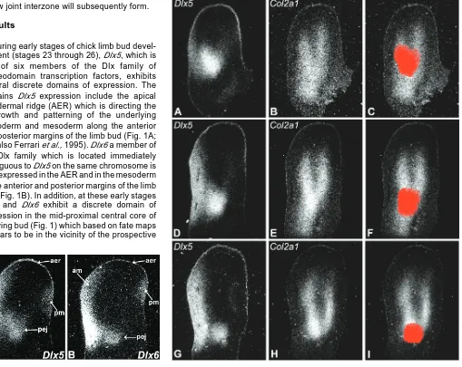
Fig. 2 (Right). Dlx5 is expressed at the prospective elbow joint during early stages of limb bud development. Expression of Dlx5 and the cartilage marker Col2a1 in adjacent sections of the same limb buds at stage 24 (A,B), stage 25 (D,E) and stage 26 (G,H). The images in (C,F,I) were generated by superimposing and aligning the images of adjacent Dlx5- and Col2a1-expressing sections (A,B) (D,E) (G,H) using Adobe Photoshop, after which the Dlx5 expression domain in the vicinity of the prospective elbow joint was selected, pseudocolored red and superimposed on the image of the adjacent Col2a1 section. Note that Dlx5 is expressed by a discrete group of cells that encompass the prospective elbow joint where the radius and ulna branch from the humerus.
In the present study we show that during early stages of chick limb development well before the onset of overt joint formation the homeobox-containing genes Dlx5 and Dlx6 are highly expressed by a discrete group of cells that encompass the prospective elbow joint where the differentiating humerus rudiment branches into the radius and ulna anlage. The onset of Dlx5 expression in the region of the presumptive elbow joint is shortly followed by the initiation of expression amongst the Dlx5-expressing cells of Gdf5, which encodes a secreted sig-naling molecule that is involved in regulating the onset of joint formation (Storm and Kingsley, 1996; Settle et al., 2003). These observations suggest that Dlx5 and Dlx6 may provide positional information that specifies the site at which the
elbow joint interzone will subsequently form.
Results
During early stages of chick limb bud devel-opment (stages 23 through 26), Dlx5, which is one of six members of the Dlx family of homeodomain transcription factors, exhibits several discrete domains of expression. The domains Dlx5 expression include the apical ectodermal ridge (AER) which is directing the outgrowth and patterning of the underlying mesoderm and mesoderm along the anterior and posterior margins of the limb bud (Fig. 1A; see also Ferrari et al., 1995). Dlx6 a member of the Dlx family which is located immediately contiguous to Dlx5 on the same chromosome is also expressed in the AER and in the mesoderm at the anterior and posterior margins of the limb bud (Fig. 1B). In addition, at these early stages Dlx5 and Dlx6 exhibit a discrete domain of expression in the mid-proximal central core of the wing bud (Fig. 1) which based on fate maps appears to be in the vicinity of the prospective
elbow joint. Dlx5 is also expressed in the vicinity of the prospec-tive knee joint in the leg bud (data not shown).
joint formation Dlx5 does appear to be expressed by a discrete group of cells that encompass the region of the limb bud where the elbow joint will subsequently form (Fig. 2A, D, G). The Dlx5-expressing cells in the area of the presumptive elbow joint are located where the differentiating humerus branches into the radius and ulna (Fig. 2B, E, H). To more accurately confirm the localization of this Dlx5 expression domain at the presumptive elbow joint, the images of adjacent Dlx5- and Col2a1-express-ing sections were superimposed in Abode Photoshop and precisely aligned, after which the Dlx5 expression domain in the vicinity of the prospective elbow joint was selected, psuedocolored red and superimposed on the adjacent Col2a1-expressing image. As shown in Figs. 2C, F, I, this analysis confirms that this domain of Dlx5 expression indeed encom-passes the prospective elbow joint where the humerus bifur-cates into the radius and ulna.
The Col2a1 probe (Nah et al., 1988) used to monitor the formation of the Y-shaped humerus, radius and ulna rudiment does not distinguish between two alternatively spliced forms of Col2a1 transcripts, Col IIA which is typically expressed by prechondrogenic as well as chondrogenic cells and Col IIB which is expressed by more differentiated chondrocytes (Ryan and Sandell, 1990; Nah and Upholt, 1991; Ng et al., 1993). However, the Y-shaped humerus, radius and ulna rudiment at these early stages of cartilage differentiation is also character-ized by the expression of other cartilage markers such as aggrecan (Mallein-Gerin et al., 1988).
Our results indicate that Dlx5 is expressed in the presump-tive elbow joint during the period of development when the elbow joint is being specified. To further pursue the association of Dlx5 with elbow joint formation, we next compared the temporal expression in adjacent sections of the same limb buds of Dlx5 with that of the secreted signaling molecule Gdf5, which
regulated in the elbow joint interzone (data not shown and Lizarraga et al., 2002).
Discussion
The conversion of differentiated chondrocytes at sites of incipient joint formation into the densely packed cells of the joint interzone is the first morphological indication of joint formation in the skeletal elements of the developing limb and several of the genes and signals that regulate interzone formation have been identified (see Pacifici et al., 2002, 2005 for reviews). However, little or nothing is known about the genes, signals, or mechanisms that specify the positions within cartilage rudi-ments at which joint interzones will form.
The experimental studies of Holder (1977) indicate that the elbow joint is specified very early in limb development by a distinctive population of precursor cells located at the site at which the elbow joint interzone will subsequently form. When the presumptive elbow joint is removed at an early stage (stage 24) of chick limb bud development well before elbow joint interzone formation (stage 27), the elbow joint fails to form. This indicates that joint formation is dependent on cells in the presumptive elbow joint region that had been specified to form a joint very early in limb development. Furthermore, the studies of Holder (1977) suggest that the presumptive elbow joint may be capable of autonomously differentiating into joint cells, suggesting that the interzone may be derived from a distinct precursor cell type that differentiates according to information acquired at earlier stages. Interestingly, the limbs ofnGli3/Plzf double knockout mouse embryos lack proximal (stylopod and zeugopod) skeletal elements, but nevertheless possess a single proximal cartilaginous ball of cells resembling a joint that expresses joint markers including Gdf5, suggesting the joint
Fig. 3.Expression of Dlx5 and the joint marker Gdf5 in adjacent sections of the same limb buds at stage 23 (A,B) and stage 24 (C,D). The image in (E) was generated by superimposing and aligning the images of adjacent Dlx5- and Col2a1 -expressing sections (C,D) using Adobe Photoshop, after which the Dlx5 expression domain in the vicinity of the prospective elbow joint was selected, pseudocolored red and superimposed on the image of the adjacent Gdf5 section. Dlx5 (A), but not Gdf5 (B) is expressed in the region of the prospective elbow joint at stage 23. At stage 24, Gdf5 expression is initiated amongst the Dlx5-expressing cells in the vicinity of the prospective elbow joint (C-E).
down-may be derived from a distinct population of precursor cells (Barna et al., 2005).
In the present study we demonstrate that at early stages of chick limb development well before the onset of overt joint formation the homeodomain transcription factors Dlx5 and Dlx6 are highly expressed by a discrete group of cells that encom-pass the region of the limb bud at which the elbow joint will subsequently form. The Dlx5- and Dlx6-expressing cells at the prospective elbow joint are located where the differentiating humerus branches into the radius and ulna. Thus, Dlx5 and Dlx6 are the earliest molecular markers of the prospective elbow joint yet described. The Dlx-5- and Dlx6-expressing cells we have identified correspond quite well to the presumptive elbow joint cells whose removal at an early stage results in subsequent fusion of the humerus with the radius and ulna and failure of elbow joint formation (Holder, 1977). This striking correspondence suggests the possibility that the Dlx-express-ing cells located at the presumptive elbow joint constitute a distinctive population of cells that play an important role in the specification of the elbow joint and/or in providing positional information that specifies the site at which the elbow joint will form.
One possible mechanism by which Dlx genes may specify elbow joint formation is by regulating the expression of signal-ing molecules that control the onset of joint interzone formation. Indeed, we have found that shortly after the onset of expression of Dlx5 at the presumptive elbow joint, the expression of Gdf5, a secreted signaling molecule that regulates the onset of joint interzone formation, is initiated amongst the Dlx5-expressing cells. This suggests the possibility that Dlx genes may be involved in regulating the initiation of Gdf5 expression and/or in specifying the site at which Gdf5 expression initiates.
It has previously been suggested that homeobox genes may determine the position at which joints form (Yokouchi–et al., 1991; Brunet et al., 1998; Pacific et al., 2002, 2005). Interest-ingly, the expression domain of Dlx genes at the prospective elbow joint corresponds to the site where the expression do-mains of members of the HoxA and HoxD clusters intersect (Yokouchi et al., 1991). Thus, Dlx genes may act in conjunction with HoxA and HoxD genes in providing positional cues which determine the site of elbow joint formation and/or the site of bifurcation of the humerus.
Experimental Procedures
In situ hybridization was done as previously described (Ferrari et al.,
1999, 2002) on serially sectioned limb buds using the following 33 P-labeled probes: a 323 bp Dlx5-specific probe from the 3’ untranslated
region (UTR) of chicken Dlx5 (Ferrari et al., 1995); a 1 kb Dlx6 probe
consisting of 240 bp of exon 3 and 760 bp of 3’ UTR from a chicken Dlx6
genomic clone; a 328 bp Gdf5-specific probe prepared as previously
described (Lizarraga et al., 2002) and a chicken Col2a1 probe (Nah et al.,
1988). In order to correlate the domains of expression of different genes, adjacent sections (within 20 µm of one another) of the same limb buds were mounted on separate slides and hybridized with different probes.
Acknowledgements
The technical assistance of Gail Lizarraga is acknowledged. Supported by NIH grants HD041448 and HD022610.
References
BARNA, N., PANDOLFI, P. P. and NISWANDER, L. (2005). Gli3 and Plzf cooperate
in proximal limb patterning at early stages of limb development. Nature 436:
277-81.
BRUNET, L. J., MCMAHON, J. A., MCMAHON, A. P. and HARLAND, R. M. (1998). Noggin, cartilage morphogenesis and joint formation in the mammalian
skel-eton. Science 280: 1455-7.
CRAIG, F. M., BENTLEY, G. and ARCHER, C. W. (1987). The spatial and temporal pattern of collagens I and II and keratan sulphate in the developing chick
metatarsophalangeal joint. Development 99: 383-91.
FERRARI, D., HARRINGTON, A., DEALY, C. N. and KOSHER, R. A. (1999).
Dlx-5 in limb initiation in the chick embryo. Dev Dyn 216: 10-Dlx-5.
FERRARI, D. and KOSHER, R. A. (2002). Dlx5 is a positive regulator of chondro-cyte differentiation during endochondral ossification. Dev Biol 252: 257-70.
FERRARI, D., SUMOY, L., GANNON, J., SUN, H., BROWN, A. M. C., UPHOLT, W.
B. and KOSHER, R. A. (1995). The expression pattern of the Distal-less
homeobox-containing gene Dlx-5 in the developing chick limb bud suggests its
involvement in apical ectodermal ridge activity, pattern formation and cartilage
differentiation. Mech Dev 52: 257-64.
GUO, X., DAY, T. F., JIANG, X., GARRET-BEAL, L., TOPOL, L. and YANG, Y. (2004). Wnt/β-catenin signaling is sufficient and necessary for synovial joint
formation. Genes Dev 18: 2404-17.
HARTMANN, C. and TABIN, C. J. (2001). Wnt-14 plays a pivotal role in inducing synovial joint formation in the developing appendicular skeleton. Cell 104: 341-51.
HOLDER, N. (1977). An experimental investigation into the early development of
the chick elbow joint. J Embryol Exp Morph 39: 115-27.
IWAMOTO, M., HIGUCHI, Y., KOYAMA, E., ENOMOTO-IWAMOTO, M., KURISU, K., YEH, H., ABRAMS, W. R., ROSENBLUM, J. and PACIFICI, M. (2000). Transcription factor ERG variants and functional diversification of chondrocytes
during limb long bone development. J Cell Biol 150: 27-40.
KOYAMA, E., LEATHERMAN, J. L., SHIMAZU, A., NAH, H.-D. and PACIFICI, M. (1995). Syndecan-3, tenascin-C and the development of cartilaginous skeletal
elements and joints in chick limbs. Dev Dyn 203: 152-62.
LIZARRAGA, G., LICHTLER, A., UPHOLT, W. B. and KOSHER, R. A. (2002). Studies on the role of Cux1 in regulation of the onset of joint formation in the
developing limb. Dev Biol 242: 44-54.
MALLEIN-GERIN, F., KOSHER, R. A., UPHOLT, W. B. and TANZER, M. L. (1988). Temporal and spatial analysis of cartilage proteoglycan core protein gene expression during limb development by in situ hybridization. Dev Biol 126: 337-45.
MERINO, R., MACIAS, D., GANAN, Y., ECONOMIDES, A. N., WANG, X., WU, Q., STAHL, N., SAMPATH, K. T., VARONA, P. and HURLE, J. M. (1999). Expres-sion and function of Gdf-5 during digit skeletogenesis in the embryonic chick leg bud. Dev Biol 206: 33-45.
MITROVIC, D. (1977). Development of the metatarsophalangeal joint of the chick
embryo: morphological, ultrastructural and histochemical studies. Am J Anat
150: 333-48.
MITROVIC, D. (1978). Development of the diarthrodial joints in the rat embryo. Am
J Anat 151: 475-86.
NAH, H.-D., RODGERS, B. J., KULYK, W. M., KREAM, B. E., KOSHER, R. A. and UPHOLT, W. B. (1988). In situ hybridization analysis of the expression of the type II collagen gene in the developing chicken limb bud. Collagen Relat Res 8: 277-94.
NAH, H.-D. and UPHOLT, W. B. (1991). Type II collagen mRNA containing an alternatively spliced exon predominates in the chick limb prior to
chondrogen-esis. J Biol Chem 266: 23446-52.
NG, L. J, TAM, P. P. L. and CHEAH, K. S. E. (1993). Preferential expression of alternatively spliced mRNAs encoding type II procollagen with a cysteine-rich amino-propeptide in differentiating cartilage and nonchondrogenic tissues
during early mouse development. Dev Biol 159: 403-17.
PACIFICI, M., KOYAMA, E. and IWAMOTO, M. (2005). Mechanisms of synovial joint and articular cartilage formation: recent advances, but many lingering
PACIFICI, M., LIU, M. and KOYAMA, E. (2002). Joint formation: new findings shed more light on this critical process in skeletogenesis. Curr Opin Orthop 13: 339-44.
RYAN, M. C. and SANDELL, L. J. (1990). Differential expression of a cysteine-rich domain in the amino-terminal propeptide of type II (cartilage) procollagen by
alternative splicing of mRNA. J Biol Chem 265: 10334-9.
SETTLE, S. H., Jr., ROUNTREE, R. B., SINHA, A., THACKER, A., HIGGINS, K. and KINGSLEY, D. M. (2003). Multiple joint and skeletal patterning defects caused
by single and double mutations in the mouse Gdf6 and Gdf5 genes. Dev Biol
254: 116-30.
STASKO, S. E. and WAGNER, G. F. (2001). Possible roles for stanniocalcin during early skeletal patterning and joint formation in the mouse. J Endocrinol 171: 237-48.
STORM, E. E. and KINGSLEY, D. M. (1996). Joint pattering defects caused by
single and double mutations in members of the bone morphogenetic protein
(BMP) family. Development 122: 3969-79.
TAKAGI, T., MORIBE, H., KONDOH, H. and HIGASHI, Y. (1998). δEF1, a zinc finger and homeodomain transcription factor, is required for skeleton patterning
in multiple lineages. Development 125: 21-31.
YOKOUCHI, Y., SASAKI, H. and KUROIWA, A. (1991). Homeobox gene expres-sion correlated with the bifurcation process of limb cartilage development. Nature 353: 443-5.