Short Communication
NUDC expression during amphibian development
NICOLE MOREAU
1, JONATHAN P. AUMAIS
2, CHRISTELLE PRUDHOMME
1,
SHELLI M. MORRIS
2,#and LI-YUAN YU-LEE
2,3,4,*
Groupe Développement des Vertébrés1, UMR 7622 CNRS-Université Pierre et Marie Curie, Paris, France and Departments of Molecular and
Cellular Biology2, Immunology3 and Medicine4, Baylor College of Medicine, Houston, Texas, USA
ABSTRACT To identify gene products important for gastrulation in the amphibian Pleurodeles waltl, a screen for regional differences in new protein expression at the early gastrula stage was performed. A 45 kDa protein whose synthesis was specific for progenitor endodermal cells was identified. Microsequencing and cDNA cloning showed that P45 is highly homologous to rat NUDC, a protein suggested to play a role in nuclear migration. Although PNUDC can be detected in all regions of the embryo, its de novo synthesis is tightly regulated spatially and temporally through-out oogenesis and embryonic development. New PNUDC synthesis in the progenitor endodermal cells depends on induction by the mesodermal cells in the gastrula. During development, PNUDC is localized in the egg cortical cytoplasm, at the cleavage furrow during the first embryonic division, around the nuclei and cortical regions of bottle cells in the gastrula, and at the basal region of polarized tissues in the developing embryo. These results show for the first time the expression and compartmentalization of PNUDC at distinct stages during amphibian development.
KEY WORDS: NUDC, nuclear migration, LIS-1, oogenesis, gastrulation
0214-6282/2001/$25.00 © UBC Press
Printed in Spain
www.ijdb.ehu.es
* Address correspondence to: Dr. Li-Yuan Yu-Lee. Department of Medicine, Baylor College of Medicine, One Baylor Plaza, Houston, Texas 77030, USA. Fax: +1-713-798-5780. e-mail: [email protected]
# Present address: Fred Hutchinson Cancer Research Center, 1100 Fairview Ave. N., P.O. Box 19024, Seattle, WA 98109, USA.
Abbreviations used in this paper: Met, methionine; MTOC, microtubule orga-nizing center; NUDC, nuclear distribution; ORF, open reading frame; PNUDC, Pleurodeles NUDC.
Nuclear migration is an important process that occurs through-out development. A number of nuclear distribution (nud) genes that regulate nuclear movement have been identified in the filamentous fungus Aspergillus nidulans (Morris, 1976; Osmani et al., 1990). Several nud genes, including nudC and nudF, encode components and regulators of the minus-end directed molecular motor complex dynein (Morris, 2000). Aspergillus nudC is re-quired for viability and is involved in cell wall deposition, nuclear distribution and colony growth, and is thought to act by modulating the levels of NUDF (Osmani et al., 1990; Xiang et al., 1995; Chiu et al., 1997). Mammalian NudC interacts in vitro and in vivo with the NUDF homologue called Lis1, which is involved in neuronal migration during embryonic brain development (Morris S.M. et al., 1998; Reiner et al., 1993), suggesting that the NUDC/Lis1 com-plex participates in the development of the mammalian cortex (Morris S.M. et al., 1998; Matsumoto and Ledbetter, 1999; Morris, 2000; Wynshaw-Boris and Gambello, 2001). Recent studies show that NUDC is found in a complex with dynein and dynactin (Aumais et al., 2001), suggesting its involvement in dynein functions, such as positioning of the Golgi apparatus, microtubule organizing center (MTOC) and other organelles (Morris S.M. and Yu-Lee, 1998).
Pleurodeles NUDC was cloned in a search for new proteins that are synthesized during gastrulation in the amphibian Pleurodeles
waltl. The animal cap, dorsal blastopore lip and endodermal region were excised from [35S]-Met-labeled embryos. By 2-D gel
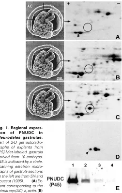
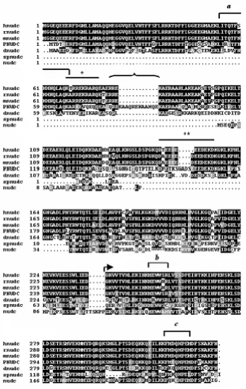
electrophoresis, a 45 kDa protein with a pI @ 5.0 (P45) was found to be synthesized only in the progenitor endodermal cells but not in the animal cap or dorsal blastopore lip (Fig. 1). Three P45 peptides, isolated from 500 gastrulae, were microsequenced and found to show a high degree of identity to rat NUDC (RNUDC) (Morris S.M. et al., 1997) (Fig. 2). By screening a Pleurodeles cDNA library, a cDNA encoding a 346 amino acid ORF was cloned. The ORF exhibits a 74% identity and 83% similarity with human, rat and mouse NUDC and a 50% identity and 67% similarity to DNUDC (Cunniff et al., 1997). The C-terminal 94 amino acid NUDC-homology region is highly conserved across all species, suggesting a functional conservation over evolution. Immunoblotting with anti-RNUDC C peptide antibodies (Morris S.M. and Yu-Lee, 1998) identified three isoforms of P45, the most basic of which corresponded with the 35S-Met-labeled P45 (Fig.
anti-A
B
C
D
E
RNUDC antibodies in detecting a NUDC-like protein in Pleurodeles (Fig. 1E). P45 is therefore renamed as Pleurodeles NUDC or PNUDC.
PNUDC is a maternal protein that is briefly synthesized during early oogenesis and stored throughout early embryogenesis (Table 1). PNUDC is synthesized again as a zygotic protein as early as Stage 8a of gastrulation. To determine whether mesodermal signals are involved in inducing new synthesis of PNUDC in endodermal cells, explants of endodermal progenitor cells were labeled with [35S]-Met and incubated over 4 hr to progress to the beginning of
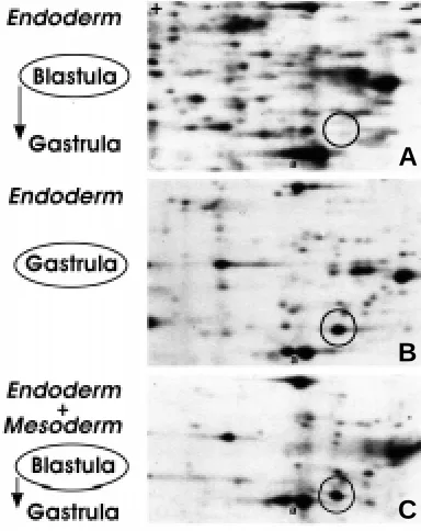
gastrulation. Endodermal cells explanted during the blastula stage and cultured in isolation did not synthesize PNUDC (Fig. 3A), while endodermal cells explanted just after the beginning of gastrulation showed robust new synthesis of PNUDC (Fig. 3B). Mesodermal
TABLE 1
DE NOVO SYNTHESIS AND STEADY STATE EXPRESSION OF PNUDC OVER PLEURODELES WALTL DEVELOPMENT
OOGENESIS DE NOVO SYNTHESIS STEADY-STATE LEVEL
Stage II + +
Stage 5 (early blastula) — +
Stage 7 (late blastula) — +
Stage 8a (early gastrula) + +
Stage 9 (gastrula) + +
Dorsal blastopore lip — +
NEURULA
Neural plate + +
Endoderm + +
The de novo synthesis [presence (+) or absence (-)] of PNUDC is determined by 2-D gel electrophoresis followed by autoradiography of 10,000 g supernatant of defolliculated [35
S]-Met-labeled oocytes, embryos or explants. The same blots were then incubated with anti-RNUDC C antibodies to detect steady state levels of PNUDC.
cells explanted before or during gastrulation did not synthesize PNUDC (data not shown). When the endodermal cells explanted at the blastula stage were juxtaposed with mesodermal cells also explanted from the blastula stage, new synthesis of PNUDC was reproducibly observed (Fig. 3C), suggesting that PNUDC synthesis in the endodermal cells requires a mesodermal signal.
The spatial expression of PNUDC during Pleurodeles oogenesis and embryogenesis was analyzed by immunocytochemistry. In previtellogenic Stage II oocytes, elevated PNUDC staining was found in the cytoplasm (Fig. 4A). In vitellogenic Stages III and IV oocytes, PNUDC staining was concentrated between the yolk plate-lets and the perinuclear area (Fig. 4B). In late Stages V and VI oocytes, the cytoplasm exhibited a low level of fluorescence, as illustrated by a Stage VI oocyte (Fig. 4A), although steady-state PNUDC proteins were readily detected by immunoblotting (Table 1). In all oocyte stages, the follicle cells showed cytoplasmic PNUDC staining.
In the egg just beneath the plasma membrane, concentrated PNUDC staining was found in the cortical cytoplasm, where it was thicker at the vegetal (Fig. 4D) than the animal (Fig. 4C) pole. During the first embryonic cleavage, PNUDC staining was enhanced along the cleavage furrow at both the animal (Fig. 4E) and vegetal (Fig. 4F) poles. At the animal pole, just beneath the cleavage furrow, intense PNUDC staining was observed in an area without yolk platelets (Fig. 4E). PNUDC staining was observed throughout the cytoplasm, but could also be detected over the cell nucleus (Fig. 4 G,H). In late blastula stages, strong PNUDC staining was observed in the cortical cytoplasm as well as in cytoplasmic droplets that were protruding into the blastocoel roof (Fig. 4 I,J).
At the gastrula stage, prominent PNUDC staining was again observed in cytoplasmic droplets in the animal cap (Fig. 4 K,L), although no new PNUDC synthesis was detected in this region
Fig. 1. Regional expres-sion of PNUDC in Pleurodeles gastrulae.
Part of 2-D gel autoradio-graphs of explants from [35S]-Met-labeled gastrula derived from 10 embryos. P45 is indicated by a circle. Scanning electron micro-graphs of gastrula sections on the left are from Shi and Boucaut (1995). (A) Ex-plant corresponding to the animal cap (AC). a, actin. (B)
(Fig. 1A and Table 1). In the vegetal part just below the equator, the local endodermal cells invaginate to form a slit-like blastopore. These cells, called bottle cells, change their shape dramatically and line the initial archenteron (Holtfreter, 1943). The bottle cells exhibited elevated PNUDC staining over their nuclei as well as their cell periphery (Fig. 4 M,N). During organogenesis, PNUDC staining was detected primarily in the cytoplasm, especially along the basal region of cells in different tissues (Fig. 4 O-R). PNUDC staining can be discerned as a bright line at the interface between neuroectoderm and mesoderm, mesoderm and endoderm, and at
the basal limits of the neural tube. The specificity of the PNUDC staining was demonstrated by blocking with excess NUDC pep-tides (Fig. 4S). These studies show that PNUDC is expressed at distinct sites and developmental stages during Pleurodeles oo-genesis and embryooo-genesis.
Although the precise function of PNUDC is unknown, the spatio-temporal expression pattern of PNUDC is in agreement with our working model that NUDC is a component of the Lis1/ dynein/dynactin motor (Aumais et al., 2001), which is important for nuclear and vesicular transport within the cell and for the
Fig. 2. Amino acid sequence analysis of PNUDC. Amino acid sequence comparison of PNUDC with NUDC proteins from other species. The PNUDC peptides identified by microsequencing are indicated as a - c. The basic (single asterisk) and acidic (double asterisk) re-gions are overlined. Unique insertions, com-prised of the tandemly-repeated QREKAA se-quence, are indicated by a bracket. The highly conserved carboxyl-terminus NUDC-homology region is indicated by an arrow. hnudc, human; rnudc, rat; mnudc, mouse; pnudc, Pleurodeles waltl; dnudc Drosophila melanogaster; spnudc, Schizosacharomyces pombe; nudC, Aspergillus nidulans.
extensive cellular migration that occur during oogenesis, gastrulation and orga-nogenesis.
Experimental Procedures
Collection and labeling of oocytes and em-bryos
Analysis of [35S]-polypeptide pattern and immunodetection
Ten pieces each of oocytes, embryos or explants were pooled and homogenized in Tris-EDTA buffer (Chen and Stumm-Zollinger, 1986) con-taining 10-4mM Pefabloc (protease inhibitor) (Interchim, Amresco, Solon, OH). After centrifugation for 10 min at 10,000 g, proteins in the supernatant were precipitated overnight at – 20°C in 9 volumes of absolute ethanol, centrifuged for 30 min at 5,000 g and resolved by two-dimensional (2-D) gel electrophoresis. The 2-D gel was performed with equilibrium pH gradient electrophoresis using ampholins 3-11 in the first dimension, followed by electrophoresis on an SDS-10 % polyacrylamide slab gel in the second dimension. The gels were loaded with equal acid precipitable cpm from oocytes or explants from embryos and with extract from one embryo for each developmental stage. The 2-D separated polypeptides were transferred to nitrocellulose membranes (Optitran BA.S 85; Schleicher & Schuell, D-37582 Dassel), autoradiographed, immunoblotted with rabbit RNUDC C anti-bodies directed against a conserved carboxyl-terminus peptide (Morris S.M. and Yu-Lee, 1998) (1:500 dilution), followed by donkey anti-rabbit immuno-globulin G (IgG) antibodies conjugated to horseradish peroxidase (Amersham) (1:1000 dilution), and detected by enhanced chemiluminescence (Amersham).
Purification and microsequencing of P45 (PNUDC)
Protein extracts from 500 gastrulae were separated by electrophoresis on 10 % SDS-PAGE gels. The P45 region containing actin was stained with Coomassie brilliant blue R-250, excised, electroeluted (Electroeluter 422; BioRad) and resolved on 2-D gels as described above. P45 was digested in 200µl of 0.1 M Tris-HCl pH 8.6, 0.5 mM EDTA, 0.03 % SDS for 18 h at 35°C with 0.4µg endoprotease. Ten spots were microsequenced (Institut Pasteur, Paris). The peptides were separated by reverse-phase high-pressure liquid chromatography (HPLC) using DEAE-C18 columns and trifluoroacetic acid-acetonitrile gradient solutions. Sequence homology was searched for in the GenBank CDS data bank.
A
B
C
Fig. 3.␣ Endoderm-mesoderm interaction is necessary for PNUDC synthesis. Part of 2-D gel autoradiographs of explants from [35 S]-Met-labeled blastula or gastrula. The endodermal explants from the blastula were treated in vitro as follows. (A) Progenitor endodermal cells explanted at the blastula stage and cultured alone until control embryos reached the gastrula stage. (B) Progenitor endodermal cells explanted at the gastrula stage. (C) Progenitor endodermal cells explanted at the blastula stage, incubated together with mesodermal area from the same blastula stage. The position of PNUDC is indicated by a circle. a, actin.
Cloning of Pleurodeles NudC cDNA
A Pleurodeles late-tadpole stage λZAPII cDNA library (a gift from Dr. J.F. Riou, UMR7622, Université P. & M. Curie, Paris) was screened with a murine NudC cDNA probe (Morris S.M. et al., 1998), using low stringency hybridiza-tion condihybridiza-tions. Two phage clones (λ4A and λ4B) were obtained which contained the most distal two-thirds of PNudC cDNA. A 5’ sequence was generated from clone 4B to re-screen the Pleurodeles cDNA library. Thirty potential clones were identified. PCR was carried out with Pfu-Turbo (Stratagene), using T3 as the 5' PCR primer and a 23 bp PNudC-specific internal antisense primer containing an XhoI cloning site (lowercase) (5'-GATCctcgagTCCCTGTTGACTCTCGGATGCC-3') as the 3' primer. Three PCR products were obtained (cl-3, 400 bp; cl-6, 700 bp and cl-8, 520 bp) and sequenced. The PNUDC ORF from the complete 1.5 kb cDNA was deter-mined using the ORF Finder program (http://www.ncbi.nlm.nih.gov/gorf), and was verified to be the Pleurodeles NudC homologue by performing a WU-BLASTX-2.0+BEAUTY search. The PNudC cDNA sequence is under GenBank accession number AF259800.
Immunocytochemistry
Pieces of ovary or embryos were fixed in MEMFA (0.5 M MOPS [pH 7.4], 100 mM EGTA, 1 mM MgSO4, 4 % formaldehyde), dehydrated in methanol and stored at -20°C until needed. Cryostat sections (10µm) were prepared from fixed ovary or embryos rehydrated in PBS, infiltrated with PBS contain-ing 15 % cold water fish gelatin (FLUKA Biochemika) and 15 % sucrose (Fagotto and Gumbiner, 1994), and embedded in O.C.T. compound (Miles Inc.). Sections were blocked for 30 min in PBS containing 1 % BSA and incubated with rabbit anti-RNUDC C (1:100 dilution) or affinity-purified rabbit anti-RNUDC C (1:20 dilution) antibodies (Morris S.M. and Yu-Lee, 1998) for 3 h at room temperature. Sections were incubated with fluorescein isothiocyanate-conjugated donkey anti-rabbit IgG antibodies (Interchim) and mounted in Mowiol.
Acknowledgments
The authors wish to thank Dr. J.F. Riou (Université P. & M. Curie, Paris) for providing the λZAPII late-tadpole stage cDNA library, Colette Montmory (Université P. & M. Curie, Paris) for excellent technical assistance, Dr. Paula Burch (Baylor College of Medicine, Houston) for assistance with sequence analyses, and Dr. Greg May (M.D. Anderson Cancer Center, Houston) and Dr. Kathleen Mahon (Baylor) for helpful comments and critical reading of the manuscript. This work was supported by N.I.H. grant DK53176 to L.-y. Yu-Lee.
References
AUMAIS, J.P., TUNSTEAD, J.R., MCNEIL, R.S., SCHAAR, B.T., MCCONNELL, S.K., LIN, S.-H., CLARKE, G.D. and YU-LEE, L.-Y. (2001) NudC associates with Lis1 and the dynein motor at the leading pole of neurons. J. Neurosci., in press.
BONNANFANT-JAIS, M.L. and MENTRE, P. (1983). Study of oogenesis in the newt Pleurodeles waltlii M. I: Ultrastructural study of the different stages of oocyte development. J. Submicrosc. Cytol. 15: 453-478.
CHEN, P.S. and STUMM-ZOLLINGER, E. (1986). Patterns of protein synthesis in oocytes and early embryos of Rana esculenta complex. Roux’s Arch. Dev. Biol. 195: 1-9.
CHIU, Y.-H., XIANG, X., DAWE, A.L. and MORRIS, N.R. (1997). Deletion of nudC, a nuclear migration gene of Aspergillus nidulans, causes morphological and cell wall abnormalities and is lethal. Mol. Biol. Cell 8: 1735-1749.
CUNNIFF, J., CHIU, Y.H., MORRIS, N.R. and WARRIOR, R. (1997). Characterization of DnudC, the Drosophila homologue of an Aspergillus gene that functions in nuclear motility. Mech. Dev. 66: 55-68.
FAGOTTO, F. and GUMBINER, B.M. (1994). β-catenin localization during Xenopus embryogenesis: accumulation at tissue and somite boundaries. Development 120: 3667-3679.
GURDON, J.B. (1976). Injected nuclei in frog oocytes: Fate, enlargement and chroma-tin dispersal. J. Embryol. Exp. Morphol. 36: 523-540.
HOLTFRETER, J. (1943). A study of the mechanisms of gastrulation: Part I. J. Exp. Zool. 94: 261-318.
A
B
C
E
G
I
D
F
H
J
K
M
O
Q
L
N
P
R
S
MOREAU, N. and BOUCHER, D. (1981). Une méthode rapide d’extraction en masse des noyaux d’ovocytes de Pleurodèle. Biol. Cell. 42: 185-188.
MORRIS, N.R. (1976). Mitotic mutants in Aspergillus nidulans. Genet. Res. 26: 237-254.
MORRIS, N.R. (2000). Nuclear migration: From fungi to the mammalian brain. J.Cell Biol. 148: 1097-1101.
MORRIS, S.M., ANAYA, P., XIANG, X., MORRIS, N.R., MAY, G.S. and YU-LEE, L.-Y. (1997). A prolactin-inducible T cell gene product is structurally similar to the Aspergillus nidulans nuclear movement protein NUDC. Mol.Endocrinol. 11: 229-236.
MORRIS, S.M. and YU-LEE, L.-Y. (1998). Expression of RNUDC, a potential nuclear movement protein, in mammalian cells: localization to the Golgi apparatus. Exp. Cell Res. 238: 23-32.
MORRIS, S.M., ALBRECHT, U., REINER, O., EICHELE, G. and YU-LEE, L.-Y. (1998). LIS-1, a protein involved in neuronal migration, interacts with a nuclear movement protein NUDC. Curr. Biol. 8: 603-606.
Fig. 4.␣ Immunolocalization of PNUDC over Pleurodeles waltl oogenesis and embryo-genesis. (A) Previtellogenic oocyte (Stage␣ II). Arrowhead, cytoplasmic PNUDC staining. Arrow, background staining in the nucleus. Double arrowhead, faint fluorescence in the cytoplasm of the neighboring Stage␣ VI oocyte. (B) Stage␣ IV oocyte. Cytoplasmic staining between the yolk platelets (double arrowheads) and around the nucleus (arrow). Arrow-head, cytoplasm of follicular cells surrounding the oocyte. (C,D) Meridian section of an egg. Arrow, PNUDC staining in the cortical cytoplasm in the animal (C) and vegetal (D) poles.
(E,F,G,H) Meridian section of a two-cell embryo. Arrows, PNUDC fluorescence in the cortical area along the cleavage furrow in the animal (E) and vegetal (F) poles. Arrowhead, cytoplasmic area without yolk platelets. (G) Arrow, staining in and around the nucleus of one
of the embryonic cells. (H) Hoechst nuclear counterstain. (I,J) Meridian section of a late stage blastula. (I)␣ Animal part: blastocoel roof. Double arrowhead, PNUDC staining over some nuclei. Arrow, strong polar cytoplasmic staining. Arrowhead, cytoplasmic droplet protruding into the blastocoel cavity. (J)␣ Hoechst nuclear counterstain. K,L,M,N) Sagittal section of gastrula stage. (K,L) ␣ Animal cap. Arrowhead, PNUDC staining over the nuclei and cell periphery. Arrow, cytoplasmic droplets as in (I). (L)␣ Hoechst nuclear counterstain. (M,N) ␣Bottle cells in the vegetal part. Arrowhead, nucleus; arrowhead, periphery of bottle cells. (N)␣ Hoechst nuclear counterstain. (O,P) Transverse section of mid-neurula stage. (O)␣ Arrow, basal limit of neuroectoderm; arrowhead, mesoderm. NG, neural groove. (P)␣ Hoechst nuclear counterstain. (Q,R) Frontal section of tailbud (stage ␣ 21). (Q)␣ Arrowhead, mesoderm; double arrowhead, ectoderm; arrow, basal limit of the neural tube (NT). (R)␣ Hoechst nuclear counterstain. (S) Transversal section of tailbud (stage ␣ 21). PNUDC immunofluorescence staining is blocked by NUDC peptide competition (4 µg). Scale bar, 100 µm in all panels.
OSMANI, A.H., OSMANI, S.A. and MORRIS, N.R. (1990). The molecular cloning and identification of a gene product specifically required for nuclear movement in Aspergillus nidulans. J. Cell Biol. 111: 543-551.
REINER, O., CARROZZO, R., SHEN, Y., WEHNERT, M., FAUSTINELLA, F., DOBYNS, W.B., CASKEY, C.T. and LEDBETTER, D.H. (1993). Isolation of a Miller-Dieker lissencephaly gene containing G protein β-subunit-like repeats. Nature 364: 717-721.
SHI, D.L. and BOUCAUT, J.C. (1995). The chronological development of the Urodele Amphibian Pleurodeles waltl. Int. J. Dev. Biol. 39: 427-441.
WYNSHAW-BORIS, A. and GAMBELLO, M.J. (2001). Lis1 and dynein motor function in neuronal migration and development. Genes & Dev. 15: 639-651.
XIANG, X., OSMANI, A.H., OSMANI, S.A., XIN, M. and MORRIS, N.R. (1995). nudF, a nuclear migration gene in Aspergillus nidulans, is similar to the human LIS-1 gene required for neuronal migration. Mol. Biol. Cell 6: 297-310.