Original Article
Endocytosis and transcytosis in growing astrocytes in primary
culture. Possible implications in neural development
LUÍS MEGÍAS
1, CONSUELO GUERRI
2, EUGENIO FORNAS
3, INMACULADA AZORIN
3, ELENA BENDALA
2,
MARÍA SANCHO-TELLO
4, JUAN M. DURÁN
3, MÓNICA TOMÁS
3,4, MARÍA J. GOMEZ-LECHON
3and JAIME RENAU-PIQUERAS
3,41Departamento de Ciencias Morfológicas, Universidad de Granada, Granada, 2Instituto de Investigaciones Citológicas, Valencia, 3Centro de Investigación, Hospital La Fe, Valencia and 4Departamento de Biología Celular, Universidad de Valencia, Valencia, Spain
ABSTRACT Endocytosis constitutes an essential process in the regulation of the expression of cell surface molecules and receptors and, therefore, could participate in the neural-glial interactions occurring during brain development. However, the relationship between endocytic pathways in astroglial cells under physiological and pathological conditions remains poorly understood. We analyzed the endocytosis and transcytosis processes in growing astrocytes and the possible effect of ethanol on these processes. Evidence demonstrates that ethanol affects endocytosis in the liver and we showed that ethanol exposure during brain development alters astroglial development changing plasma membrane receptors and surface glycoprotein composition. To study these processes we use several markers for receptor-mediated endocytosis, fluid phase endocytosis and non-specific endocy-tosis. These markers were labeled for fluorescence microscopy and electron microscopy. 125I-BSA was used to study the effect of ethanol on the internalization and recycling of this macromolecule. The distribution of several proteins involved in endocytosis (caveolin, clathrin, rab5 and β-COP) was analyzed using immunofluorescence, immunoelectron microscopy and immunoblotting. Our results indicate that growing astrocytes have a developed endocytic system mainly composed of caveolae, clathrin coated pits and vesicles, tubulo-vesicular and spheric endosomes, multivesicular bodies and lysosomes. Ethanol exposure induces a fragmentation of tubular endosomes, decreases the internali-zation of 125I-BSA, alters the processing of internalized BSA, and decreases the levels of caveolin, clathrin, rab5 and β-COP. These results indicate that ethanol alters the endocytosis and transcytosis processes and impairs protein trafficking in astrocytes, which could perturb astrocyte surface expression of molecules involved in neuronal migration and maturation during brain development.
KEY WORDS:
astrocytes, endocytosis, transcytosis, ethanol
0214-6282/2000/$20.00 © UBC Press
Printed in Spain
www.ehu.es/ijdb
*Address for reprints: Departamento de Biologia y Patología Celular, Centro de Investigación, Hospital La Fe, Avda. Campanar 21, 46009-Valencia, Spain. FAX: +34-963868718. e-mail: renau_jai@ correo.gva.es
Abbreviations used in this paper: BDNF, Brain-derived Neurotrophic Factor; BSA, Bovine Serum Albumin; CF, Cationized Ferritin; CNS, Central Nervous System; DMEM, Dulbecco’s Modified Eagle’s Medium; FCS, Fetal Calf Serum; GFAP, Glial Fibrillary Acidic Protein; HRP, Horseradish Peroxidase; MVBs, Multivesicular Bodies; PBS, Phosphate Buffered Solution; SDS, Sodium Dodecyl Sulphate; SFDM, Serum Free DMEM Medium; Tf, Transferrin; Vv, Volume Density.
Introduction
Astroglial cells constitute one of the most abundant cell types in the brain and play critical roles during development, functional maintenance and regeneration of the nervous system. It has been suggested that contacts between neurons and glial cells in the embryo modulate the number of various kinds of cells and deter-mine their correct proportion. In addition, these contacts are essential for radial and tangential neuronal migration and in the guidance of growth cones (see review by Vernadakis, 1996). Moreover, recently it has been suggested that subventricular zone astrocytes are also neural stem cells (Barres, 1999; Doetsch et al., 1999). These functions appear to be mediated by several glial factors such as neuronal adhesion molecules (NCAM), neural
responsible for the internalization, recycling and degradation of extracellular material, and the turnover of cell surface components (Mukherjee et al., 1997; Gu and Gruenberg, 1999). In addition, astrocytes facilitate the transport of macromolecules in the brain from one cell to another by a ‘tunnel’ or translocation mechanism (Juurlink and Devon, 1990). However, despite the wealth of infor-mation about the membrane receptors involved in signal sorting and metabolic functions in astrocytes, the relationship between these processes and endocytic pathways in glial cells under normal and pathological conditions remains unclear.
Previous results demonstrate that ethanol exposure during astroglia development alters the integrity of the astrocyte
cytosk-eleton, affects the release of growth factors, and changes plasma membrane receptors and glyco-protein composition (Renau-Piqueras et al., 1992; Vallés et al., 1994; Guerri and Renau-Piqueras, 1997). Alteration in the synthesis, sorting and/or recycling of growth factors or its receptors as well as other plasma membrane glycoproteins in astrocytes is one mechanism that could participate in CNS abnormalities associated with ethanol-induced ter-atogenesis in brain such as the production of ectopic neurons in the brain cortex (Miller, 1992; Guerri and Renau-Piqueras, 1997). This possibility is supported by findings demonstrating that prenatal ethanol ex-posure (Renau-Piqueras et al., 1997) and chronic ethanol consumption affect the hepatic secretion of glycoproteins (Tuma and Sorrell, 1981; Guasch et al., 1992; Larkin et al., 1996), and impair the vesicu-lar transport of proteins in the hepatocyte (Tuma et al., 1991a; Casey et al., 1992; Torok et al., 1997). The mechanism(s) involved in these alterations remain(s) unclear, although several studies suggest that hepatocytes exposed to ethanol are able to synthesize proteins within the endoplasmic reticu-lum, but are impaired in subsequent steps of pro-cessing, trafficking and plasma membrane assem-bly (Tuma et al., 1986; Renau-Piqueras et al., 1989; Guasch et al., 1992). In addition, endocytosis ap-pears to be especially susceptible to ethanol (Tuma et al., 1990). This raises the question of whether ethanol also affects this process in astrocytes, thus impairing the intracellular traffic of neurotrophic fac-tors and other macromolecules during brain devel-opment.
The objective of this study was to broaden our knowledge of endocytosis and transcytosis in astro-cytes and to investigate whether ethanol impairs these processes in growing astrocytes in primary culture (7 days of culture). This period was selected because it has been reported that actively growing glial cells in the developing brain regulate neuronal growth through the elaboration of neurotrophic fac-tors (Lu et al., 1991; Vallés et al., 1994). We have used several markers to analyze the different types of endocytosis. In addition, the effect of ethanol on several proteins associated with the endocytic path-ways, including caveolin, clathrin, rab5, and β-COP, was also analyzed. Our results demonstrate that growing astrocytes have a developed endocytic system and that exposure to ethanol alters endocytosis and transcytosis in these cells thereby inducing an accumulation of endocytosed mol-ecules in these cells.
Results
Ultrastructural analysis of cellular elements involved in the endocytic pathway
Ultrathin sections showed, as previously reported (Juurlink and Devon, 1990; Mayordomo et al., 1992), that control and ethanol-exposed cultures were formed by large flat astrocytes. These
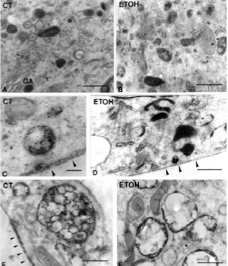
cultures consisted of 1-4 layers of overlapping cytoplasmic sheets. Both the control and treated cells displayed most of the structures which have been associated in other cell types with the endocytic and recycling pathways (Fig. 1). In many cells and mainly in the processes, flask-shaped invaginations of the plasma membrane corresponding to caveolae were observed (Fig. 1A,B,C and F). A large number of vesicles resembling caveolae were also seen in the cytoplasm (Fig. 1A). Clathrin coated pits and vesicles were also found in most of the cells examined (Fig. 1E). When the cells were cut “in face”, a large number of caveolae, tubulo-vesicular forma-tions lacking ribosomes, lysosomes and multivesicular bodies (MVBs) were observed. The main ultrastructural difference be-tween control and ethanol- exposed astrocytes with respect to the endocytic route was a significant increase in the volume density (Vvi) of lysosomes and MVBs in ethanol-treated cells (1.09±0.09% in control cells and 4.20±0.71% in cells exposed to ethanol, p< 0.05). MVBs in treated cells, in addition, contained fewer vesicles than the controls (see Fig. 4E and F).
125I-BSA internalization
To study the internalization and translocation of BSA in control and alcohol-treated astrocytes, cells were incubated at 4°C for 60 min with 125I-BSA. Then, cells were washed and reincubated with warm unlabeled culture medium at 37°C, and the time-courses of internalized and released 125I-BSA were studied after 1,5,15,30 and 60 min. As shown in Figure 2A, at 1.0 µCi/ml in control astrocytes, BSA was rapidly internalized, reaching maximal cell binding (acid resistant) after 5 min and then declining gradually over the next 60 min. These changes were accompanied by a progressive increase in the release of BSA in to the medium (Fig. 2B). Two main effects were observed in ethanol-exposed astro-cytes: first, the initial rate of BSA internalization (5 and 15 min of chase periods) was slightly affected and, second, the BSA re-leased was markedly affected in cells exposed to ethanol (Fig. 2B). These results suggest that ethanol impairs the endocytosis and
recycling processes. Finally, the results obtained using 125I-BSA at 0.05 and 0.1 µCi/ml were similar to those described for 1.0 µCi/ml (data not shown).
Fluorescence microscopy of endocytosis
We used pulse-chase experiments and fluorescence micros-copy to analyze, first of all, the capacity of proliferating control and ethanol-exposed astrocytes to internalize different macromolecules and, secondly, to assess the cellular structures involved in the endocytic process. Transferrin (Tf) is a useful marker of early endosomes, and after internalization it recycles back to the cell surface and there is no background from the biosynthetic pathway. Moreover, only a small proportion of internalized Tf is transported to other endocytic compartment elements such as lysosomes (Mukherjee et al., 1997). After 60 min of incubation at 4°C, and 10 min of chase, fluorescence corresponding to endocytosed Tf appeared concentrated in the perinuclear region and in long tubular structures (Fig. 3A,B) that irradiated from the cellular body toward the processes, where they often appeared parallel to the cell surface (Fig. 3B). In contrast, in astrocytes exposed to ethanol, the fluorescence was found in small tubular structures of more irregular morphology than in control cells. These structures ap-peared as small loops, circles and spirals randomly distributed in the cell body and processes (Fig. 3E). They were not stained with MitoFluor Green in either the control or treated cells.
When FITC-BSA was assessed using the same approach and after a chase of 10 min, we observed that endocytosed BSA appeared as bright fluorescent spots located in the perinuclear region and as tubules of different length dispersed in the cytoplasm or aligned near the cell surface of the control astrocytes (Fig. 3C,D). These tubular structures were smaller than those observed after Tf incubation. A similar FITC-BSA distribution pattern was observed in astrocytes exposed to ethanol, although the fluorescence spots located in the perinuclear region appeared more densely concen-trated than in the control astrocytes (Fig. 3F). In some cells, an
increase in the number of BSA-containing tubular endosomes was also observed. Finally, the endocytic compartment in control and ethanol-exposed astrocytes after incubation with labeled horse-radish peroxidase (HRP) showed a morphology similar to that described for BSA (data not shown).
Electron microscopy analysis of the endocytic process
Endocytosis of cationized ferritin (CF)
The uptake of CF was used as a general tracer to mark the structures involved in the endocytic pathway in astrocytes (Fig. 4). In control astrocytes, CF particles were evenly distributed after the cells were incubated at 4oC or after fixation of cells (data not shown). After 60 min incubation at 4oC and a 1 min chase, CF particles were found on the membrane surface in uncoated and
coated pits as well as in some tubular and vesicular endosomes. After a 15 min chase, CF was detected in the apical plasma membrane, in small sized vesicles and tubules and in vesicular and tubular endosomes; some MVBs were also labeled. However, neither lyso-somes nor elements of the Golgi apparatus contained the tracer. Endosomes were distinguishable from lyso-somes because in endolyso-somes the marker adhered to the membrane and there was no acid phosphatase activity (Herzog and Farquhar, 1983). In some cells, CF particles located in the deepest basal plasma membrane, between the cell and the substratum, appeared as a continuous layer. After a chase period of 30 min the distribution of CF was similar to that in cells chased for 15 min. However, an increase in labeled MVBs and cells displaying CF over the deep-est basal membrane was observed (Fig. 4C). In addi-tion, lysosomes and Golgi apparatus vesicles containing CF particles were also seen (Fig. 4A). Finally, after 60 min of chase, CF accumulated mainly in MVBs (Fig. 4E), lysosomes, some vesicles and in the basal-most plasma membrane, where the thickness of the CF layer de-creased as compared with 30 min of chase (Fig. 4E). This labeling over the most basal surface of the cells is interpreted as the result of a transcytosis process be-cause monolayers of astrocytes displayed abundant tight junctions which prevent the traffic of CF from the apical to the basal-most surface throughout the intercel-lular space (see Fig. 5H). Astrocytes exposed to ethanol showed a distribution of CF similar to that of the respec-tive controls (Fig. 4B). However, an increase in labeled endosomes (Fig. 4B), MVBs (Fig. 4F) and lysosomes and a dramatic reduction in CF particles over the basal plasma membrane (Fig. 4D) were observed in most of the cells after 30 and 60 min chases. These findings were confirmed by stereological procedures (Fig. 6). In addition, the appearance of MVBs from ethanol-ex-posed astrocytes was clear and showed fewer luminal vesicles than those of the respective control cells (Fig. 4F). The number of cells with CF over the basal-most plasma membrane was also reduced after ethanol treat-ment, which suggests that ethanol exposure affects transcytosis in astrocytes. In these cells the CF layer was absent or discontinuous and thinner than in the controls (Fig. 4D).
Endocytosis of BSA and HRP
Since astrocytes possess receptors for serum albumin (Juurlink and Devon, 1990), we also assessed the endocytosis and transcytosis pattern of BSA-gold complexes in control and ethanol-exposed astrocytes in primary culture (Fig. 5). After 15 min of chase, gold particles were seen in the different cell layers and the basal-most membrane (Fig. 5A,B and G). Within the cells, the particles appeared in vesicular and tubular endosomes and in lysosomes (Fig. 5A,D,E).When the cells were chased for 30 and 60 min, no labeling over the apical membrane was observed and the marker was distributed in the same structures as after a 15 min chase, but the number of gold-particle containing structures was diminished. However, this decrease was more pronounced in control that in ethanol-exposed cells, as was demonstrated by
stereological analysis (Fig. 6). Finally, at no chase period were gold particles observed between the basal-most astrocytic process and the substratum of ethanol-exposed cultures.
Electron microscopy showed that the qualitative and quantitative endocytosis pattern of HRP was similar to that described for BSA in control and treated astrocytes (Figs. 5F and 6). However, in double or triple uptake experiments there was a dissociation of the BSA and HRP-gold complexes (Fig. 5C,H).
Subcellular distribution of proteins involved in the endocytic pathway
To further ascertain whether ethanol affects the vesicular transport events involved in the sorting and endocytic pathways, we analyzed the pattern and distribution of some of the most important proteins associated with these processes, including caveolin, clathrin, rab5 and βCOP (Figs. 7-11).
Caveolin
It is the principal structural protein of caveolae and is involved in the nonclathrin mediated endocytosis events (Parton, 1996). Immunofluorescence analy-sis revealed, as previously described (Cameron et al., 1997), that caveolin is distributed as fluorescent points scattered throughout the cytoplasm, perinuclear space and cell surface. In some astrocytes, immu-noreactivity was also distributed along linear arrays. Labeling was also accumulated over some areas of the cell surface (Fig. 7A,B). Analysis of these fluores-cence patterns (percentage of cells displaying a particular pattern) indicated significant differences between the control and ethanol-exposed astrocytes (data not shown). Immunogold studies (Fig. 8A,C) revealed that gold particles corresponding to anti-caveolin binding sites appeared isolated or as small aggregates distributed over the cytoplasm and plasma membrane. Some of these aggregates displayed a circular or semi-circular shape and could correspond to the labeling of small vesicles. These structures appeared mainly located near the plasma mem-brane, where they were associated with the cell cortex. Gold particle density analysis demonstrates that ethanol exposure decreases the number of anti-caveolin binding sites (A/C ratio= 0.65) (Fig. 9). However, no changes in the gold particle distribution
plasm (Fig. 7C). In some cells, clathrin has a punctate diffuse cytoplasmic distribution. Although this pattern was similar in con-trol and treated cells, the labeling appeared to be more pronounced in control astrocytes. Using immunoelectron microscopy, we found that gold particles corresponding to anti-clathrin binding sites were located over or near the plasma membrane, small vesicles, endosomes and as individual particles scattered over the cyto-plasm (Fig. 8B-C). Double labeling with caveolin and anti-clathrin antibodies demonstrated that the two proteins do not co-localize in the same cell structures (Fig. 8C). Quantification of the anti-clathrin binding-site in alcohol-treated astrocytes revealed that although this treatment does not alter the distribution pattern, were observed. Finally, immunoblotting analysis also indicates that
ethanol significantly reduces the content of caveolin in astrocytes (Fig. 10).
Clathrin
It is the main structural component of the coated vesicles involved in membrane budding receptor-mediated endocytosis, and other steps in the cycle of vesicle assembly, uncoating and fusion (Kirchhausen et al., 1997; Mukherjee et al., 1997; Gaidarov et al., 1999). Immunofluorescence localization of clathrin in control and ethanol-exposed astrocytes showed the presence of labeling over the plasma membrane, juxtanuclear (Golgi) region and
it significantly decreases the gold particle density (A/C ratio= 0.58) (Fig. 9). The alcohol-induced reduction in the clathrin content in astrocytes was also confirmed by western blotting (Fig. 10).
Rab5
It is a low molecular weight GTP-binding protein that plays a role in endocytic vesicle trafficking and is found primarily on early endosomes (Mukherjee et al., 1997; Novick and Zerial, 1997). Immunofluorescence of rab5 revealed that this protein appeared as fluorescent spots scattered over the cytoplasm, mainly in the cell body (Fig. 7D). Similarly, immunogold studies demonstrate the presence of anti-rab5 binding sites over cytoplasm and endosomes (Fig. 8D-E). This localization pattern was similar in both cell types, although a significant decrease in the gold particle density was observed after ethanol exposure (A/C ratio= 0.55) (Fig. 9). Moreover, as demonstrated by particle distribution analysis, whereas in controls
the cytoplasmic gold particles showed a contagious distribution, in alcohol-treated astrocytes the particles were randomly distributed (Fig. 11). Immunoblotting also indicates that ethanol exposure in-duces a significant decrease in the rab5 content (Fig. 10).
β-COP
It is a protein involved in many steps of intracellular transport, including during endocytosis (Aniento et al., 1996; Cosson and Letourneur, 1997). Immunofluorescence of β-COP in astrocytes revealed that this protein is mainly located in the juxtanuclear (Golgi apparatus) region (Fig. 7E). Weak labeling was also observed as a diffuse staining over the cytoplasm. Astrocytes exposed to ethanol showed a similar labeling pattern, although fluorescence appeared to be less intense than in controls. Moreover, in some cells the juxtanuclear labeling appeared fragmented and/or surrounding the nucleus (Fig. 7F). Immunoelectron microscopy confirms that gold particles appeared over the Golgi area, some vesicles and cytoplasm (Fig. 8F), as has been described in other cell types (Griffiths et al., 1995). Gold particle density analysis indicated that ethanol induces a significant decrease in total β-COP labeling (A/C ratio= 0.49), although these differences between the two cell types decreased when quantification was carried out excluding the Golgi apparatus area (A/C ratio= 0.75) (Fig. 9). By western blotting we also found small differences between control and ethanol-exposed growing astrocytes (4 and 7 days of culture) (Fig. 10).
Discussion
Despite the number of studies on astrocyte-neuronal interac-tions and the involvement of astroglial cells in a variety of signaling and metabolic functions in the developing and adult brain (see review by Vernadakis, 1996), the relationship between these functions and the traffic of protein pathways, including
endocyto-Fig. 5. Endocytosis of BSA-gold and HRP-gold by control astrocytes. (A) After a chase period of 15 min BSA-gold particles were seen in the different cell layers (1-3), but were absent in the most apical membrane. (B) After longer chase periods, some BSA-gold particles were seen between the basal-most layer and the substratum (arrowheads, see also panel G). (C) Double labeling experiments (BSA-gold and HRP-gold) where 10 and 20 nm particles correspond to BSA and HRP, respectively. (D) BSA-gold particles are located in clathrin coated vesicles. (E) Endosome containing BSA-gold. (F) Micrographs showing a late endosome contain-ing HRP-gold. (H) Ten and 20 nm gold particles appear accumulated in the intercellular space. The presence of a tight-junction blocks the traffic of particles between two processes. Control and ethanol-exposed astro-cytes show similar structures involved in the endocytosis of BSA and HRP. The main difference between the two cell groups is the absence of gold particles between the basal-most astrocytic process and the substratum. Bars, A,B,C,E and G 0.5 µm ; D, 0.1 µm; F, 0.7 µm; H, 0.1 µm.
sis, in astrocytes remains poorly understood. In the present study, in which a combination of biochemical and morphological methods is used, we show in detail that growing cortical rat astrocytes in primary culture have a developed, active endocytic system and that exposure to low levels of ethanol alters the processes of endocytosis and transcytosis and impairs the protein trafficking in these cells.
Internalization of BSA
Our results suggest that the internalization of BSA-receptor complex is affected by ethanol. This is sup-ported by the findings demonstrating that during the initial rate of 125I-BSA internalization (up to 15 min) the levels of 125I-BSA were lower in astrocytes exposed to ethanol than in controls. Alterations in the process of internalization of several proteins have also been reported in hepatocytes exposed to ethanol (Tuma et al., 1991a,b, 1996; Tworek et al., 1996; Thiele et al., 1999). Although it is unclear how ethanol affects these processes, it has been demonstrated that ethanol changes the properties of some receptors (Casey et al., 1993). An impairment of BSA internalization in astrocytes exposed to ethanol could also be the result of the effect of this compound on cytoskeleton integrity (Sáez et al., 1991; Vallés et al., 1997) or plasma membrane composition (Sun et al., 1987; Renau-Piqueras et al., 1992). Alternatively, changes in plasma membrane composition might also affect membrane caveolae domains and/or clathrin adaptor complexes leading to an alteration in both ligand interaction with its receptor and endocytosis processes via clathrin-coated pits or nonclathrin pathways (Mukherjee et al., 1997). Indeed, we show that ethanol exposure signifi-cantly decreases the amount of caveolin and clathrin.
Early endosomes
We have used Tf as a marker of early endosomes because after its internalization in coated vesicles the Tf-receptor complex is first delivered to the sorting endosome and then enters the recycling compart-ment, where it remains stored or is recycled to the cell
that a modification in the levels of this protein could also contribute to the morphological changes observed in early endosomes after ethanol exposure. Ethanol-induced disruption of early endosomes could also explain the alteration in the distribution and content of rab5 in astrocytes exposed to ethanol. Rab5 is a small GTPase involved in the regulation of membrane docking and fusion of early endosomes (Gorvel et al., 1991; Stenmark et al., 1994; Novick and Zerial, 1997). Interestingly, it has also been proposed that this protein functionally links regulation of membrane trans-port, motility and intracellular distribution of early endosomes (Nielsen et al., 1999; Pfeffer, 1999). Recent studies have demon-strated that chronic ethanol intake induces redistribution of other rab proteins such as rab2 (Larkin et al., 1996) in hepatocytes. Since rab proteins have been implicated as regulators of vesicle-mediated trafficking events, one of the mechanisms involved in ethanol-induced impairment of endocytosis could be probably an alteration of rab proteins.
surface (Mukherjee et al., 1997). Our results indicate that, after a chase period of 10 min, fluorescent Tf appeared mainly in the recycling compartment, as is demonstrated by its localization in tubular elements which are similar to those described in other cell types (Mukherjee et al., 1997). We also found that ethanol induces a fragmentation of the tubular structures of early endosomes which might relate to an effect of ethanol on microtu-bules. Indeed, it has been shown that ethanol disrupts the microtubules of hepatocytes and astrocytes (Sáez et al., 1991; Yoon et al., 1998) and that a disruption of microtubules results in a fragmentation of the recycling compartment (McGraw et al., 1993; Marsh et al., 1995). Moreover, it has been reported that calmodulin regulates the morphology of tubular endosomes in the recycling compartment, modulating the endocytic transport (Co-lombo et al., 1997; Mukherjee et al., 1997), and we have shown that ethanol alters the content of calmodulin and calmodulin binding-proteins in astrocytes (Faura et al., 1992). This suggests
An important question is the role played by the recycling compartment in astrocytes. It has been reported that astrocytes can endocytose brain-derived neurotrophic factor (BDNF) using the truncated receptor, TrkB. Then internalized BDNF is stored and, without degradation, is released in a receptor-bound active form and can support neuronal survival (Rubio, 1997). This mecha-nism would be relevant during the ontogeny of the nervous system and in injured brain, where neuron survival becomes neurotrophin-dependent. Indeed, we have found an increase in the expression of the truncated form of TrkB in the brain of rats prenatally exposed to ethanol (Guerri et al., 1998). Therefore, one role of early endosomes in growing astrocytes may be to store different sub-stances, including growth factors, which could be delivered under specific physiological and pathological conditions.
Late endosomes
In contrast with Tf and as occurs with other ligands, after internalization BSA dissociates from its receptor and accumu-lates in sorting endosomes, which mature and give rise to late endosomes, including MVBs. Finally, BSA accumulates in lysos-omes by fusing with preexisting lysoslysos-omes (Keller and Simons, 1997; Mukherjee et al., 1997). Our results using FITC- labeled BSA indicate that after 10 min, fluorescence accumulates in perinuclear spots and in small tubular structures which are differ-ent from the early tubular endosomes observed with the labeled Tf. No alterations in the pattern of fluorescent BSA internalization were observed in astrocytes exposed to alcohol, but the increase in the intensity of perinuclear fluorescence that was observed in these cells suggests an accumulation of BSA in late endosomes and/or in lysosomes. This interpretation is supported by two findings. First of all, in the experiments with 125I-BSA, after
prolonged periods of chase (15 to 60 min) the intracellular levels of 125I-BSA were higher in ethanol-treated cells than in controls. Secondly, stereological data indicate an accumulation of vesicles and endosomes containing CF or BSA as well as an increase in lysosomes in ethanol-exposed astrocytes which probably is the result of the ethanol-induced damage to different cell compo-nents. Our results suggest that ethanol probably impairs the later steps of the endocytic pathway. Interestingly, impaired
endocyto-Fig. 8. Immunogold of caveolin, clathrin, rab5, and β -COP in control growing astrocytes. Caveolin. (A) Gold particles appear isolated or as small clusters displaying a circular shape (arrowheads) near the plasma membrane. Clathrin. (B) Gold particles corresponding to anti-clathrin binding sites are located over or near the plasma mem-brane, small vesicles and endosomes. Some particles appeared scattered over the cytoplasm. In the figure, an endosome containing CF and labeled with anti-clathrin is seen. (C) Double labeling with caveolin and anti-clathrin antibodies indicates that the two proteins do not co-localize. (D and E) Rab5 is localized in the cytoplasm and over the membrane of endosomes. (F) β-COP ap-pears mainly over the cisternae (arrows) and vesicles (arrowheads) of the Golgi apparatus area or scattered over the cytoplasm (circles). Ethanol-exposed astrocytes show a labeling pattern for these proteins similar to that de-scribed for the controls. Bar, A,C-F, 0.2 µm; B, 0.1 µm.
sis and/or degradation of asialoglycoprotein, epidermal growth factor and insulin have been observed in hepatocytes from ethanol-fed animals (Dalke et al., 1990; Tuma et al., 1991b; Tworek et al., 1996). In addition, despite the increase in the number of endosomes containing CF or BSA in treated cells, the number of vesicles and marker particles contained in MVBs in these cells decreased with respect to control cells. This suggests that ethanol also impairs the fusion between endosomes and MVBs and in fact we have observed a decrease in the amount of β-COP which is involved in the biogenesis of these MVBs (Aniento et al., 1996; Gu et al., 1997). In addition, changes in the lipid composition of MVBs membranes and endosomes or alterations in microtubules could also contribute to a decrease in the number of vesicles in these organelles (Gu and Gruenberg, 1999). How-ever, studies on other proteins involved in later steps of endocy-tosis, such as rab7, are needed to clarify the effect of ethanol on late endosomes.
Transcytosis
Transcytosis of CF and BSA has been widely studied in polarized and non-polarized cells, including differentiated astro-cytes (Juurlink and Devon, 1990; Romagnoli and Herzog, 1991; Antohe et al., 1997). The interstitial space of CNS, especially in the grey matter, is compartmentalized into a large number of interconnecting narrow tortuous channels by the processes aris-ing from astrocytes (Kosaka and Hama, 1986). This geometry does not facilitate the ready diffusion of macromolecules from one region to another of the brain. Therefore, as previously suggested (Juurlink and Devon, 1990), the process of transcytosis may represent a shortcut for rapid and direct transport of macromol-ecules across the cells and could be an important mechanism in macromolecule transport in the brain. Interestingly, endocytosis and transcytosis are processes showing important differences, even in their the microtubular requirements (Hunziker et al., 1990). We show that ethanol inhibits the transcytosis of BSA and CF in astrocytes which occurs by the opposite sorting of these molecules throughout the several layers of cells. Therefore, a consequence of this effect could be a defective astrocyte-medi-ated regulation of the macromolecular composition of the extra-cellular space in the brain.
Mechanisms involved in the effect of ethanol on the endocytosis processes
Recent research in liver has demonstrated that short and long-term ethanol exposure induces a retention of nascent proteins (Larkin et al., 1996), impairs vesicle transport (Torok et al., 1997) and protein trafficking and alters the assembly of the plasma membrane (Tuma et al., 1986, 1991a). Traffic along the receptor-mediated endocytosis pathway appears to be especially susceptible to ethanol in the liver (Tuma et al., 1990, 1991a,b). We show that in non-hepatic cells such as growing astrocytes, ethanol also impairs the endocyto-sis and transcytoendocyto-sis processes and alters intracellular protein traffick-ing. It would therefore seem that these effects could constitute a general mechanism of ethanol toxicity in the cell.
Although it is unclear how ethanol affects these processes, it is widely accepted that acetaldehyde, the first product of ethanol metabolism in the liver, can impair vesicle transport by disrupting microtubules (Yoon et al., 1998). Although there is evidence supporting this hypothesis with respect to the liver (Smith et al.,
1992), it cannot be the only mechanism involved in the effect of ethanol on the cytoskeleton, because astroglial cells have low capacity to metabolize ethanol and the production of acetalde-hyde in these cells is very low compared with hepatocytes (Eysseric et al., 1997). Moreover, we have found that ethanol alters cytoskeletal elements in astrocytes, including microtubules (Sáez et al., 1991; Vallés et al., 1997). Our results agree with the idea that a number of separate mechanisms targeted by ethanol participate in the overall action of ethanol in the endocytic pro-cess. Some mechanisms probably relate to changes in the plasma membrane (Sun et al., 1987; Renau-Piqueras et al., 1992) and, therefore, ethanol-induced alterations in the plasma and intracellular membrane composition could interfere with the abil-ity of the cell to respond to the external microenvironment and could affect with various transport processes, including binding of proteins such as clathrin, caveolin, β-COP and rab5 to mem-branes and fusion between endocytic elements (Slomiany et al., 1999). Alternatively, ethanol may target enzymes involved in the vesicle fusion and budding events (e.g. ATPases, GTPases) by altering rab GTPase functions, which seem to participate as specific regulators of membrane transport and fusion (Novick and Zerial, 1997).
In summary, our results indicate that ethanol alters membrane traffic in growing astrocytes, which could lead to defective transport and altered disposal of important physiological proteins during brain development such as NGF and its receptors (Vallés et al., 1994) as well as NCAM (Miñana et al., 1998a). Therefore, ethanol-induced alterations in endocytosis in growing astrocytes could be an important mechanism underlying the adverse effect of ethanol on the developing brain, including the fetal alcohol syndrome.
Materials and Methods
Astrocyte cultures
Primary cultures of astrocytes from 21-day-old rat fetuses were prepared from brain hemispheres as described in detail (Renau-Piqueras et al., 1989; Gomez-Lechón et al., 1992). Fetuses were obtained under sterile conditions from rats, and the cerebral hemispheres were dissected free of meninges and mechanically dissociated by pipetting in Dulbecco´s modified Eagle´s medium (DMEM, Gibco BRL, Eggenstein, Germany). The cell suspension was vortexed at maximum speed for 1 min and filtered through a nylon mesh with a pore size of 80 mm. Cells were plated on 35-mm Nunc plastic tissue culture dishes (1x10⁶ cells/dish, 2 ml/dish) and maintained in the same medium containing 20% fetal calf serum (FCS) and 1% antibiotics. Cultures were grown in a humidified atmosphere of 5% CO2 and 95% air at 37ºC. The medium was changed every 2 days. Under these conditions, the cells grow rapidly for 7-10 days (proliferative period) (Guerri et al., 1990). In all the experiments we used astrocytes at 7 days because, as is mentioned above, during the proliferation period astrocytes release several neurotrophic factors and express their recep-tors (Lu et al., 1991; Vallés et al., 1994). Some cells were grown in the presence of ethanol in the culture medium (Renau-Piqueras et al., 1989). The ethanol concentration in the medium was checked daily and adjusted to a final concentration of 30 mM (ethanol evaporation after 24 h was 10-20%).This concentration is similar to the blood level others have observed in pregnant chronic female drinkers or when 3 to 5 drinks are consumed by a 60-kg woman within 1 h (Eckardt et al., 1998). Ethanol was quantified by head-space gas chromatography as previously described (Guerri and
Sanchis, 1985). Finally, the purity of the astrocyte cultures was assessed by immunofluorescence using a mouse anti-GFAP monoclonal antibody (Boheringer Manheim, Cat# 814369), and the possible contamination by neurons was assessed by the same procedure using an anti-neurofilament 160-kDa monoclonal antibody (Sigma Chemicals Co., Cat# N5264).
Electron microscopy
Cell monolayers growing in plastic culture dishes were randomly selected (5 dishes per treatment, 3 different cultures), washed three times in PBS, fixed in 1.5% glutaraldehyde+1.0% formaldehyde in 0.1 mM cacodylate buffer, ph 7.4, for 60 min at 4ºC and embedded in Epon 812 (10 blocks per culture dish) (Mayordomo et al., 1992). In some cases, monolayers were embedded “in flat” and fragments of the Epon dish containing cells were glued on Epon blocks.
Analysis of endocytosis
Studies with fluorescent probes
Receptor-mediated endocytosis was analyzed by incubating the cells in serum-free DMEM medium (SFDM) containing bovine serum albumin (BSA) conjugated with FITC (Sigma Chemicals Co., Spain) or BODIPY-FL labeled transferrin (Tf) (Molecular Probes Europe BV, Leiden) diluted 1:5 and 1:250, respectively (Juurlink and Devon, 1990; Tooze and Hollinshead; 1991; Ghosh and Maxfield, 1995; Mukherjee et al., 1997). Cells growing on glass coverslips were washed with SFDM and incubated at 4°C for 60 min in SFDM containing the labeled probe. At the end of the incubation, astrocytes were washed twice with warm SFDM to eliminate unbound marker and incubated in serum containing DMEM medium for several chase periods (5,10,30 and 60 min) at 37°C. This low-temperature binding procedure is widely used to increase the amount of label internalized in a pulse-chase experiment. At low temperature (4°C), cells are capable of binding extracellular ligands but internalization is prevented. Thus, the population of surface receptors can be saturated with ligand conjugated during 4°C incubation (Dunn and Maxfield, 1990). After the period of chase, the cells were fixed in cold 2% glutaraldehyde in 0.1 M cacodylate buffer (pH 7.4) for 15 min, washed, mounted and examined with an Olympus fluores-cence microscope. Although we used several chase periods, we have analyzed mainly the chase period of 10 min, because at this time most of the endocytic elements appeared labeled in astrocytes as occurs in other cell types (Mukherjee et al., 1997).
For fluid-phase endocytosis, cells were washed three times in warm SFDM and incubated with horseradish peroxidase (HRP) conjugated with FITC (Sigma Chemicals Co.) (1:3 dilution) at 37°C for 5,15,30 and 60 min (Marsh et al., 1986; Mukherjee et al., 1997). After the different incubation times, cells were rinsed and fixed as described above.
In some experiments we used the Hoechst dye (molecular probes) to stain nuclei, because it makes it possible to individualize the cells. The fluorescent mitochondrial marker MitoFluor Green (molecular probes, cat # M-7502) was used in some experiments.
Electron microscopy
Uptake of cationized ferritin (CF) was analyzed to study nonspecific endocytosis using also pulse-chase experiments (Herzog and Farquhar, 1983; Klumperman et al., 1991; Lindo et al., 1993). The cells were rinsed and incubated in SFDM containing 800 mg/ml of CF (Sigma Chemicals Co.) (Lindo et al., 1993) for 60 min at 4°C. Then the cells were washed and incubated with DMEM at 37°C for 0,1,15,30 and 60 min. At the end of the different chase periods, cells were embedded as monolayers either in Epon 812 (5 plates per time period and treatment) or in Lowicryl K4M for immunocytochemical studies (see below).
Receptor-mediated endocytosis in control and ethanol-exposed astro-cytes was assessed by incubating the cells with SFDM containing BSA conjugated with 10 nm gold particles (1:10 dilution, Sigma Chemicals Co., A520= 5.5; particles per A520 per ml = 5.9x10¹²) SFDM (Juurlink and Devon, 1990; Deng et al ., 1991) at 4°C for 60 min. After this incubation, cells were washed in DMEM and maintained in the same culture medium at 37°C for Fig. 11. Graph showing the effect of ethanol-exposure on the gold
0,1,15,30 and 60 min in DMEM at 37°C. After each chase period, cells were washed, fixed as monolayers (1.5% glutaraldehyde + 1.0% formaldehyde in 0.1M cacodylate buffer, pH 7.4 at 4°C) and embedded in Epon 812 (5 plates per time period and treatment).
For fluid-phase endocytosis the cells were washed three times in SFDM and incubated for 0,1,15,30 and 60 min at 37°C in DMEM containing HRP conjugated with 20 nm gold particles (1:10 Sigma Chemicals Co., A520= 5.4; particles per A520 per ml = 6.2x10¹¹, work dilution, 1:10) (Marsh et al., 1986). After washing, the cells were embedded in Epon 812 (5 plates per time period and treatment). In some experiments, the cells were simultaneously loaded with BSA and HRP, BSA and CF, HRP and CF, or BSA, HRP and CF (Rabinowitz et al., 1992). BSA and CF were also used to assess the transcytosis process in growing astrocytes (Juurlink and Devon, 1990; Romagnoli and Herzog, 1991; Antohe et al., 1997).
125I-BSA internalization
Control and ethanol-exposed astrocytes were incubated for 60 min at 0ºC in 1.5 ml SFDM containing 125I-BSA (ICN, Spain, concentration: 1.91 mCi/ml; specific activity: 1.41 mCi/mg) at a final concentration of 0.05, 0.1 or 1.0 mCi/ ml. After this period, the medium was collected and the cells were incubated in 1.5 ml DMEM containing unlabeled BSA at 37°C for 0,1,5,15,30 and 60 min. After each chase period, the medium was collected and the cells were resuspended in DMEM. In some experiments, astrocytes were treated with 0.2 M acetic acid and 0.5 M NaCl (pH 3.5) for 10 min to remove surface-associated BSA (Vallés et al., 1994). After acid treatment the cells were resuspended in DMEM. Radioactivity was determined in a gamma counter (Packard-Cobra). The amount of radioactivity in acid-treated precipitates represents internalized BSA. Radioactivity in cells without acid treatment corresponds to total bound BSA (surface-bound and internalized BSA). Finally, radioactivity in the medium represents the release BSA-degrading activity into the medium, the release back into the medium of intact BSA or the translocated BSA. Nonspecific binding was determined in parallel incubations in presence of an excess of non-labeled BSA and amounted to 4-10% of the total surface-bound or internalized radioactivity.
Acid phosphatase cytochemistry
For cytochemical demonstration of acid phosphatase (AcPase) as a lysosomal marker, monolayers of cells previously incubated with CF were processed as described in detail using cerium ions as a capturing agent (Mayordomo et al., 1992; Lindo et al., 1993). The incubation medium was composed of 0.1 M acetate buffer (pH 5.0), 1 mM b-glycerophosphate (disodium salt) and 2 mM CeCl3.
Stereology
The volume density (Vv) of the endocytic vesicles, endosomes and lysosomes containing BSA, HRP and CF as well as the Vv of AcPase positive lysosomes was determined using point counting and stereological proce-dures (Steinman et al., 1983; Sancho-Tello et al., 1987; De Paz et al.,1990; Babia et al., 1999). The minimum sample size in each case was determined by the progressive mean technique (confidence limit, ±10%) (Williams, 1977).
Primary antibodies
Polyclonal anti-caveolin 1-a (Transduction Labs., Cat #C13630). Working dilution 1:500 for immunofluorescence, immunocytochemistry and 1:1000 for Western blotting.
Monoclonal anti-clathrin HC (N-19) (Transduction Labs., Cat #C43820). This antibody recognized the heavy chain of the clathrin. Working dilution 1:50 for immunofluorescence and immunocytochemistry and 1:200 for Western blotting. In some cases we also used a monoclonal anti-clathrin HC (clone 23) obtained from Transduction Lab.
Anti-rab5 monoclonal antibody generously provided by Dr. M. Zerial (EMBL, Heidelberg). Working dilution 1:200 for immunofluorescence and immunocytochemistry and 1:500 for Western blotting.
Monoclonal anti- β-COP (clone maD, Sigma Chemical Co., G6160)
recognizing an epitope shared by Golgi β-COP protein (110 kDa). Working dilution 1:80 for immunofluorescence and immunocytochemistry and 1:200 for western blotting.
Sodium dodecyl sulphate (SDS)-polyacrylamide gel electrophoresis and Western blot
Astrocytes were washed with PBS, harvested from tissue culture wells, homogenized in extraction buffer (PBS, 2% Nonidet P-40, 1.25 mM phenylmethylsulfonyl fluoride, 40 mM leupeptin, aprotinin 10Fg ml-1, 1 mM sodium ortovanadate) and incubated on ice for 15 min. Cell lysates were frozen and thawed three times and then centrifuged for 20 min at 15.000xg. The resulting supernatants were mixed with SDS sample buffer 6x [1x0.125 M Tris-HCl, pH 6.8, 2% SDS, 2 mM EDTA, 5% (vol/vol) 1-mercaptoethanol] and boiled for 3 min. Proteins were separated in SDS-polyacrylamide slab gels using the discontinuous gel and buffer system of Laemmli (1970), employing 12.5 and 7.5% (wt/vol) polyacrylamide in the separation gel. After electrophoresis, the proteins were transferred to nitrocellulose paper using a Semidry Electroblotter apparatus. The nitrocellulose paper was incubated overnight with a primary antibody and then incubated for 1 h with anti-mouse or anti-rabbit-IgG-alkaline phosphatase conjugate (Promega). After 10-20 min of color develop-ment, the nitrocellulose sheets were washed and photographed (Miñana et al., 1998b).
Immunofluorescence
Astrocyte monolayers growing on 16-mm glass coverslips were used for immunofluorescence studies following procedures previously described (Sancho-Tello et al., 1995). Briefly, cells were washed with phosphate-buffered saline (PBS, 1.5 mM MgCl2 and 1 mM CaCl2) and fixed for 10 min in -20°C methanol. After blocking for 10 min in 3% BSA in PBS, cells were incubated with the primary antibody diluted in BSA-PBS. The incubation was carried out at 37°C for 2 h in a moist chamber. After washing with 0.1% Nonidet P-40 in PBS, cells were incubated for 1 h at room temperature with the secondary antibody (FITC-conjugated goat anti-goat or anti-mouse IgG, diluted 1:100 in 1% BSA-PBS). The coverslips were then washed with PBS and mounted in FA Mounting Fluid (Difco).
Immunocytochemistry
Control and ethanol-exposed astrocytes in primary culture were fixed as monolayers with 0.5% glutaraldehyde -4% formaldehyde in 0.1M cacody-late buffer, pH 7.4, for 60 min at 4°C, detached from the plastic with a rubber policeman, washed in the buffer, incubated for 60 min in 50 mM NH4Cl, dehydrated in methanol and embedded in Lowicryl K4M as previously described (Renau-Piqueras et al., 1989, 1997).
Immunolocation of caveolin, clathrin, rab5, and β-COP was performed with the immunogold procedure as previously described (Renau-Piqueras et al., 1989, Iborra et al., 1992; Miñana et al., 1998b). In some ultrathin sections, double labeling using anti-clathrin and anti-caveolin (Mab, Trans-ductions Labs., Cat #C43420) antibodies was performed as previously described (Renau-Piqueras et al., 1989; Iborra et al., 1992; Bosser et al., 1995). In addition, clathrin, caveolin and rab-5 were analyzed on sections of cells previously incubated with CF. Quantitative analysis of immunogold micrographs was performed as described (Iborra et al., 1992) and the results expressed as the density of particles in ethanol-exposed cells/ density of gold particles in control cells (A/C ratio). Statistical analysis of data was done using the Student’s t test (p<0.05).The distribution of gold particles was assessed using a test for clumping or regularity consisting of calculating the variance: mean ratio (Williams, 1977).
Acknowledgments
References
ANIENTO, F., GU, F., PARTON, R.G. and GRUENBERG, J. (1996). An endosmal β -COP is involved in the pH-dependent formation of transport vesicles destined for late endosomes. J. Cell. Biol. 133: 29-41.
ANTOHE, F., SERBAN, G., RADULESCU, L. and SIMIONESCU, M. (1997). Transcytosis of albumin in endothelial cells is brefeldin A - independent. Endothe-lium 5: 125-136.
AULETTA, M., NIELSEN, F.C. and GAMMELTOFT, S. (1992). Receptor-mediated endocytosis and degradation of insulin-like growth factor I and II in neonatal rat astrocytes. J. Neurosci. Res. 31: 14-20.
BABIA, T., AYALA, I., VALDERRAMA, F., MATO, E., BOSCH, M., SANTARÉN, J.F., RENAU-PIQUERAS, J., KOK, J.W., THOMSON, T.M. and EGEA, G. (1999). N-Ras induces alterations in Golgi complex architecture and in constitutive protein transport. J. Cell. Sci. 112: 477-489.
BARRES, B.A. (1999). A new role for glia: Generation of neurons! Cell 97: 667-670.
BOSSER, R., FAURA, M., SERRATOSA, J., RENAU-PIQUERAS, J., PRUSCHY, M. and BACHS, O. (1995). Phosphorylation of rat liver heterogeneous nuclear ribonucleoproteins A2 and C can be modulated by cadmodulin. Mol. Cell. Biol. 15: 661-670.
CAMERON, P.L., RUFFIN, J.W., BOLLAG, R., RASMUSSEN, H. and CAMERON, R.S. (1997). Identification of caveolin and caveolin-related proteins in the brain. J. Neurosci. 15: 9520-9535.
CASEY, C.A., CAMACHO, K.B. and TUMA, D.J. (1992). The effects of chronic ethanol administration on the rates of internalization of various ligands during hepatic endocytosis. Biochem. Biophys. Acta 1134: 96-104.
CASEY, C.A., WIEGERT, R.L. and TUMA, D.J. (1993). Chronic ethanol administra-tion impairs ATP-dependent acidificaadministra-tion of endosomes in rat liver. Biochem. Biophys. Res. Commun. 195: 1127-1133.
COLOMBO, M.I., BERON, W. and STAHL, P.D. (1997). Calmodulin regulates endosome fusion. J. Biol. Chem. 272: 7707-7712.
COSSON, P. and LETOURNEUR, F. (1997). Coatomer (COPI) -coated vesicles: role in intracellular transport and protein sorting. Curr. Opin. Cell. Biol. 9: 484-487.
DALKE, D.D., SORRELL, M.F., CASEY, C.A. and TUMA, D.J. (1990). Chronic ethanol administration impairs receptor-mediated endocytosis of epidermal growth factor by rat hepatocytes. Hepatology 12: 1085-1091.
DE PAZ, P., RENAU-PIQUERAS, J., MIRAGALL, F. and BARRIO, J.P. (1990). Stereological analysis of plasma membrane. Distribution of ligands on the cell surface components and membrane flow in endocytosis. Comput. Meth. Prog. Bio. 31: 267-268.
DENG, Y., GRIFFITHS, G. and STORRIE, B. (1991). Comparative behavior of lysosomes and the pre-lysosome compartment (PLC) in in vivo cell fusion experiments. J. Cell Sci. 99: 571-582.
DOETSCH, F., CAILLE, I., LIM, D.A., GRACIA-VERDUGO, J.M. and ALVAREZ-BUYLLA, A. (1999). Subventricular zone astrocytes are neural stem cells in the adult mammalian brain. Cell 97: 703-716.
DUNN, F.W. and MAXFIELD, F.R. (1990). Use of fluorescence microscopy in the study of receptor-mediated endocytosis. In Noninvasive Techniques in Cell Biology (Eds. J.K. Foskett and S. Grinstein). Wiley-Liss, Inc., New York, pp. 153-176.
ECKARDT, M.J., FILE, S.E., GESSA, G.L., HOFFMAN, P.L., GRANT, K.A., GUERRI, C., KALANT, H., KOOB, G.F., LI, T.K. and TABAKOFF, B. (1998). Effects of moderate alcohol consumption on the central nervous system. Alcoholism: Clin. Exp. Res. 22: 998-1040.
EYSSERIC, H., GONTHIER, B., SOUBEYRAN, A., BESSARD, G., SAXOD, R. and BARRET, L. (1997). Characterization of the production of acetaldehyde by astrocytes in culture after ethanol exposure. Alcoholism: Clin. Exp. Res. 21: 1018-1023.
FAURA, M., PORTOLÉS, M., IBORRA, F.J., RENAU-PIQUERAS, J., SÁEZ, R., SERRATOSA, J. and BACHS, O. (1992). Alterations induced by prenatal expo-sure to ethanol on p62 and fodrin, two calmodulin-binding proteins in astroglial cells. Alcohol Alcoholism 27 (Suppl. 1): 36.
GAIDAROV, I., SANTINI, F., WARREN, R.A. and KEEN, J.H. (1999). Spatial control of coated-pit dynamics in living cells. Nature Cell. Biol. 1: 1-7.
GHOSH, R.N. and MAXFIELD, F.R. (1995). Evidence for nonvectorial, retrograde transferrin trafficking in the early endosomes of HEp2 cells. J. Cell. Biol. 128: 549-561.
GOMEZ-LECHÓN, M.J., IBORRA, F.J., AZORÍN, I., GUERRI, C. and RENAU-PIQUERAS, J. (1992). Cryopreservation of rat astrocytes from primary cultures. J. Tissue Cult. Meth. 14: 73-78.
GORVEL, J.P., CHAVRIER, M., ZERIAL, M. and GRUENBERG, J. (1991). Rab5 controls early endosome fusion in vitro. Cell 64: 915-925.
GRIFFITHS, G., PEPPERKOK, R., LOCKER, J.K. and KREIS, T.E. (1995). Immunolocalization of β-COP to the ER-Golgi boundary and the TNG. J. Cell Sci. 108: 2839-2856.
GU, F. and GRUENGERG, J. (1999). Biogenesis of transport intermediates in the endocytic pathway. FEBS Lett. 452: 661-666.
GU, F., ANIENTO, F., PARTON, R.G. and GRUENBERG, J. (1997). Functional dissection of COP-I subunits in the biogenesis of multivesicular endosomes. J. Cell. Biol. 139: 1183-1195.
GUASCH, R., RENAU-PIQUERAS, J. and GUERRI, C. (1992). Chronic ethanol consumption induces accumulation of proteins in the liver Golgi apparatus and decreases galactosyltransferase activity. Alcoholism: Clin. Exp. Res. 16: 942-948.
GUERRI, C. and RENAU-PIQUERAS, J. (1997). Alcohol, astroglia, and brain devel-opment. Mol. Neurobiol. 15: 65-81.
GUERRI, C. and SANCHIS, R. (1985). Acetaldehyde and alcohol levels in pregnant rats and their fetuses. Alcohol 2: 267-270.
GUERRI, C., CLIMENT, E. and SANCHO-TELLO, M. (1998). Effects of alcohol exposure on the expression of the neurothrophin receptor trk B, during brain development. Alcoholism: Clin. Exp. Res. 22: 147A.
GUERRI, C., SÁEZ, R., SANCHO-TELLO, M., MARTIN DE AGUILERA, E. and RENAU-PIQUERAS, J. (1990). Ethanol alters astrocyte development: A study of critical periods using primary cultures. Neurochem. Res. 15: 559-565.
HERZOG, V. and FARQUHAR, M.G. (1983). Use of electron-opaque tracers for studies on endocytosis and membrane recycling. Methods Enzymol. 98: 203-225.
HUNZIKER, W., MALE, P. and MELLMAN, I. (1990). Differential microtubule require-ments for transcytosis in MDCK cells. EMBO J. 9: 3515-3525.
IBORRA, F.J., RENAU-PIQUERAS, J., PORTOLES, M., BOLEDA, M.D., GUERRI. C. and PARES, X. (1992). Immunocytochemical and biochemical demonstration of formaldehyde dehydrogenase (class III alcohol dehydrogenase) in the nucleus. J. Histochem. Cytochem. 40: 1865-1878.
JUURLINK, B.H.J. and DEVON, R.M. (1990). Macromolecular translocation - a possible function of astrocytes. Brain Res. 533: 73-77.
KELLER, P. and SIMONS, K. (1997). Post-Golgi biosynthetic trafficking. J. Cell Sci. 110: 3001-3009.
KIRCHHAUSEN, T., BONIFACINO, J.S. and RIEZMAN, H. (1997). Linking cargo to vesicle formation: receptor tail interactions with coat proteins. Curr. Opin. Cell. Biol. 9: 488-495.
KLUMPERMAN, J., BOEKESTIJN, J.C., MULDER, A.M., FRANSEN, J.A.M. and GINSEL, L.A. (1991). Intracellular localization and endocytosis of brush border enzymes in the enterocyte-like cell line Caco-2. Eur. J. Cell. Biol. 54: 76-84.
KOSAKA, T. and HAMA, K. (1986). Three-dimensional structure of astrocytes in the rat dentate girus. J. Comp. Neurol. 249: 242-260.
LAEMMLI, U.K. (1970). Cleavage of structural proteins during the assembly of the head of bacteriophage T4. Nature 227: 680-685.
LARKIN, J.M., OSWALD, B. and McNIVEN, M.A. (1996). Ethanol-induced retention of nascent proteins in rat hepatocytes is accompanied by altered distribution of the small GTP-binding protein rab2. J. Clin. Invest. 98: 2146-2157.
LINDO, L., IBORRA, F.J., AZORIN, I., GUERRI, C. and RENAU-PIQUERAS, J. (1993). Analysis of the endocytic-lysosomal system (vacuolar apparatus) in astrocytes during proliferation and differentiation in primary culture. Int. J. Dev. Biol. 37: 565-572.
LU, B., YOKOYAMA, M., DREYFUS, C.F. and BLACK, I.B. (1991). NGF gene expression in actively growing brain glia. J. Neurosci. 11: 318-326.
MARSH, E.W., LEOPOLD, P.L., JONES, N.L. and MAXFIELD, F.R. (1995). Oligomerized transferrin receptors are selectively retained by a luminal signal in a long-lived endocytic recycling compartment. J. Cell. Biol. 129: 1509-1522.
MARSH, M., GRIFFITHS, G., DEAN, G.E., MELLMAN, I. and HELENIUS, A. (1986). Three-dimensional structure of endosomes in BHK-21 cells. Proc. Natl. Acad. Sci. USA 83: 2899-2903.
MAYORDOMO, F., RENAU-PIQUERAS, J., MEGIAS, L., GUERRI, C., IBORRA, F.J., AZORIN, I. and LEDIG, M. (1992). Cytochemical and stereological analysis of rat cortical astrocytes during development in primary culture. Effect of prenatal exposure to ethanol. Int. J. Dev. Biol. 36: 311-321.
MCGRAW, T.E., DUNN, K.W. and MAXFIELD, F.R. (1993). Isolation of a tempera-ture-sensitive variant Chinese hamster ovary cell line with a morphologically altered endocytic recycling compartment. J. Cell. Physiol. 136: 389-397.
MIÑANA, R., SANCHO-TELLO, M., CLIMENT, E., SEGUÍ, J.M., RENAU-PIQUERAS, J. and GUERRI, C. (1998a). Intracellular location, temporal expression, and polysialylation of neural cell adhesion molecule in astrocytes in primary culture. Glia 24: 415-427.
MIÑANA, R., SEGUÍ, J.M., RENAU-PIQUERAS, J. and GUERRI, C. (1998b). Prena-tal alcohol exposure alters the expression pattern of neural cell adhesion molecule during brain development. Alcoholism: Clin. Exp. Res. 22: 184A.
MUKHERJEE, S., GHOSH, R.N. and MAXFIELD, F.R. (1997). Endocytosis. Physiol. Rev. 22: 759-803.
NIELSEN, E., SEVERIN, F., BACKER, J.M., HYMAN, A.A. and ZERIAL, M. (1999). Rab5 regulates motility of early endosomes on microtubules. Nature Cell Biol. 1: 376-382.
NOUEL, D., FAURE, M-P., ST. PIERRE, J-A., ALONSO, R., QUIRION, R. and BEAUDET, A. (1997). Differential binding profile and internalization process of neurotensin via neuronal and glial receptors. J. Neurosci. 17: 1795-1803.
NOVICK, P. and ZERIAL, M. (1997). The diversity of Rab proteins in vesicle transport. Curr. Opin. Cell. Biol. 9: 496-504.
PARTON, R.G. (1996). Caveolae and caveolins.Curr. Opin. Cell. Biol. 8: 542-548.
PFEFFER, S.R. (1999). Motivating endosome motility. Nature Cell Biol. 1: E145-E147.
RABINOWITZ, S., HORSTMANN, H., GORDON, S. and GRIFFITHS, G. (1992). Immunocytochemical characterization of the endocytic and phagolysosomal compartments in peritoneal macrophages. J. Cell. Biol. 116: 95-112.
RENAU-PIQUERAS, J., GUASCH, R., AZORIN, I., SEGUI, J.M. and GUERRI, C. (1997). Prenatal alcohol exposure affects galactosyltransferase activity and glyconjugates in the Golgi apparatus of fetal rat hepatocytes. Hepatology 25: 343-350.
RENAU-PIQUERAS, J., GUERRI, C., BURGAL, M., DE PAZ, P., SÁEZ, R. and MAYORDOMO, F. (1992). Prenatal exposure to ethanol alters plasma membrane glycoproteins of astrocytes during development in primary culture as revealed by concanavalin A binding and 5'-nucleotidase activity. Glia 5: 65-74.
RENAU-PIQUERAS, J., ZARAGOZA, R., DE PAZ, P., BAGUENA-CERVELLERA, R., MEGIAS, L. and GUERRI, C. (1989). Effects of prolonged ethanol exposure on the glial fibrillary acidic protein-containing intermediate filaments of astrocytes in primary culture: a quantitative immunofluorescence and immunogold electron microscopic study. J. Histochem. Cytochem. 37: 229-240.
ROMAGNOLI, P. and HERZOG, V. (1991). Transcytosis in thyroid follicle cells: regulation and implications for thyroglobulin transport. Exp. Cell. Res. 194: 202-209.
RUBIO, N. (1997). Mouse astrocytes store and deliver brain-derived neurotrophic factor using the non-catalytic gp97trkB receptor. Eur. J. Neurosci. 9: 1847-1853.
SÁEZ, R., BURGAL, M., RENAU-PIQUERAS, J., MARQUÉS, A. and GUERRI, C. (1991). Evolution of several cytoskeletal proteins of astrocytes in primary culture: Effect of prenatal alcohol exposure. Neurochem. Res. 16: 737-747.
SANCHO-TELLO, M., RENAU-PIQUERAS, J., BÁGUENA-CERVELLERA, R. and GUERRI, C. (1987). A biochemical and stereological study of neonatal rat hepatocyte subpopulations. Effect of pre- and postnatal exposure to ethanol. Virchows Arch. B 54: 170-181.
SANCHO-TELLO, M., VALLÉS, S., MONTOLIU, C., RENAU-PIQUERAS, J. and GUERRI, C. (1995). Developmental pattern of GFAP and vimentin gene expres-sion in rat brain and in radial glial cultures. Glia 15: 157-166.
SLOMIANY, A., PIOTROWSKI, E., GRABSKA, M., PIOTROWSKI, J. and SLOMIANY, B.L. (1999). Chronic ethanol-initiated apoptosis in hepatocytes is induced by changes in membrane biogenesis and intracellular transport. Alcoholism: Clin. Exp. Res. 23: 334-343.
SMITH, S., JENNETT, R., SORRELL, M. and TUMA, D. (1992). Substoichiometric inhibition of microtubule formation by acetaldehyde-tubulin adducts. Biochem. Pharmacol. 44: 65-72.
STEINMAN, R.M., MELLMAN, E.R., MULLER, W.A. and COHN, Z.A. (1983). Endocy-tosis and the recycling of plasma membrane. J. Cell. Biol. 96: 1-27.
STENMARK, H., PARTON, R.G., STEELE-MORTIMER, A., LUTCKE, J., GRUENBERG, J. and ZERIAL, M. (1994). Inhibition of rab5 GTPase activity stimulates membrane fusion in endocytosis. EMBO J. 13: 1287-1296.
SUN, S.H., FU, Y.H., JOU, T.C., SUN, G.Y. and SUN, A.Y. (1987). Ethanol effects on phospholipids and triacylglycerols of rat brain astrocyte cell line. Alcohol Alcohol-ism (Suppl.) 1: 691-695.
THIELE, G.M., MILLER, J.A., KLASSEN, L.W. and TUMA, D.J. (1999). Chronic ethanol consumption impairs receptor-mediated endocytosis of formaldehyd-treated albumin by isolated rat liver endothelial cells. Hepatology 29: 1511-1517.
TOOZE, J. and HOLLINSHEAD, M. (1991). Tubular-early endosomal network in AtT20 and other cells. J. Cell. Biol. 115: 635-653.
TOROK, N., MARKS, D., HSIAO, K., OSWALD, B.J. and McNIVEN, M.A. (1997). Vesicle movement in rat hepatocytes is reduced by ethanol exposure: alterations in microtubule-based motor enzymes. Gastroenterology 113: 1938-1948.
TUMA, D.J. and SORRELL, M.F. (1981). Effects of ethanol on the secretion of glycoproteins by rat liver slices. Gastroenterology 80: 273-278.
TUMA, D.J., CASEY, C.A. and SORRELL, M.F. (1990). Effects of ethanol on hepatic protein trafficking: Impairment of receptor-mediated endocytosis. Alcohol Alcohol-ism 25: 117-125.
TUMA, D.J., CASEY, C.A. and SORRELL, M.F. (1991a). Effect of alcohol on hepatic protein metabolism and trafficking. Alcohol Alcoholism (Suppl.) 1: 297-303.
TUMA, D.J., CASEY, C.A. and SORRELL, M.F. (1991b). Chronic ethanol-induced impairments in receptor-mediated endocytosis of insulin in rat hepatocytes. Alcoholism: Clin. Exp. Res. 15: 808-813.
TUMA, D.J., MAILLIARD, M.E., CASEY, C.A., VOLENTINE, G.D. and SORRELL, M.F. (1986). Ethanol-induced alterations of plasma membrane assembly in the liver. Biochem. Biophys. Acta 856: 571-577.
TUMA, D.J., TODERO, S.L., BARAK-BERNHAGEN, M. and SORRELL, M.F. (1996). Effects of chronic ethanol administration on the endocytosis of cytokines by rat hepatocytes. Alcoholism: Clin. Exp. Res. 20: 579-583.
TWOREK, B.L., TUMA, D.J. and CASEY, C.A. (1996). Decreased binding of asialoglycoproteins to hepatocytes from ethanol-fed rats. J. Biol. Chem. 271: 2531-2538.
VALLÉS, S., LINDO, L., MONTOLIU, C., RENAU-PIQUERAS, J. and GUERRI, C. (1994). Prenatal exposure to ethanol induces changes in the nerve growth factor and its receptor in proliferating astrocytes in primary culture. Brain Res. 656: 281-286.
VALLÉS, S., PITARCH, J., RENAU-PIQUERAS, J. and GUERRI, C. (1997). Ethanol exposure affects glial fibrillary acidic protein gene expression and transcription during rat brain development. J. Neurochem. 69: 2484-2493.
VERNADAKIS, A. (1996). Glia-neuron intercommunications and synaptic plasticity. Progress Neurobiol. 49: 185-214.
WILLIAMS, M. (1977). Stereological techniques. In Practical methods in electron microscopy (Ed. A.M. Glauert). Amsterdam, North Holland/American Elsevier, pp 1-83.
YOON, Y., TÖRÖK, N., KRUEGER, E., OSWALD, B. and McNIVEN, M.A. (1998). Ethanol-induced alterations of the microtubule cytoskeleton in hepatocytes. Am. J. Physiol. 274: G757-G766.