Wnt signalling during limb development
VICKI L. CHURCH and PHILIPPA FRANCIS-WEST*
Department of Craniofacial Development, King’s College London, Guy’s Hospital, London, UK
ABSTRACT Wnts control a number of processes during limb development - from initiating outgrowth and controlling patterning, to regulating cell differentiation in a number of tissues. Interactions of Wnt signalling pathway components with those of other signalling pathways have revealed new mechanisms of modulating Wnt signalling, which may explain how different responses to Wnt signalling are elicited in different cells. Given the number of Wnts that are expressed in the limb and their ability to induce differential responses, the challenge will be to dissect precisely how Wnt signalling is regulated and how it controls limb development at a cellular level, together with the other signalling pathways, to produce the functional limb capable of co-ordinated precise movements.
KEY WORDS:
Wnt, limb, development, chondrogenesis, myogenesis
0214-6282/2002/$25.00
© UBC Press Printed in Spain
www.ijdb.ehu.es
*Address correspondence to: Dr. Philippa Francis-West. Department of Craniofacial Development, King’s College London, Guy’s Hospital, London Bridge, London, SE1 9RT, U.K. Fax: + 44-207-955-2704. e-mail: [email protected]
Abbreviations used in this paper: AER, apical ectodermal ridge; BMP, bone morphogenetic protein; CamKII, calcium/calmodulin-dependent kinase II; CTGF, connective tissue growth factor; Dkk, dickkopf; Dsh, dishevelled; EGF, epidermal growth factor; Fgf, fibroblast growth factor; GSK3β, glycogen synthase kinase 3β; HSPG, heparan sulphate proteoglycan; Ihh, Indian hedgehog; JNK, c-Jun N-terminal kinase; LDL, low density lipoprotein; LRP, LDL-related protein; MyHC, myosin heavy chain; PKC, protein kinase C; PTHrP, parathyroid hormone-related peptide; RA, rheumatoid arthritis; RTK, receptor tyrosine kinase; Sfrp, secreted frizzled-related protein; WIF, Wnt inhibitory factor; WISP, Wnt induced secreted protein.
The Wnt Gene Family
The Wnt family of secreted glycosylated factors consists of 22 members in vertebrates which have a range of functions during development from patterning individual structures to fine tuning at a cellular level controlling cell differentiation, proliferation and survival. The founding members of this family are the Drosophila segment polarity gene Wingless (Wg), required for wing develop-ment, together with Wnt1 (originally named int-1) in the mouse. The latter was identified due to integration of the mouse mammary tumour virus into the Wnt1 locus, which resulted in epithelial hyperplasia and increased susceptibility to mammary carcinomas (Nusse and Varmus, 1982; Nusse et al., 1984; Tsukamoto et al., 1988). Initially, based on their ability to induce a secondary axis in Xenopus embryos and to transform the mammary epithelial cell line C57MG, the family was divided into two subgroups. The Wnt1 class, which comprises Wnt1, -3a and -8, can induce a secondary axis in Xenopus, whilst the Wnt5a class, consisting of Wnt4, -5a and –11, cannot. Similarly, Wnt1, -3a and -7a have transforming activity whilst Wnt4 and -5a do not (www.stanford.edu/~rnusse/ wntwindow.html).
The Wnt family signals through the frizzled receptors which are seven pass transmembrane receptors, like the smoothened recep-tors involved in transduction of the hedgehog signalling pathway. The frizzled protein family consists of 10 genes characterised by an N-terminal cysteine-rich domain responsible for ligand binding. The family can be divided simplistically on the basis of the intrac-ellular motif S/T-X-V proposed to be involved in binding PDZ domains such as that found in the Wnt signalling component dishevelled. The S/T-X-V motif is not present in Fz3, -6 and -9 but
is found in the others (Cadigan and Nusse, 1997). The frizzled receptors can function together with the LRP co-receptors, which are single transmembrane proteins containing LDL receptor re-peats, two frizzled motifs and four EGF type repeats in the extracellular domain (reviewed by Pandur and Kühl, 2001; also see Roszmusz et al., 2001). The LRPs, which include the vertebrate genes LRP4, -5 and -6 and the Drosophila gene arrow, form a complex with frizzled in a Wnt-dependent manner and signal in the canonical pathway (Tamai et al., 2000; Wehrli et al., 2000; re-viewed by Pandur and Kühl, 2001).
Indeed, mutations in the Drosophila genes sugarless (also called kiwi and suppenkasper) and sulphateless, which encode homo-logues of UDP-glucose dehydrogenase and N-deacetylase/N-sulphotransferase respectively, have revealed the necessity of these glycosaminoglycan biosynthetic enzymes in Wingless sig-nalling (Binari et al., 1997; Häcker et al., 1997; Haerry et al., 1997; Lin and Perrimon, 1999). These proteins are needed for heparan sulphate modification of the co-receptors dally and dally-like. Similarly, the avian extracellular sulphatase QSulf1 can regulate heparan-dependent Wnt signalling and is required in the Wnt regulation of MyoD expression in myogenic C2C12 cells (Dhoot et al., 2001).
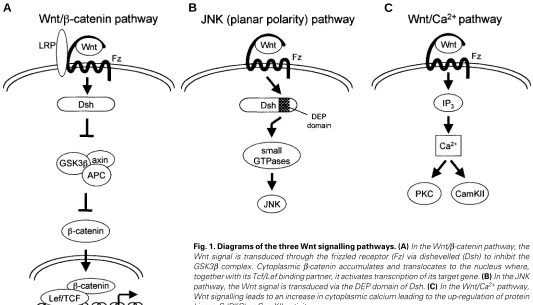
In the classical Wnt signalling pathway, Wnts signal via β -catenin (see Fig. 1). Briefly, Wnt signalling represses the axin/ glycogen synthase kinase-3β (GSK3β) complex, which normally stimulates the degradation of β-catenin via the ubiquitin pathway (reviewed by Kikuchi 2000). Therefore, in Wnt-activated cells, cytoplasmic β−catenin accumulates and is translocated to the nucleus where, in conjunction with T cell-specific factor/lymphoid enhancer binding factor 1 (Tcf/Lef1) transcription factors, it acti-vates the transcription of Wnt target genes. A second pathway activated in response to Wnt signalling signals via the small GTPases Rho and Cdc42 to c-Jun N-terminal kinase (JNK) (Fig. 1; Boutros et al., 1998; Li et al., 1999; reviewed by McEwen and Peifer, 2000). This pathway is utilised in Drosophila planar cell polarity and by Wnt11 in convergent extension movements during gastrulation. Both of these pathways utilise dishevelled (Dsh): the
β−cateninpathway is dependent on all three domains present in Dsh (DIX, PDZ and DEP) whilst the JNK pathway only requires the
DEP domain. Thirdly, some Wnts, such as Wnt5a, can stimulate the release of intracellular Ca2+, activating protein kinase C (PKC)
and Ca2+/calmodulin-dependent kinase II (CamKII) (see Fig. 1;
Sheldahl et al., 1999; reviewed by Kühl et al., 2000). The pathway that is utilised appears to depend on the receptor profile and intracellular signalling molecules and is not determined by the specificity of the ligand. For example, Wnt1, which has classically been shown to signal through the β-catenin pathway, has also been implicated in the PKC pathway and more recently has been shown to signal through a novel GTPase in the JNK pathway (Tao et al., 2001; Ziemer et al., 2001). Similarly, Wnt5a and Wnt3a, which have distinct effects in Xenopus embryos, can both induce
β-catenin accumulation in cardiac myocytes (Toyofuku et al., 2000; Yamanaka et al., 2002). Differentiation between the β-catenin and JNK pathway is controlled by the Dsh-associated kinase PAR-1, which promotes signalling via β-catenin (Sun et al., 2001).
The Wnt Antagonists
As with other growth factors such as members of the TGF-β and fibroblast growth factor (Fgf) families, Wnt signalling can be antagonised by secreted factors. These antagonists include the
secreted frizzled related proteins (Sfrps), cerberus, dickkopfs
(Dkks) and Wnt inducible factor (WIF-1). However, unlike the other antagonists, cerberus has not been reported to be expressed in the developing limb. The Sfrp family consists of at least five members and contains the frizzled related N-terminal domain, which can bind to Wnts, but lacks the intracellular sequence found in the frizzled receptors. They antagonise Wnt function by binding to the Wnt
A
B
C
Fig. 1. Diagrams of the three Wnt signalling pathways. (A) In the Wnt/β-catenin pathway, the Wnt signal is transduced through the frizzled receptor (Fz) via dishevelled (Dsh) to inhibit the GSK3β complex. Cytoplasmic β-catenin accumulates and translocates to the nucleus where, together with its Tcf/Lef binding partner, it activates transcription of its target gene. (B) In the JNK pathway, the Wnt signal is transduced via the DEP domain of Dsh. (C) In the Wnt/Ca2+ pathway,
molecule and preventing receptor activation. However, they have also been proposed to act as agonists, thus facilitating Wnt function – one possible mechanism may be to increase the solubility of the Wnt molecule to aid its diffusion (Lin et al., 1997). Alternatively, as Sfrps are membrane bound, like the HSPG core protein dally, they may help to localise Wnts to the cell surface, effectively increasing the concentration of Wnt ligand for the receptor. WIF-1 has an extracellular “WIF” domain, five EGF-like repeats and a small hydrophilic C-terminal domain (Hsieh et al., 1999). The WIF-1 domain alone can block Wnt activity suggesting that it binds Wnts. Interestingly, this domain shares homology with the extracellular sequence present in the orphan tyrosine kinase Ryk receptors but whether there is any functional significance of this is at present unknown (Patthy, 2000). However, Ryk, like Wnts, has been suggested to have transforming activity whilst the Ryk mouse knockout bears resemblances to the Wnt5a mouse null mutant (Katso et al., 1999; Yamaguchi et al., 1999; Halford et al., 2000). In both mutants, there is overall growth retardation characterised by a shorter snout and limbs. In contrast to the other antagonists, Dkk1 and -4 bind directly to the Wnt co-receptor LRP6 and block Wnt signalling through the β-catenin pathway (Mao et al., 2001; Zorn, 2001). Surprisingly however, the related molecules Dkk2 and -3 do not antagonise Wnt signalling and the former actually promotes Wnt signalling at least in Xenopus (Krupnik et al., 1999; Wu et al., 2000).
Limb Outgrowth and Patterning
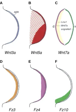
Members of the Wnt family are differentially expressed either within the ectoderm or mesenchyme where they play a number of roles (see Fig. 2). Analogous to their role in Drosophila and emphasising the importance of this gene family, members of the Wnt family are expressed in two key signalling centres in the limb – the apical ectodermal ridge (AER), which controls outgrowth, and the dorsal ectoderm, which controls dorso-ventral patterning. Wnt8c and Wnt2b (also known as Wnt13 ), which are transiently expressed in the lateral plate mesoderm, initiate outgrowth of the leg and wing respectively (Kawakami et al., 2001). Thus, ectopic overexpression of these Wnts in the interflank region of a stage 13/ 14 chick embryo, prior to limb outgrowth, induces ectopic Fgf10 expression and limb formation via the β-catenin pathway. Fgf10 subsequently induces Wnt3a expression in the AER, which in turn switches on the expression of Fgf8, again via the β-catenin pathway, and hence promotes AER formation (Kengaku et al., 1998; Kawakami et al., 2001). The Wnt antagonists Sfrp3 and Dkk1 are expressed in the lateral plate mesoderm where they may limit the limb- and AER-inducing activity of Wnt2b, -8c and –3a, either temporally or spatially (Ladher et al., 2000a; Mukhopadhyay et al., 2001). Misexpression of Dkk1 in the chick results in the loss of or irregularities in the AER whilst loss of Dkk1 in the mouse broadens the AER along the dorso-ventral axis (Mukhopadhyay et al., 2001). The defects seen in the Dkk studies may reflect the role of Wnt signalling in both limb initiation and subsequent limb outgrowth.
In contrast to the chick, Wnt3a in the mouse is not expressed in the AER. However, consistent with the essential role of Wnts in limb outgrowth, gene-inactivation of both Lef1 and Tcf1, which are expressed in the early developing limb, prevents limb outgrowth (Galceran et al., 1999). In these mice, the AER does not form as shown by the lack of Fgf8 expression and the absence or
down-regulation of the distal mesenchymal markers Msx1 and Wnt5a. In addition, dorso-ventral patterning is disrupted: expression of the ventral ectodermal marker engrailed1 is absent whilst expression of Lmx1b, normally restricted to the dorsal mesenchyme, is found ventrally indicating that a double dorsal limb has formed. This defect possibly indicates a very early role of Lef1/Tcf1 during the establishment of the dorso-ventral axis. In the chick, Wnt3a has been shown to induce mesenchymal expression of Lef1 (Kengaku et al., 1998). However, as with other progress zone markers such as Fgf10 and Msx1, it is possible that Wnt3a acts via Fgf8 in the ectoderm and that Lef1 is not directly induced by Wnt3a (Kengaku et al., 1998). Indeed Fgf8 can substitute for the AER and induce/ maintain Lef1 expression in the limb mesenchyme (Grotewold and Rüther, 2002).
In the chick, Wnt3a expression persists in the AER throughout limb development where it maintains Fgf8 expression and AER function (Kengaku et al., 1998). The regulation of Fgf8 expression in the ectoderm suggests that Wnt signalling within the AER can act
A
B
C
D
E
F
cell-autonomously. Consistent with this is the expression of β -catenin, Tcf1, Lef1 and the receptor Fz4 in the AER (Oosterwegel et al., 1993; Kengaku et al., 1998; Lu et al., 1997; Nohno et al., 1999). Another possible role for cell-autonomous Wnt function within the AER would be to regulate the expression of gap junc-tions, which are needed to maintain the integrity of the AER (Becker et al., 1999; Makarenkova and Patel, 1999). Wnt signalling via the
β-catenin pathway has been shown to transcriptionally activate Cx43, a component of gap junctions, in P19 cells and cardiomyocytes and to increase gap junction communication in Xenopus (Olson et al., 1991; van der Heyden et al., 1998; Ai et al., 2000). Misexpression of Wnt1 in the mesenchyme has also been associated with a change in Cx43 expression – Cx43 is down-regulated in the Wnt1-expressing cells, but is up-regulated in the surrounding mesen-chyme (Meyer et al., 1997). Whether these are direct effects or due to changes in patterning/cell differentiation is unclear, although it is possible that Wnts, together with Fgf signalling from the AER, also regulate gap junctional expression in the limb mesenchyme (Makarenkova et al., 1997). Several other Wnts, such as Wnt5a and –12, together with the Wnt antagonists Sfrp1 and Dkk1, are also expressed in the AER, although their functions are unknown (Christiansen et al., 1995; Esteve et al., 2000; Mukhopadhyay et al., 2001; Grotewold and Rüther, 2002).
In addition to controlling Fgf8 expression, in the chick Wnt3a and –7a signal in conjunction with Fgfs to induce/maintain the expression of Csa1, a gene mutated in the human Townes-Brocks syndrome which is characterised by pre-axial polydactyly (Farrell and Münsterberg, 2000). Neither Wnt nor Fgf signalling alone can induce/maintain Csa1 expression in the progress zone. This shows that in addition to acting alone, Wnts may also act in synergy with other factors, again highlighting the complexity of Wnt signalling in limb bud development. This synergy is also seen during otic and neural induction in chicks, and in antero-posterior patterning of the neuroectoderm in Xenopus, suggesting that it may be a fundamental process of many aspects of development, which to date, has not been investigated (McGrew et al., 1997; Ladher et al., 2000b; Wilson et al., 2001). This in part may be due to the ability of Fgfs to substitute almost wholeheartedly for the AER making investigation into the role of other AER factors almost redundant.
Another Wnt required for limb outgrowth is Wnt5a. Gene inactivation of Wnt5a in mice results in a shortening of the limb reflecting an overall retardation in development (Yamaguchi et al., 1999). All skeletal structures are affected but in general the severity increases in a proximal to distal direction. Thus, the distal phalanges are missing whilst the proximal elements are short-ened. However, patterning of the elements is normal. This trunca-tion is also observed in other regions of the body – the rostro-caudal axis, the jaws and the genitalia - suggesting that a common mechanism mediated by Wnt5a controls the development of all these structures. This hypothesis, at least for the genitals and limbs, has previously been put forward on the basis of gene-inactivation of members of the Hox gene complex which affects both structures (Kondo et al., 1997). In both the paraxial meso-derm and the limb bud mesenchyme, loss of Wnt5a results in a decrease in proliferation (Yamaguchi et al., 1999). Whether a similar proximo-distal progression of defects occurs in the face is unclear but in light of the parallels that are often drawn between limb and face development, it will be of interest to compare.
Analysis of the regulation of Wnt5a expression has shown that, as with many progress zone markers, Wnt5a is regulated by Fgf signalling from the AER (Kawakami et al., 1999).
In contrast, Wnt7a controls dorso-ventral patterning. In the absence of Wnt7a (i.e. following gene-inactivation of Wnt7a in mice, and the naturally occurring Wnt7a mutant, postaxial hemimelia, which is due to a splicing defect in Wnt7a ) the sesamoid bones and foot pads are duplicated, being found both ventrally and dorsally, and the dorsal tendons assume a ventral pattern (Parr and McMahon, 1995; Parr et al., 1998). However, there is not a complete transformation of a dorsal to ventral fate as the dorsal ectodermal derivatives, the nails and hair, are still present, although they are abnormal (Parr and McMahon, 1995). Wnt7a maintains Shh expression which is necessary for antero-posterior patterning (Parr and McMahon, 1995; Yang and Niswander, 1995). Thus, in the Wnt7a mutants, the posterior digits are lost consistent with the loss of Shh expression (Parr and McMahon, 1995; Parr et al., 1998). Wnt7a signalling may depend on LRP6 as loss of LRP6 function in the mouse also results in dorso-ventral patterning and anterior-posterior patterning defects similar to that seen in the Wnt7a mouse mutant (Pinson et al., 2000). In addition, in LRP6 mutants, the AER is not maintained possibly reflecting a role in Wnt3a signalling. As Wnt7a is initially expressed throughout the dorsal limb ectoderm, only later be-coming confined to the distal dorsal ectoderm, it is unclear why the proximal structures are unaffected. There may be redundancy with other Wnts substituting for Wnt7a function or this may reflect the differences in patterning mechanisms between the autopod and the zeugopod/stylopod.
Wnt7a regulates the expression of the LIM-homeobox-con-taining gene Lmx1 in the chick and its homologue Lmx1b in the mouse. Indeed, the onset of Lmx1/1b expression is coincident with Wnt7a expression (Riddle et al., 1995; Cygan et al., 1997). In the chick and mouse limb bud, Lmx1/1b is expressed in the dorsal subectodermal mesenchyme over a distance of 9-12 cell layers (Riddle et al., 1995; Vogel et al., 1995; Cygan et al., 1997). This hints, but does not definitively prove, how far ectodermal Wnts diffuse or signal through the mesenchyme and is similar to that seen in Drosophila. Misexpression of Lmx1 dorsalises the ventral distal limb bud showing that it is a key downstream mediator of Wnt7a signalling (Riddle et al., 1995; Vogel et al., 1995). In Wnt7a mutants, Lmx1b expression in the limb bud is initiated but is down-regulated distally by E11.5 showing that Wnt7a is not required for the early and proximal expression of Lmx1b, and consistent with the absence of proximal defects in the Wnt7a-/- limb bud (Cygan et al., 1997).
A
B
C
D
E
myogenic differentiation. Once within the limb bud, they become committed switching on the expression of the myogenic determi-nation factors MyoD and Myf5. During migration and within the limb bud they come into contact or close proximity with a number of Wnt signals, including Wnt5a and -11 in the mesenchyme and ectodermal Wnts such as Wnt3 and -4. Whether Wnt signalling initiates myogenesis in the limb bud is unclear. However, the ability of Wnts or β-catenin to activate myogenesis in the somite and in P19 cells respectively, together with the expression of the Wnt antagonist Sfrp2 in uncommitted myogenic precursors, which is down-regulated as the myogenic cells differentiate, suggests that this is possible (Cossu et al., 2000; Ladher et al., 2000a; Petropoulos and Skerjanc, 2002).
In addition, Wnt signalling may modulate other aspects of myogenic development such as migration and cell morphology. In the limb bud, N-cadherin is essential for myogenic migration and, during chondrogenesis, Wnt7a has been shown to maintain the expression of this cadherin (Brand-Saberi et al., 1996; Tufan and Tuan, 2001). Thus, if parallels between muscle and skeletal development can be drawn, this may suggest an additional potential role of Wnts during limb muscle development. Finally, work in our laboratory has shown that Wnt signalling can regulate the terminal stages of muscle differentiation and can alter the number of myoblasts expressing either slow or fast Myosin heavy chains (MyHCs) (Anakwe et al., submitted). Each muscle is composed of a unique combination of slow and fast fibres which will determine how it will function. Slow fibres have oxidative metabolism, contract slowly and in general are required for posture whilst fast fibres contract rapidly with high force, have glycolytic metabolism and are necessary for movement. There-fore, our data suggest that Wnt signals determine the intricate pattern of slow/fast MyHC expression in the musculature. expression of Fgf8, Fgf10, Wnt3a, Wnt7a
and Lmx1. The normal expression of Wnt7a and Lmx1 suggests that their expression is not autoregulated. In humans, mutation of LMX1B results in the autosomal dominant nail-patella syndrome characterised by dor-sal limb defects (Dreyer et al., 1998). The knee caps are hypoplastic or absent, the elbows are abnormal, and the fingernails may be brittle or absent.
Wnt7a appears to signal through Fz10, which is expressed in the dorsal-posterior-distal mesenchyme underlying Wnt7a ex-pression in the ectoderm and colocalising with Shh (Kawakami et al., 2000). Assays in Xenopus have shown that Fz10 and Wnt7a synergise when co-injected showing that Wnt7a may signal through this receptor (Kawakami et al., 2000). In the limb bud Fz10 expression can be induced by Shh and Wnt7a providing a positive feedback loop to main-tain the expression of these genes in the distal mesenchyme (Kawakami et al., 2000). Wnt7a expression is regulated negatively by engrailed1, a homeobox-containing gene which is essential for formation of the ventral structures. In the absence of engrailed1,
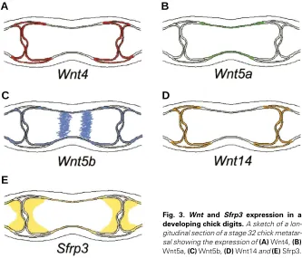
Fig. 3. Wnt and Sfrp3 expression in a developing chick digits. A sketch of a lon-gitudinal section of a stage 32 chick metatar-sal showing the expression of (A) Wnt4, (B)
Wnt5a, (C) Wnt5b, (D) Wnt14 and (E) Sfrp3.
Wnt7a is ectopically expressed in the ventral ectoderm resulting in the formation of bi-dorsal distal limbs (Loomis et al., 1996; Cygan et al., 1997). Conversely, misexpression of engrailed1 in the chick results in a down-regulation of Wnt7a in the dorsal ectoderm (Logan et al., 1997).
Finally, Wnt signalling has been implicated in the sculpting of the limb bud, removing “excess” tissue by programmed cell death. Indeed, the ability of BMP4 to induce cell death in the developing limb appears to be mediated by Dkk1 (Grotewold and Rüther, 2002). Loss of function of Dkk1 in mice results in the down-regulation of Msx1, a component of the cell death pathway, in the anterior and posterior necrotic zones and the interdigital mesen-chyme, whilst gain of Dkk1 function in chicks causes excessive cell death via activation of the c-jun pathway (Mukhopadhyay et al., 2001; Grotewold and Rüther, 2002). The decrease in cell death in the mouse mutants contributes to the polydactyly and fusion of digits that occurs in Dkk1 mouse mutants (Mukhopadhyay et al., 2001). In addition, Fz2, Fz3 and Fz4, and Dkk-2,and –3, are expressed in the interdigital mesenchyme suggesting that a fine balance of Wnt signalling controls cell death/survival in this region (Monaghan et al., 1999; Nohno et al., 1999).
Other Wnts that are expressed in the limb bud include Wnt3, -4, -6 and -7b in the ectoderm (Parr et al., 1993). Their role during patterning and outgrowth of the limb is unknown although Wnt-4, -7a and -7b can induce neurotrophin-3 expression and hence survival of sensory neurons (Patapoutian et al., 1999).
Muscle Differentiation
Skeletal Development
The expression patterns and putative functions of several of the Wnts and components of the signalling pathway have been de-scribed during skeletal development. Their particular spatiotemporal expression patterns, together with functional analyses in the chick, suggest that different members of the Wnt family possess distinct roles during chondrogenesis and joint development.
During the early stages of chondrogenesis, Sfrp3 (Frzb) is ex-pressed in the central mesenchymal condensation where it encom-passes the domain of Sox9 expression which marks chondrogenic commitment (Wada et al., 1999; Baranski et al., 2000; Esteve et al., 2000; Ladher et al., 2000a). Wnt5a, -5b, -11 and –14, together with the receptors Fz4 and –6, and the Wnt antagonists Dkk2 and –3, are also expressed in the mesenchyme surrounding the developing cartilage elements (Christiansen et al., 1995, 1996; Tanda et al., 1995; Kawakami et al., 1999; Monaghan et al., 1999; Nohno et al., 1999; Hartmann and Tabin, 2000; 2001). Wnt1, -7a and –14 have been shown to inhibit chondrogenesis in micromass culture and, therefore, the presence of the antagonists, particularly Sfrp3, may allow chondrogenesis to proceed (Rudnicki and Brown, 1997; Stott et al., 1999; Hartmann and Tabin, 2001). The inhibition by Wnt7a has been correlated with the maintenance of N-cadherin expression and hence cell adhesion which halts chondrogenesis just after the initiation stage (Tufan and Tuan, 2001).
As chondrocytes proceed through the differentiation pathway, the expression profile of Wnts, receptors and antagonists alters (see Fig. 3). For instance, Sfrp3 becomes confined to the epiphy-seal (i.e. relatively undifferentiated) chondrocytes (Hoang et al., 1996; Wada et al., 1999; Baranski et al., 2000; Ladher et al., 2000a). Wnt5b is expressed in a sub-population of the diaphyseal chondrocytes whilst β-catenin and Tcf4 are more widely expressed with transcripts of both overlapping in the articular, prehypertrophic and hypertrophic chondrocytes (Hartmann and Tabin, 2000; Church et al., in press).
Misexpression of Wnt5a results in the formation of shorter skeletal elements due to a delay in chondrocyte differentiation. Thus, there is a reduction in the number of hypertrophic chondrocytes and a smaller Ihh-expressing (i.e. prehypertrophic) domain (Kawakami et al., 1999; Hartmann and Tabin, 2000). Similarly, misexpression of dominant-negative versions of Fz1 and –7, two of the receptors expressed in developing elements, has a similar effect to Wnt5a in that misexpression results in shorter elements: the hypertrophic domain is absent and the prehypertrophic zone is smaller (Hartmann and Tabin, 2000). In contrast, in vitro micromass studies have shown that these two receptors can have different effects when overexpressed: Fz7, but not Fz1, inhibits chondrogenesis (Tufan et al., 2002).
Several Wnts and their signalling pathway components are expressed in the perichondrium, a layer of fibroblasts, surround-ing the cartilage element. The perichondrium inhibits chondrocyte differentiation by a PTHrP-Ihh signalling loop, contributes to appositional growth and also ultimately will be become the site of bone collar formation. Other molecular players that control peri-chondrial development and mediate signalling pathway interac-tions are uncharacterised. However, candidate factors include Wnt5a, -5b, -11, Fz1, Fz2, β-catenin and Dkk1 which are ex-pressed here (Christiansen et al., 1995; Tanda et al., 1995; Lako et al., 1998; Monaghan et al., 1999; Hartmann and Tabin, 2000; Church et al., in press).
In contrast to the other Wnts, Wnt4 and -14 have been associ-ated with joint formation. During early joint development, both genes are expressed in the presumptive joint regions (Kawakami et al., 1999; Hartmann and Tabin, 2000; 2001). Additionally, cWnt14 is expressed in the mesenchyme adjacent to the joint regions (Hartmann and Tabin, 2001). As the joint undergoes differentiation, both genes are expressed in the synovial mem-brane and the connective tissue of the joint capsule (Church et al., in press; Hartmann and Tabin, 2000; 2001). Mouse Sfrp2 (in contrast to chick Sfrp2) is also expressed at sites of joint develop-ment (Leimeister et al., 1998). Misexpression of Wnt14 in the developing chick limb has shown that Wnt14 can induce the joint phenotype as indicated by the absence of cartilage matrix and loss of chondrogenic markers concomitant with the acquisition of joint markers (Hartmann and Tabin, 2001). The study also implicated Wnt14 as a key factor in defining joint spacing in development: in addition to the induction of ectopic joints, Wnt14 seemed to prevent the formation of endogenous joints in the adjacent region (Hartmann and Tabin, 2001). Furthermore, Wnt14 can induce dedifferentia-tion of chondrocytes in micromass cultures, a process that occurs during normal joint development (Hartmann and Tabin, 2001). In contrast to Wnt14, overexpression of Wnt4 does not induce joint formation but instead accelerates chondrocyte differentiation and bone collar formation as suggested by an expanded hypertrophic zone and ultimate shortening of the elements, as well as a thicker osteoid layer with increased expression of osteogenic markers and calcification (Hartmann and Tabin, 2000). It has been proposed that Wnt4 signals from the developing joint to control chondrocyte differentiation (Hartmann and Tabin, 2000).
At present it is unclear which pathways are utilised by Wnt signalling during skeletal differentiation. β-catenin can mimic the effect of Wnt1, -7a and –14 in inhibiting chondrogenic differentiation in vitro whilst overexpression of β-catenin in vivo mimics the effect of Wnt4 to accelerate terminal chondrocyte differentiation (Hartmann and Tabin, 2000). It is interesting to note that Wnt4 and –5a are in the same class of Wnts yet have opposing effects on chondrocyte differentiation (Hartmann and Tabin, 2000). These different effects may result from the activation of different pathways.
Degenerative Skeletal Disorders
Classically, misregulation of the Wnt pathway has been associ-ated with carcinogenesis. However, recent evidence has emerged to show that regulation of Wnt function is essential for both skeletal development and maintenance of the skeleton in the adult. There are currently three skeletal dsyplasias which are linked to the Wnt signalling pathway. These are progressive pseudorheumatoid dys-plasia, characterised by the loss of the articular cartilage and joint space starting during early childhood; Robinow syndrome, characterised by a severe skeletal dysplasia with short bones, brachydactyly, spinal defects and facial anomalies; and osteoporo-sis-pseudoglioma syndrome, which results in decreased bone mass in adults (Hurvitz et al., 1999; Afzal et al., 2000; Gong et al., 2001). In the former, Wnt-Induced Secreted Protein 3 (WISP3) is mutated
syndrome brachydactyly type B, possibly reflecting a Wnt-mediated patterning defect (Schwabe et al., 2000). In addition, as other players that modulate Wnt signalling emerge, it is possible that other human syndromes may also be linked to deregulation of the Wnt signalling pathways. For example, β−catenin can interact with Sox proteins which antagonise Wnt function (Zorn et al., 1999; Takash et al., 2001). Mutations in human Sox9 give rise to the syndrome campomelic dysplasia, a bone dysmorphogenesis. Although not tested it is possible that this syndrome is in part due to increased Wnt activity. Similarly, the paired-type homeobox protein Alx4 can bind Lef1 and may modulate the β-catenin/Lef1 signalling pathway (Boras and Hamel, 2002). In humans, haploinsufficiency of ALX4 results in a bone mineralisation defect in the skull (Wuyts et al., 2000; Mavrogiannis et al., 2001). Likewise, notch signalling, which is important for skeletal development, interacts with components of the Wnt pathway (Axelrod et al., 1996; Ross and Kadesch, 2001). Thus, a number of signalling pathways that control skeletogenesis may converge on or modify the Wnt signalling pathway.
Wisps are members of the CCN (CTGF/Cyr61/Nov) family of growth factors which participate in a multitude of processes, including chondrogenesis (reviewed by Perbal, 2001; Lau and Lam, 1999). The subfamily of Wisps comprises three proteins: Wisp1 and –2 were initially identified as proteins that are up-regulated in Wnt1-transformed mammary epithelial (C57MG) cells and Wisp3 was identified by screening EST databases for homolo-gous genes (Pennica et al., 1998). Further analysis showed that the Wisp1 promoter contains Tcf/Lef1 binding sites and is directly activated by Wnt1 signalling (Xu et al., 2000). Furthermore, like Wnt1, Wisp1 also has oncogenic activity and, therefore, appears to mediate some of the effects of Wnt1 signalling. Whilst Wisp1 is under the control of Wnt1/β-catenin signalling, connective tissue growth factor (CTGF), the founder member of the family, is under the control of TGF-β (Grotendorst et al., 1996). Therefore, distinct sets of growth factor signals appear to converge on different CCN proteins to mediate their effects. Wisp3 is expressed in synoviocytes, chondrocytes and bone marrow stem cells undergoing chondro-genesis in vitro, and Wisp2 is expressed by chondrocytes and osteoblasts (Hurvitz et al., 1999; Kumar et al., 1999). These data, together with the Wisp3 mutation in progressive pseudorheumatoid dysplasia, strongly suggest that Wisps may mediate Wnt regula-tion of the formaregula-tion and maintenance of cartilage and joints.
The 120 residue cysteine-rich Frizzled motif is found not just in frizzed receptors and Sfrps, but is also present in type XVIII collagen, carboxypeptidase Z and receptor tyrosine kinases (RTKs), which include the ROR family (Rehn et al., 1998). Thus it is possible that Wnts may bind to these molecules and, in the case of the receptors, activate or block (by steric competition for the “legitimate” ligand) the signalling pathway. ROR2 is ex-pressed during face and limb skeletal development in the mouse (Matsuda et al., 2001). For example, ROR2 transcripts are found in the perichondrium of the digits. ROR1 is expressed in the anterior and posterior necrotic zones of the limbs and in interdigital areas, in a similar region to the Wnt antagonist Dkk1, and thus may control cell death (Matsuda et al., 2001). Like the human Robinow syndrome, mice null for ROR2 have a severe skeletal phenotype which is also characterised by misshapen bones and abnormal growth plate morphology (Afzal et al., 2000; Takeuchi et al., 2000). Thus, ROR2 has essential roles in chondrocyte differentiation and ossification. As yet, the ligands for the RORs
are not known but the presence of a frizzled domain suggests that Wnts may modify/induce ROR signalling.
Finally, mutation of LRP5, which is expressed in osteoclasts, results in osteoporosis-pseudoglioma syndrome (Gong et al., 2001). This syndrome is characterised by a low bone mass and increased vulnerability to fractures and deformation. A dominant negative LRP5 blocks the ability of bone morphogenetic proteins (BMPs) to induce alkaline phosphatase activity in C3H10T1/2 and ST2 cell lines showing that Wnt signalling acts downstream of BMPs (Gong et al., 2001).
Rheumatoid arthritis (RA) is a debilitating disease characterised by inflammatory responses resulting in the erosion of the articular cartilage. A number of Wnts and frizzled receptors (namely Wnt1, -5a, -10b, -11 and -13, and Fz2, -5 and -7) are expressed in synovial tissue from patients with RA, with a notable upregulation of Wnt5a and Fz5 expression (Sen et al., 2000). Importantly, overexpression of Wnt5a in normal synovial cells in vitro stimulates the production of interleukin-6, -8 and -15, cytokines that can contribute to the eventual destruction of the joint in RA, whilst blocking Wnt5a and Fz5 function abolishes these responses (Sen et al., 2000; 2001). Wnt signalling has also been implicated in osteoarthritis where Sfrp4 expression has been shown to correlate with apoptotic chondrocytes within the degenerating articular cartilage (James et al., 2000). Another member of the Sfrp family, Sfrp2, sensitises MCF7 cells to apoptotic stimuli suggesting that Sfrp4 may directly contribute to the chondrocyte apoptosis (Melkonyan et al., 1997). These are the first reports implicating Wnt signalling in arthritic disease and are sure to be followed by many important discoveries.
References
AFZAL, A.R., RAJAB, A., FENSKE, C.D., OLDRIDGE, M., ELANKO, N., TERNES-PEREIRA, E., TUYSUZ, B., MURDAY, V.A., PATTON, M.A., WILKIE, A.O. and JEFFERY, S. (2000). Recessive Robinow syndrome, allelic to dominant brachy-dactyly type B, is caused by mutation of ROR2. Nat Genet 25: 419-22.
AI, Z., FISCHER, A., SPRAY, D.C., BROWN, A.M. and FISHMAN, G.I. (2000). Wnt-1 regulation of connexin43 in cardiac myocytes. J Clin Invest Wnt-105: Wnt-16Wnt-1-7Wnt-1.
ANAKWE, K., ROBSON, L., BUXTON, P., CHURCH, V.L., ALLEN, S., HARTMANN, C., NOHNO, T., EVANS, D.J.R. and FRANCIS-WEST, P.H. (Submitted). Antago-nistic Wnt signals specify muscle fibretypes in the developing limb.
AXELROD, J.D., MATSUNO, K., ARTAVANIS-TSAKONAS, S. and PERRIMON, N. (1996). Interaction between Wingless and Notch signaling pathways mediated by dishevelled. Science 271: 1826-32.
BAEG, G.H., LIN, X., KHARE, N., BAUMGARTNER, S. and PERRIMON, N. (2001). Heparan sulfate proteoglycans are critical for the organization of the extracellular distribution of Wingless. Development 128: 87-94.
BARANSKI, M., BERDOUGO, E., SANDLER, J.S., DARNELL, D.K. and BURRUS, L.W. (2000). The dynamic expression pattern of frzb-1 suggests multiple roles in chick development. Dev Biol 217: 25-41.
BECKER, D.L., MCGONNELL, I., MAKARENKOVA, H.P., PATEL, K., TICKLE, C., LORIMER, J. and GREEN, C.R. (1999). Roles for alpha 1 connexin in morphogen-esis of chick embryos revealed using a novel antisense approach. Dev Genet 24: 33-42.
BINARI, R.C., STAVELEY, B.E., JOHNSON, W.A., GODAVARTI, R., SASISEKHARAN, R. and MANOUKIAN, A.S. (1997). Genetic evidence that heparin-like glycosaminoglycans are involved in wingless signaling. Development 124: 2623-32.
BORAS, K. and HAMEL, P.A. (2002). Alx4 binding to LEF-1 regulates N-CAM promoter activity. J Biol Chem 277: 1120-7.
BRAND-SABERI, B., GAMEL, A.J., KRENN, V., MULLER, T.S., WILTING, J. and CHRIST, B. (1996). N-cadherin is involved in myoblast migration and muscle differentiation in the avian limb bud. Dev Biol 178: 160-73.
CADIGAN, K.M. and NUSSE, R. (1997). Wnt signaling: a common theme in animal development. Genes Dev 11: 3286-305.
CHEN, H., LUN, Y., OVCHINNIKOV, D., KOKUBO, H., OBERG, K.C., PEPICELLI, C.V., GAN, L., LEE, B. and JOHNSON, R.L. (1998). Limb and kidney defects in Lmx1b mutant mice suggest an involvement of LMX1B in human nail patella syndrome. Nat Genet 19: 51-5.
CHRISTIANSEN, J.H., DENNIS, C.L., WICKING, C.A., MONKLEY, S.J., WILKINSON, D.G. and WAINWRIGHT, B.J. (1995). Murine Wnt-11 and Wnt-12 have temporally and spatially restricted expression patterns during embryonic development. Mech Dev 51: 341-50.
CHRISTIANSEN, J.H., MONKLEY, S.J. and WAINWRIGHT, B.J. (1996). Murine WNT11 is a secreted glycoprotein that morphologically transforms mammary epithelial cells. Oncogene 12: 2705-11.
CHURCH, V.L., NOHNO, T., LINKER, C., MARCELLE, C. and FRANCIS-WEST, P.H. Wnt regulation of chondrocyte differentiation. J Cell Sci. In press.
COSSU, G., DE ANGELIS, L., BORELLO, U., BERARDUCCI, B., BUFFA, V., SONNINO, C., COLETTA, M., VIVARELLI, E., BOUCHE, M., LATTANZI, L., TOSONI, D., DI DONNA, S., BERGHELLA, L., SALVATORI, G., MURPHY, P., CUSELLA-DE ANGELIS, M.G. and MOLINARO, M. (2000). Determination, diversification and multipotency of mammalian myogenic cells. Int J Dev Biol 44: 699-706.
CYGAN, J.A., JOHNSON, R.L. and MCMAHON, A.P. (1997). Novel regulatory interactions revealed by studies of murine limb pattern in Wnt-7a and En-1 mutants. Development 124: 5021-32.
DHOOT, G.K., GUSTAFSSON, M.K., AI, X., SUN, W., STANDIFORD, D.M. and EMERSON, C.P., JR. (2001). Regulation of Wnt signaling and embryo patterning by an extracellular sulfatase. Science 293: 1663-6.
DREYER, S.D., ZHOU, G., BALDINI, A., WINTERPACHT, A., ZABEL, B., COLE, W., JOHNSON, R.L. and LEE, B. (1998). Mutations in LMX1B cause abnormal skeletal patterning and renal dysplasia in nail patella syndrome. Nat Genet 19: 47-50.
ESTEVE, P., MORCILLO, J. and BOVOLENTA, P. (2000). Early and dynamic expression of cSfrp1 during chick embryo development. Mech Dev 97: 217-21.
FARRELL, E.R. and MUNSTERBERG, A.E. (2000). csal1 is controlled by a combina-tion of FGF and Wnt signals in developing limb buds. Dev Biol 225: 447-58.
GALCERAN, J., FARINAS, I., DEPEW, M.J., CLEVERS, H. and GROSSCHEDL, R. (1999). Wnt3a-/—like phenotype and limb deficiency in Lef1(-/-)Tcf1(-/-) mice. Genes Dev 13: 709-17.
GONG, Y. et al. (2001). LDL receptor-related protein 5 (LRP5) affects bone accrual and eye development. Cell 107: 513-23.
GROTENDORST, G.R., OKOCHI, H. and HAYASHI, N. (1996). A novel transforming growth factor beta response element controls the expression of the connective tissue growth factor gene. Cell Growth Differ 7: 469-80.
GROTEWOLD, L. and RUTHER, U. (2002). The Wnt antagonist Dickkopf-1 is regulated by Bmp signaling and c-Jun and modulates programmed cell death. Embo J 21: 966-75.
HACKER, U., LIN, X. and PERRIMON, N. (1997). The Drosophila sugarless gene modulates Wingless signaling and encodes an enzyme involved in polysaccha-ride biosynthesis. Development 124: 3565-73.
HAERRY, T.E., HESLIP, T.R., MARSH, J.L. and O’CONNOR, M.B. (1997). Defects in glucuronate biosynthesis disrupt Wingless signaling in Drosophila. Develop-ment 124: 3055-64.
HALFORD, M.M., ARMES, J., BUCHERT, M., MESKENAITE, V., GRAIL, D., HIBBS, M.L., WILKS, A.F., FARLIE, P.G., NEWGREEN, D.F., HOVENS, C.M. and STACKER, S.A. (2000). Ryk-deficient mice exhibit craniofacial defects associated with perturbed Eph receptor crosstalk. Nat Genet 25: 414-8.
HARTMANN, C. and TABIN, C.J. (2000). Dual roles of Wnt signaling during chondro-genesis in the chicken limb. Development 127: 3141-59.
HARTMANN, C. and TABIN, C.J. (2001). Wnt-14 plays a pivotal role in inducing synovial joint formation in the developing appendicular skeleton. Cell 104: 341-51.
HOANG, B., MOOS, M., JR., VUKICEVIC, S. and LUYTEN, F.P. (1996). Primary structure and tissue distribution of FRZB, a novel protein related to Drosophila frizzled, suggest a role in skeletal morphogenesis. J Biol Chem 271: 26131-7.
HSIEH, J.C., KODJABACHIAN, L., REBBERT, M.L., RATTNER, A., SMALLWOOD, P.M., SAMOS, C.H., NUSSE, R., DAWID, I.B. and NATHANS, J. (1999). A new
secreted protein that binds to Wnt proteins and inhibits their activities. Nature 398: 431-6.
HURVITZ, J.R., SUWAIRI, W.M., VAN HUL, W., EL-SHANTI, H., SUPERTI-FURGA, A., ROUDIER, J., HOLDERBAUM, D., PAULI, R.M., HERD, J.K., VAN HUL, E.V., REZAI-DELUI, H., LEGIUS, E., LE MERRER, M., AL-ALAMI, J., BAHABRI, S.A. and WARMAN, M.L. (1999). Mutations in the CCN gene family member WISP3 cause progressive pseudorheumatoid dysplasia. Nat Genet 23: 94-8.
JAMES, I.E., KUMAR, S., BARNES, M.R., GRESS, C.J., HAND, A.T., DODDS, R.A., CONNOR, J.R., BRADLEY, B.R., CAMPBELL, D.A., GRABILL, S.E., WILLIAMS, K., BLAKE, S.M., GOWEN, M. and LARK, M.W. (2000). FrzB-2: a human secreted frizzled-related protein with a potential role in chondrocyte apoptosis. Osteoarthri-tis Cartilage 8: 452-63.
KATSO, R.M., MANEK, S., BIDDOLPH, S., WHITTAKER, R., CHARNOCK, M.F., WELLS, M. and GANESAN, T.S. (1999). Overexpression of H-Ryk in mouse fibroblasts confers transforming ability in vitro and in vivo: correlation with up-regulation in epithelial ovarian cancer. Cancer Res 59: 2265-70.
KAWAKAMI, Y., CAPDEVILA, J., BUSCHER, D., ITOH, T., RODRIGUEZ ESTEBAN, C. and IZPISUA BELMONTE, J.C. (2001). WNT signals control FGF-dependent limb initiation and AER induction in the chick embryo. Cell 104: 891-900.
KAWAKAMI, Y., WADA, N., NISHIMATSU, S. and NOHNO, T. (2000). Involvement of frizzled-10 in Wnt-7a signaling during chick limb development. Dev Growth Differ 42: 561-9.
KAWAKAMI, Y., WADA, N., NISHIMATSU, S.I., ISHIKAWA, T., NOJI, S. and NOHNO, T. (1999). Involvement of Wnt-5a in chondrogenic pattern formation in the chick limb bud. Dev Growth Differ 41: 29-40.
KENGAKU, M., CAPDEVILA, J., RODRIGUEZ-ESTEBAN, C., DE LA PENA, J., JOHNSON, R.L., BELMONTE, J.C. and TABIN, C.J. (1998). Distinct WNT pathways regulating AER formation and dorsoventral polarity in the chick limb bud. Science 280: 1274-7.
KIKUCHI, A. (2000). Regulation of beta-catenin signaling in the Wnt pathway. Biochem Biophys Res Commun 268: 243-8.
KONDO, T., ZAKANY, J., INNIS, J.W. and DUBOULE, D. (1997). Of fingers, toes and penises. Nature 390: 29.
KRUPNIK, V.E., SHARP, J.D., JIANG, C., ROBISON, K., CHICKERING, T.W., AMARAVADI, L., BROWN, D.E., GUYOT, D., MAYS, G., LEIBY, K., CHANG, B., DUONG, T., GOODEARL, A.D., GEARING, D.P., SOKOL, S.Y. and MCCARTHY, S.A. (1999). Functional and structural diversity of the human Dickkopf gene family. Gene 238: 301-13.
KUHL, M., SHELDAHL, L.C., PARK, M., MILLER, J.R. and MOON, R.T. (2000). The Wnt/Ca2+ pathway: a new vertebrate Wnt signaling pathway takes shape. Trends Genet 16: 279-83.
KUMAR, S., HAND, A.T., CONNOR, J.R., DODDS, R.A., RYAN, P.J., TRILL, J.J., FISHER, S.M., NUTTALL, M.E., LIPSHUTZ, D.B., ZOU, C., HWANG, S.M., VOTTA, B.J., JAMES, I.E., RIEMAN, D.J., GOWEN, M. and LEE, J.C. (1999). Identification and cloning of a connective tissue growth factor-like cDNA from human osteoblasts encoding a novel regulator of osteoblast functions. J Biol Chem 274: 17123-31.
LADHER, R.K., CHURCH, V.L., ALLEN, S., ROBSON, L., ABDELFATTAH, A., BROWN, N.A., HATTERSLEY, G., ROSEN, V., LUYTEN, F.P., DALE, L. and FRANCIS-WEST, P.H. (2000a). Cloning and expression of the Wnt antagonists Sfrp-2 and Frzb during chick development. Dev Biol 218: 183-98.
LADHER, R.K., ANAKWE, K.U., GURNEY, A.L., SCHOENWOLF, G.C. and FRANCIS-WEST, P.H. (2000b). Identification of synergistic signals initiating inner ear development. Science 290: 1965-7.
LAKO, M., STRACHAN, T., BULLEN, P., WILSON, D.I., ROBSON, S.C. and LIND-SAY, S. (1998). Isolation, characterisation and embryonic expression of WNT11, a gene which maps to 11q13.5 and has possible roles in the development of skeleton, kidney and lung. Gene 219: 101-10.
LAU, L.F. and LAM, S.C. (1999). The CCN family of angiogenic regulators: the integrin connection. Exp Cell Res 248: 44-57.
LEIMEISTER, C., BACH, A. and GESSLER, M. (1998). Developmental expression patterns of mouse sFRP genes encoding members of the secreted frizzled related protein family. Mech Dev 75: 29-42.
LIN, K., WANG, S., JULIUS, M.A., KITAJEWSKI, J., MOOS, M., JR. and LUYTEN, F.P. (1997). The cysteine-rich frizzled domain of Frzb-1 is required and sufficient for modulation of Wnt signaling. Proc Natl Acad Sci USA 94: 11196-200.
LIN, X. and PERRIMON, N. (1999). Dally cooperates with Drosophila Frizzled 2 to transduce Wingless signalling. Nature 400: 281-4.
LOGAN, C., HORNBRUCH, A., CAMPBELL, I. and LUMSDEN, A. (1997). The role of Engrailed in establishing the dorsoventral axis of the chick limb. Development 124: 2317-24.
LOOMIS, C.A., HARRIS, E., MICHAUD, J., WURST, W., HANKS, M. and JOYNER, A.L. (1996). The mouse Engrailed-1 gene and ventral limb patterning. Nature 382: 360-3.
LU, J., CHUONG, C.M. and WIDELITZ, R.B. (1997). Isolation and characterization of chicken beta-catenin. Gene 196: 201-7.
MAKARENKOVA, H., BECKER, D.L., TICKLE, C. and WARNER, A.E. (1997). Fibroblast growth factor 4 directs gap junction expression in the mesenchyme of the vertebrate limb Bud. J Cell Biol 138: 1125-37.
MAKARENKOVA, H. and PATEL, K. (1999). Gap junction signalling mediated through connexin-43 is required for chick limb development. Dev Biol 207: 380-92.
MAO, B., WU, W., LI, Y., HOPPE, D., STANNEK, P., GLINKA, A. and NIEHRS, C. (2001). LDL-receptor-related protein 6 is a receptor for Dickkopf proteins. Nature 411: 321-5.
MATSUDA, T., NOMI, M., IKEYA, M., KANI, S., OISHI, I., TERASHIMA, T., TAKADA, S. and MINAMI, Y. (2001). Expression of the receptor tyrosine kinase genes, Ror1 and Ror2, during mouse development. Mech Dev 105: 153-6.
MAVROGIANNIS, L.A., ANTONOPOULOU, I., BAXOVA, A., KUTILEK, S., KIM, C.A., SUGAYAMA, S.M., SALAMANCA, A., WALL, S.A., MORRISS-KAY, G.M. and WILKIE, A.O. (2001). Haploinsufficiency of the human homeobox gene ALX4 causes skull ossification defects. Nat Genet 27: 17-8.
MCEWEN, D.G. and PEIFER, M. (2000). Wnt signaling: Moving in a new direction. Curr Biol 10: R562-4.
MCGREW, L.L., HOPPLER, S. and MOON, R.T. (1997). Wnt and FGF pathways cooperatively pattern anteroposterior neural ectoderm in Xenopus. Mech Dev 69: 105-14.
MELKONYAN, H.S., CHANG, W.C., SHAPIRO, J.P., MAHADEVAPPA, M., FITZPATRICK, P.A., KIEFER, M.C., TOMEI, L.D. and UMANSKY, S.R. (1997). SARPs: a family of secreted apoptosis-related proteins. Proc Natl Acad Sci USA 94: 13636-41.
MEYER, R.A., COHEN, M.F., RECALDE, S., ZAKANY, J., BELL, S.M., SCOTT, W.J., JR. and LO, C.W. (1997). Developmental regulation and asymmetric expression of the gene encoding Cx43 gap junctions in the mouse limb bud. Dev Genet 21: 290-300
MONAGHAN, A.P., KIOSCHIS, P., WU, W., ZUNIGA, A., BOCK, D., POUSTKA, A., DELIUS, H. and NIEHRS, C. (1999). Dickkopf genes are co-ordinately ex-pressed in mesodermal lineages. Mech Dev 87: 45-56.
MUKHOPADHYAY, M., SHTROM, S., RODRIGUEZ-ESTEBAN, C., CHEN, L., TSUKUI, T., GOMER, L., DORWARD, D.W., GLINKA, A., GRINBERG, A., HUANG, S.P., NIEHRS, C., BELMONTE, J.C. and WESTPHAL, H. (2001). Dickkopf1 is required for embryonic head induction and limb morphogenesis in the mouse. Dev Cell 1: 423-34.
NOHNO, T., KAWAKAMI, Y., WADA, N., KOMAGUCHI, C. and NISHIMATSU, S. (1999). Differential expression of the frizzled family involved in Wnt signaling during chick limb development. Cell Mol Biol 45: 653-9.
NUSSE, R., VAN OOYEN, A., COX, D., FUNG, Y.K. and VARMUS, H. (1984). Mode of proviral activation of a putative mammary oncogene (int-1) on mouse chromosome 15. Nature 307: 131-6.
NUSSE, R. and VARMUS, H.E. (1982). Many tumors induced by the mouse mammary tumor virus contain a provirus integrated in the same region of the host genome. Cell 31: 99-109.
OLSON, D.J., CHRISTIAN, J.L. and MOON, R.T. (1991). Effect of wnt-1 and related proteins on gap junctional communication in Xenopus embryos. Science 252: 1173-6.
OOSTERWEGEL, M., VAN DE WETERING, M., TIMMERMAN, J., KRUISBEEK, A., DESTREE, O., MEIJLINK, F. and CLEVERS, H. (1993). Differential expres-sion of the HMG box factors TCF-1 and LEF-1 during murine embryogenesis. Development 118: 439-48.
PANDUR, P. and KUHL, M. (2001). An arrow for wingless to take-off. Bioessays 23: 207-10.
PARR, B.A., SHEA, M.J., VASSILEVA, G. and MCMAHON, A.P. (1993). Mouse Wnt genes exhibit discrete domains of expression in the early embryonic CNS and limb buds. Development 119: 247-61.
PARR, B.A., AVERY, E.J., CYGAN, J.A. and MCMAHON, A.P. (1998). The classical mouse mutant postaxial hemimelia results from a mutation in the Wnt 7a gene. Dev Biol 202: 228-34.
PARR, B.A. and MCMAHON, A.P. (1995). Dorsalizing signal Wnt-7a required for normal polarity of D-V and A-P axes of mouse limb. Nature 374: 350-3.
PATAPOUTIAN, A., BACKUS, C., KISPERT, A. and REICHARDT, L.F. (1999). Regulation of neurotrophin-3 expression by epithelial-mesenchymal interac-tions: the role of Wnt factors. Science 283: 1180-3.
PATTHY, L. (2000). The WIF module. Trends Biochem Sci 25: 12-3.
PENNICA, D., SWANSON, T.A., WELSH, J.W., ROY, M.A., LAWRENCE, D.A., LEE, J., BRUSH, J., TANEYHILL, L.A., DEUEL, B., LEW, M., WATANABE, C., COHEN, R.L., MELHEM, M.F., FINLEY, G.G., QUIRKE, P., GODDARD, A.D., HILLAN, K.J., GURNEY, A.L., BOTSTEIN, D. and LEVINE, A.J. (1998). WISP genes are members of the connective tissue growth factor family that are up-regulated in wnt-1-transformed cells and aberrantly expressed in human colon tumors. Proc Natl Acad Sci USA 95: 14717-22.
PERBAL, B. (2001). NOV (nephroblastoma overexpressed) and the CCN family of genes: structural and functional issues. Mol Pathol 54: 57-79.
PERRIMON, N. and BERNFIELD, M. (2000). Specificities of heparan sulphate proteoglycans in developmental processes. Nature 404: 725-8.
PETROPOULOS, H. and SKERJANC, I.S. (2002). beta -catenin is essential and sufficient for skeletal myogenesis in P19 cells. J Biol Chem 20: 20
PINSON, K.I., BRENNAN, J., MONKLEY, S., AVERY, B.J. and SKARNES, W.C. (2000). An LDL-receptor-related protein mediates Wnt signalling in mice. Nature 407: 535-8.
REHN, M., PIHLAJANIEMI, T., HOFMANN, K. and BUCHER, P. (1998). The frizzled motif: in how many different protein families does it occur? Trends Biochem Sci 23: 415-7.
RIDDLE, R.D., ENSINI, M., NELSON, C., TSUCHIDA, T., JESSELL, T.M. and TABIN, C. (1995). Induction of the LIM homeobox gene Lmx1 by WNT7a establishes dorsoventral pattern in the vertebrate limb. Cell 83: 631-40.
RODRIGUEZ-ESTEBAN, C., SCHWABE, J.W., PENA, J.D., RINCON-LIMAS, D.E., MAGALLON, J., BOTAS, J. and BELMONTE, J.C. (1998). Lhx2, a vertebrate homologue of apterous, regulates vertebrate limb outgrowth. Development 125: 3925-34.
ROSS, D.A. and KADESCH, T. (2001). The notch intracellular domain can function as a coactivator for LEF-1. Mol Cell Biol 21: 7537-44.
ROSZMUSZ, E., PATTHY, A., TREXLER, M. and PATTHY, L. (2001). Localiza-tion of disulfide bonds in the frizzled module of Ror1 receptor tyrosine kinase. J Biol Chem 276: 18485-90.
RUDNICKI, J.A. and BROWN, A.M. (1997). Inhibition of chondrogenesis by Wnt gene expression in vivo and in vitro. Dev Biol 185: 104-18.
SCHWABE, G.C., TINSCHERT, S., BUSCHOW, C., MEINECKE, P., WOLFF, G., GILLESSEN-KAESBACH, G., OLDRIDGE, M., WILKIE, A.O., KOMEC, R. and MUNDLOS, S. (2000). Distinct mutations in the receptor tyrosine kinase gene ROR2 cause brachydactyly type B. Am J Hum Genet 67: 822-31.
SEN, M., CHAMORRO, M., REIFERT, J., CORR, M. and CARSON, D.A. (2001). Blockade of Wnt-5A/frizzled 5 signaling inhibits rheumatoid synoviocyte activation. Arthritis Rheum 44: 772-81.
SEN, M., LAUTERBACH, K., EL-GABALAWY, H., FIRESTEIN, G.S., CORR, M. and CARSON, D.A. (2000). Expression and function of wingless and frizzled homologs in rheumatoid arthritis. Proc Natl Acad Sci USA 97: 2791-6.
SHELDAHL, L.C., PARK, M., MALBON, C.C. and MOON, R.T. (1999). Protein kinase C is differentially stimulated by Wnt and Frizzled homologs in a G-protein-dependent manner. Curr Biol 9: 695-8.
STOTT, N.S., JIANG, T.X. and CHUONG, C.M. (1999). Successive formative stages of precartilaginous mesenchymal condensations in vitro: modulation of cell adhesion by Wnt-7A and BMP-2. J Cell Physiol 180: 314-24.
TAKASH, W., CANIZARES, J., BONNEAUD, N., POULAT, F., MATTEI, M.G., JAY, P. and BERTA, P. (2001). SOX7 transcription factor: sequence, chromosomal localisation, expression, transactivation and interference with Wnt signalling. Nucleic Acids Res 29: 4274-83.
TAKEUCHI, S., TAKEDA, K., OISHI, I., NOMI, M., IKEYA, M., ITOH, K., TAMURA, S., UEDA, T., HATTA, T., OTANI, H., TERASHIMA, T., TAKADA, S., YAMAMURA, H., AKIRA, S. and MINAMI, Y. (2000). Mouse Ror2 receptor tyrosine kinase is required for the heart development and limb formation. Genes Cells 5: 71-8.
TAMAI, K., SEMENOV, M., KATO, Y., SPOKONY, R., LIU, C., KATSUYAMA, Y., HESS, F., SAINT-JEANNET, J.P. and HE, X. (2000). LDL-receptor-related proteins in Wnt signal transduction. Nature 407: 530-5.
TANDA, N., KAWAKAMI, Y., SAITO, T., NOJI, S. and NOHNO, T. (1995). Cloning and characterization of Wnt-4 and Wnt-11 cDNAs from chick embryo. DNA Seq 5: 277-81
TAO, W., PENNICA, D., XU, L., KALEJTA, R.F. and LEVINE, A.J. (2001). Wrch-1, a novel member of the Rho gene family that is regulated by Wnt- 1. Genes Dev 15: 1796-807.
TOYOFUKU, T., HONG, Z., KUZUYA, T., TADA, M. and HORI, M. (2000). Wnt/ frizzled-2 signaling induces aggregation and adhesion among cardiac myocytes by increased cadherin-beta-catenin complex. J Cell Biol 150: 225-41.
TSUDA, M., KAMIMURA, K., NAKATO, H., ARCHER, M., STAATZ, W., FOX, B., HUMPHREY, M., OLSON, S., FUTCH, T., KALUZA, V., SIEGFRIED, E., STAM, L. and SELLECK, S.B. (1999). The cell-surface proteoglycan Dally regulates Wingless signalling in Drosophila. Nature 400: 276-80.
TSUKAMOTO, A.S., GROSSCHEDL, R., GUZMAN, R.C., PARSLOW, T. and VARMUS, H.E. (1988). Expression of the int-1 gene in transgenic mice is associated with mammary gland hyperplasia and adenocarcinomas in male and female mice. Cell 55: 619-25.
TUFAN, A.C., DAUMER, K.M. and TUAN, R.S. (2002). Frizzled-7 and limb mesenchymal chondrogenesis: Effect of misexpression and involvement of N-cadherin. Dev Dyn 223: 241-53.
TUFAN, A.C. and TUAN, R.S. (2001). Wnt regulation of limb mesenchymal chondrogenesis is accompanied by altered N-cadherin-related functions. Faseb J 15: 1436-8.
VAN DER HEYDEN, M.A., ROOK, M.B., HERMANS, M.M., RIJKSEN, G., BOONSTRA, J., DEFIZE, L.H. and DESTREE, O.H. (1998). Identification of connexin43 as a functional target for Wnt signalling. J Cell Sci 111: 1741-9.
VOGEL, A., RODRIGUEZ, C., WARNKEN, W. and IZPISUA BELMONTE, J.C. (1995). Dorsal cell fate specified by chick Lmx1 during vertebrate limb development. Nature 378: 716-20.
WADA, N., KAWAKAMI, Y., LADHER, R., FRANCIS-WEST, P.H. and NOHNO, T. (1999). Involvement of Frzb-1 in mesenchymal condensation and cartilage differ-entiation in the chick limb bud. Int J Dev Biol 43: 495-500.
WEHRLI, M., DOUGAN, S.T., CALDWELL, K., O’KEEFE, L., SCHWARTZ, S., VAIZEL-OHAYON, D., SCHEJTER, E., TOMLINSON, A. and DINARDO, S. (2000). arrow encodes an LDL-receptor-related protein essential for Wingless signalling. Nature 407: 527-30.
WILSON, S.I., RYDSTROM, A., TRIMBORN, T., WILLERT, K., NUSSE, R., JESSELL, T.M. and EDLUND, T. (2001). The status of Wnt signalling regulates neural and epidermal fates in the chick embryo. Nature 411: 325-30.
WU, W., GLINKA, A., DELIUS, H. and NIEHRS, C. (2000). Mutual antagonism between dickkopf1 and dickkopf2 regulates Wnt/beta- catenin signalling. Curr Biol 10: 1611-4.
WUYTS, W., CLEIREN, E., HOMFRAY, T., RASORE-QUARTINO, A., VANHOENACKER, F. and VAN HUL, W. (2000). The ALX4 homeobox gene is mutated in patients with ossification defects of the skull (foramina parietalia permagna, OMIM 168500). J Med Genet 37: 916-20.
XU, L., CORCORAN, R.B., WELSH, J.W., PENNICA, D. and LEVINE, A.J. (2000). WISP-1 is a Wnt-WISP-1- and beta-catenin-responsive oncogene. Genes Dev WISP-14: 585-95.
YAMAGUCHI, T.P., BRADLEY, A., MCMAHON, A.P. and JONES, S. (1999). A Wnt5a pathway underlies outgrowth of multiple structures in the vertebrate embryo. Development 126: 1211-23.
YAMANAKA, H., MORIGUCHI, T., MASUYAMA, N., KUSAKABE, M., HANAFUSA, H., TAKADA, R., TAKADA, S. and NISHIDA, E. (2002). JNK functions in the non-canonical Wnt pathway to regulate convergent extension movements in verte-brates. EMBO Rep 3: 69-75.
YANG, Y. and NISWANDER, L. (1995). Interaction between the signaling molecules WNT7a and SHH during vertebrate limb development: dorsal signals regulate anteroposterior patterning. Cell 80: 939-47.
ZIEMER, L.T., PENNICA, D. and LEVINE, A.J. (2001). Identification of a mouse homolog of the human BTEB2 transcription factor as a beta-catenin-independent Wnt-1-responsive gene. Mol Cell Biol 21: 562-74.
ZORN, A.M. (2001). Wnt signalling: antagonistic Dickkopfs. Curr Biol 11: R592-5.