The CNS midline cells and spitz class genes are required for
proper patterning of Drosophila ventral neuroectoderm
CHUL MIN LEE
1, DAE SHIN YU
1, STEPHEN T. CREWS
2and SANG HEE KIM
1*
1Department of Chemistry, Konkuk University, Seoul, Korea and 2 Department of Biochemistry and Biophysics, School of Medicine, University of North Carolina at Chapel Hill, North Carolina, USA
ABSTRACT The Drosophila embryonic central nervous system (CNS) develops from sets of neuroblasts (NBs) which segregate from the ventral neuroectoderm during early embryogenesis. It is not well established how each individual NB in the neuroectoderm acquires its characteristic identity along the dorsal-ventral axis. Since it is known that CNS midline cells and spitz class genes (pointed, rhomboid, single-minded, spitz and Star) are required for the proper patterning of ventral CNS and epidermis originated from the ventral neuroectoderm, this study was carried out to determine the functional roles of the CNS midline cells and spitz class genes in the fate determination of ventral NBs and formation of mature neurons and their axon pathways. Several molecular markers for the identified NBs, neurons, and axon pathways were employed to examine marker gene expression profile, cell lineage and axon pathway formation in the spitz class mutants. This analysis showed that the CNS midline cells specified by single-minded gene as well as spitz class genes are required for identity determination of a subset of ventral NBs and for formation of mature neurons and their axon pathways. This study suggests that the CNS midline cells and spitz class genes are necessary for proper patterning of the ventral neuroectoderm along the dorsal-ventral axis.
KEY WORDS:
Drosophila, ventral neuroectoderm patterning, CNS midline cells, spitz class genes
0214-6282/99/$15.00
© UBC Press Printed in Spain
www.lg.ehu.es/ijdb
Original Article
*Address for reprints: Department of Chemistry, Konkuk University, Seoul 143-701, Korea. FAX: 2-444-8344. e-mail: flier@kkucc.konkuk.ac.kr
Introduction
One of the most important goals in developmental neurobiol-ogy is to understand how cellular diversity of neurons and glia is generated and how their intricate cellular networks are formed. The fruit fly, Drosophila melanogaster, has been selected to study the molecular basis of neurogenesis because of its sophisticated genetics, a relatively simple central and peripheral nervous sys-tem (CNS and PNS), and the recent advent of molecular genetic and cellular technology.
The Drosophila CNS develops from the ventral neuroectoderm (VNE), also called the neurogenic region (Campos-Ortega and Hartenstein, 1997). The neurogenic region is bilaterally symmetri-cal, and is divided into left and right hemisegments by a narrow strip of specialized midline cells, the mesectodermal cells (Crews et al., 1988; Thomas et al., 1988). Neurogenesis begins with delamination of neuroblasts (NBs) and glioblasts (GBs) from the VNE, and NBs form a two-dimensional subepidermal array com-posed of approximately 30 NBs per hemisegment. The remaining cells of the neurogenic region become the ventral epidermis (VE). Each NB divides asymmetrically to generate smaller ganglion mother cells (GMCs), and then each GMC divides once to
Abbreviations used in this paper: CNS, central nervous system, NB, neuroblast; PNS, peripheral nervous system; GB, glioblast; VNE, ventral neuroectoderm; VE, ventral epidermis; GMC, ganglion mother cells; A-P, anterior-posterior; D-V, dorsal-ventral; EGF, epidermal growth factor; MP2, median precursor 2; β-gal, β-galactosidase; VUM, ventral unpaired median; SN, segmental nerve; ISN, intersegmental nerve.
produce a pair of neurons and/or glia (Cabrera, 1992; Goodman and Doe, 1993; Bossing et al., 1996; Schmidt et al., 1997). Individual NBs undergo a characteristic temporal and spatial delamination process in each segment. NB formation occurs in five pulses (S1-S5), beginning about 30 min after gastrulation (Fig. 1; Doe, 1992; Broadus et al., 1995). The first 10 S1 NBs form an orthogonal array of four rows and three columns (medial, intermediate, and lateral) in each segment. Approximately within 30-40 min intervals, a new group of NBs delaminates: five S2 NBs, five S3 NBs and one GB, five S4 NBs and six S5 NBs, generating a total of 31 NBs and 1 GB per hemisegment. This stereotyped pattern of NB delamination suggests that each NB acquires its unique identity based on its location within the neuroectoderm.
formation: the proneural genes control the competence of a group of neuroectodermal cells to become NBs and the neurogenic genes control the cellular interactions that ensure only a single cell in the group gives rise to a NB (Campos-Ortega, 1993). Although the mechanisms for NB formation are relatively well understood, the molecular mechanisms leading to the determination of individual NB identity and its lineage along the anterior-posterior (A-P) and dorsal-ventral (D-V) axis are poorly established. Several lines of evidences have suggested that NBs are generated both by invariant positional cues within the neuroectoderm that depend on developmental history and cellular interactions between neuroectodermal cells along the A-P and D-V axis (Thomas et al., 1984; Cabrera, 1992; Goodman and Doe, 1993). It is known that a number of pair rule and segment polarity genes are involved in NB fate determination by assigning positional cues in the VNE along the A-P axis (reviewed by Bhat, 1998). The secreted Wingless protein was shown to be required non-autonomously for adjacent anterior and posterior NB fate determination and formation (Chu-LaGraff and Doe, 1993), and another segment polarity gene, gooseberry-distal, was demon-strated to be necessary and sufficient for the formation of row 5 NBs (Zhang et al., 1994; Skeath et al., 1995). It was recently shown that hedgehog and patched are involved in fate determination and formation of a subset of specific NBs (Bhat, 1996; Matsuzaki and Saigo, 1996; McDonald and Doe, 1997). Despite these advances in understanding the molecular genetic regulatory mechanisms of NB formation along the A-P axis, it is not well known how individual NBs
acquire their characteristic identities along the D-V axis. It was recently reported that the VNE is subdivided into three columns (medial, intermediate, lateral) along the D-V axis by the expression of escargot in both medial and lateral columns (Yagi and Hayashi, 1997), ventral nervous system defective (vnd)/Nkx-2 (Jimenez et al., 1995; Mellerick and Nirenberg, 1995) and muscle-specific homeodomain (msh)/Msx (D’Alessio and Frasch, 1996; Wang et al., 1996; Isshiki et al., 1997) in the medial and lateral column, respectively. It was also shown that Drosophila epidermal growth factor recep-tor (Egfr) is responsible for the formation of intermedi-ate NBs, specification of medial NBs, and repression of lateral NB fate in the medial and intermediate region of VNE, resulting in subdivision into three D-V domains of the proneural clusters during the gastrulation stage (Skeath, 1998; Udolph et al., 1998; Yagi et al., 1998). The spitz class gene family is one of the best candidates for providing positional specification along the D-V axis in the VNE. This family includes pointed (pnt), rhomboid (rho), single-minded (sim), spitz (spi) and Star (S). They show common phenotypes such as the loss of VE and decrease in spacing between the longitudinal and commissural connectives (Mayer and Nüsslein-Volhard, 1988), suggesting that they affect fate determination of both CNS and VE that are generated from the VNE. It was demonstrated using a number of VE-specific mo-lecular markers that Egfr and orthodenticle (otd) genes as well as the spitz class genes are also involved in VE-specific gene expression and differ-entiation (Kim and Crews, 1993; Raz and Shilo, 1993). It was recently shown that the CNS midline cells are required for the differentiation of lateral
Fig. 1. Expression pattern of NB markers. Summary of the consensus pattern of NB at different developmental time points with the expression of four different molecular markers indicated in different colors. S1, early stage 9; S2, stage 9; S3, stage 10; S4, stage 11; S5, late stage 11. Stages are from Campos-Ortega and Hartenstein (1997). Markers: ac, achaete; en, engrailed; ming, ming; upg, unplugged. Heading, NB stage; top, anterior; dotted line, ventral midline (Adopted from Broadus et al., 1995).
neurons and formation of specific mesodermal cells (Lüer et al., 1997; Menne et al., 1997; Zhou et al., 1997).
modulating the processing of the Spi ligand in many developmen-tal processes (Sturtevant et al., 1993). pnt encodes two related transcription factors with Ets domains and they were shown to be downstream targets of the EGF receptor signaling pathway in the formation of eye photoreceptor, ventral ectoderm and oocytes (Klämbt, 1993; Brunner et al., 1994; Gabay et al., 1996; Morimoto et al., 1996). The otd gene product is a homeodomain-containing protein and is known to be required for medial VE cell fate determination (Finkelstein et al., 1990; Wieschaus et al., 1992). In summary, the molecular structures and genetic interactions of the spitz class genes in pathways encompassing a wide variety of developmental processes strongly suggest that they play impor-tant roles in ventral NB fate determination by cell-cell interactions via an EGF receptor signaling pathway. Considering the parallel defects in both CNS and VE and the potential roles of the spitz class genes in cell signaling, it was decided to investigate how the CNS midline cells affect patterning of the VNE.
In this study, several NB and neuronal molecular markers have been employed to examine the roles of the CNS midline cells and spitz class genes in patterning of the VNE along the D-V axis. It was shown here that the CNS midline cells specified by the sim gene are required for the fate determination of ventral NBs, formation of neurons, and establishment of proper axon pathways. The other spitz class genes also play important roles in this process. These results suggest that the CNS midline cells and spitz class genes are involved in proper patterning of the VNE along the D-V axis.
Results
The CNS midline cells are necessary for the proneural gene expression
NB formation and fate determination depend on the function of the achaete-scute complex of proneural genes, achaete (ac), scute (sc) and lethal of scute (l’sc) to provide a group of neuroec-todermal cells with competence to become a NB at the specific
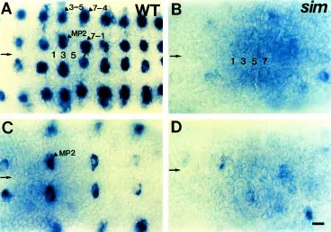
position and time (Campos-Ortega, 1993; Parras et al., 1996; Skeath and Doe, 1996). These genes are initially expressed within the neuroectoderm in partially overlapping arrays of proneural clusters of about six cells. One NB develops from each cluster and the remaining ectodermal cells of the cluster lose gene expression during the S1-S2 stages of neurogenesis (Romani et al., 1989; Martin-Bermudo et al., 1991; Skeath and Carroll, 1992). To investigate whether the CNS midline cells affect the proneural gene expression essential for NB formation and fate determina-tion, expression pattern of ac gene product, T5 RNA, was ana-lyzed by in situ hybridization. In wild-type embryos, ac is ex-pressed in the MP2 and NBs 3-5, 7-1, and 7-4 per hemisegment at stage 9 (Fig. 2A), and then in MP2 at stage 10 (Fig. 2C). In sim embryos, ac expression is absent in 95% of hemisegments (the number of hemisegments examined, n=122), although residual ac expression is occasionally observed (Fig. 2B,D). This result indicates that the CNS midline cells are required for proneural gene, ac expression in the medial and lateral NBs from the initial stage of NB formation. The absence of ac expression in the orthogonal array of NBs may reflect the change of NB identity or defect in NB formation and division.
The CNS midline cells are required for the expression of the S1-S5 NB markers that are essential for NB fate determination To determine whether the absence of ac expression in the sim mutant is due to NB fate change or defects in NB formation and division, several markers for NB 7-1 were chosen to examine expression pattern in NB 7-1. en starts to be expressed in S1 NBs 1 and 4, and then in S2-S5 NBs 1-2, 6-1, 6-2, 6-4, 2, and 7-3 located in the 6th and 7th rows in each segment (Fig. 7-3A; Doe, 1992; Broadus et al., 1995). At stage 13, en is expressed in three clusters of neurons in the posterior half of each segment: midline neuroblast cluster, bilateral clusters of 2-3 medial neurons and of 10-12 lateral neurons (Fig. 3C). At stage 16, en is expressed in the midline neuroblast cluster, bilateral clusters of 6 medial neurons
and of 6 lateral neurons (Fig. 3E). In addition, a pair of lateral neurons located at the ventral side of the ventral nerve cord show en expression. In stage 9 sim embryos, en expression in NB 7-1 is absent in 47% of hemisegments (n=103, Fig. 3B). Later in stage 13 and 16 sim embryos, the number of en-positive lateral neurons is reduced by half (Fig. 3D,F), and en expression in a bilateral pair of ventral neurons is reduced (Fig. 3F).
It was shown that ming/lacZ is a useful marker for NBs delaminating at the S3-S5 stages of neurogenesis (Cui and Doe,
1992; Broadus et al., 1995). ming/lacZ marker was used to determine whether the CNS midline cells affect marker gene expression in NB 7-1 and the other NBs delaminating at later S3-S5 stages after the initial round of neurogenesis has begun. ming/ lacZ expression starts in a subset of the CNS midline cells at stage 9, and slightly later at early stage 10 appears in a single NB 6-1. By stage 10, ming/lacZ is expressed in five S3 NBs 1-2, 5-2, 6-1, 7-2, and 7-4, then in three additional S4 NBs 2-1, 3-3, and 4-1 at stage 11, and finally in a total of 18 S5 NBs (Fig. 4A). In stage 11 sim embryos, ming/lacZ expression is severely reduced espe-cially in all the midline cells and 90% of medial NBs 2-1, 3-1, 4-1, and 5-1, intermediate NBs 1-2 and 2-2, and lateral NB 2-4 as shown in Figure 4B (n=150). On the other hand, ming/lacZ expression in NBs 5-2, 6-1, and 7-1 still remains. At stage 15, ming/lacZ-expressing neurons become fused at the ventral mid-line, resulting in a narrow ventral nerve cord (Fig. 4C,D). Another S5 NB marker, upg/lacZ expression begins in the midline NB at stage 11 and shows in S5 NBs 4-1, 5-3, 6-2, and 7-2 at late stage 11 (Chiang et al., 1995; Fig. 5A). In sim embryos, upg/lacZ expression is absent in 95% of S5 NB 4-1, and much reduced in the remaining NBs at late stage 11 (n=128; Fig. 5B) and in many neurons at stage 15 (Fig. 5D). upg/lacZ-expressing neurons appear to degenerate by stage 15 (Fig. 5C,D).
Taken together, these analyses showed that in sim mutant embryos NB 7-1 fails to express ac and en genes but still expresses ming/lacZ, indicating that the identity of NB 7-1 may have not been properly attained. These results suggest that the CNS midline cells are required for the expression of genes necessary for the proper identity determination of medial NBs during the S1-S5 stages of neurogenesis.
The CNS midline cells and spitz class genes affect the formation of GMCs and neurons during mid-stage neurogenesis
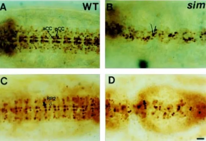
To determine whether the CNS midline cells influence the formation of the ventral GMCs and neurons derived from the NBs that underwent several rounds of cell division, eve expression patterns in many identified GMCs and neurons were analyzed by staining with the anti-Eve antibody. In wild-type embryos, eve is first expressed in five GMCs derived from at least three different NBs in each hemisegment at late stage 11; NB 1-1 gives rise to aCC and pCC neurons, NB 4-2 to RP2 and NB 7-1 to two CQ neurons (Fig. 6A, Broadus et al., 1995). Thus, examination of eve expression in the CQ neurons derived from S1 NB 7-1 would provide useful information on whether the loss of ac and en expressions in the NB 7-1 is due to NB formation or NB identity change. In stage 11 sim embryos (Fig. 6G), this analysis reveals the presence of eve-expressing CQ neurons derived from the NB 7-1 that shows no expressions of ac (Fig. 2A,B) and en (Fig. 3A,B) in stage 9 but shows weak ming/lacZ expression in stage 11 sim mutants (Fig. 4A,B). In addition, all the RP2 neurons do not express eve, but the other GMCs and neurons show normal eve expression. The aCC/pCC and CQ neurons become fused at the midline, and their cell number is similar to that of the wild-type embryos. Later in stage 15 sim embryos, eve expression in RP2 neurons remains in 45% of hemisegments (n=192). In the other spitz class mutants at stage 11, eve expression in RP2 neurons is reduced, and the number of eve-positive CQ neurons become reduced by half compared to that of the wild-type embryos especially in Egfr, otd, pnt, S and spi mutants (Fig. 6B-F,H). This
result indicates that the correct number of CQ neurons are derived from NB 7-1, and they are still present in normal position, divide and differentiate in the sim mutant. This observation suggests that NB formation or division is not severely defective, but the identity of S1 NB 7-1 is altered in the sim mutant. It also shows that the CNS midline cells and spitz class genes also affect the formation of ventral GMCs and neurons.
To examine precisely the effects of the sim gene on the formation of aCC/pCC and RP2 neurons, the enhancer trap lines A31 and rG335 which label aCC/pCC and RP neurons, respec-tively, were employed. The A31 enhancer trap line has a P[lacZ] insert in the fasciclin II locus, and shows β-gal expression in the aCC and pCC neurons from stage 11 (Grenningloh et al., 1991). The A31 enhancer trap line shows β-gal expression in the aCC and pCC per segment at stage 15 (Fig. 7A). In sim mutant embryos, the aCC and pCC neurons become fused at the midline but the number of neurons appears to remain almost the same as that of the wild-type (Fig. 7B), which is consistent with the previous result obtained with anti-Eve antibody (Fig. 6G). Another enhancer trap line rG335 was used to follow the RP neurons. The rG335 enhancer trap line shows stronger β-gal expression in the RP neurons and weaker expression in the RP2 neurons located medial to the midline cells (Fig. 7C) at stage 15. In stage 15 sim embryos, β-gal expression in the RP2 neurons disappears in more than 84% of hemisegments (n=138, Fig. 7D), confirming the previous result obtained by using anti-Eve antibody staining (Fig. 6G). This result also indicates that the identity of remaining 45% of eve-positive RP2 neurons at stage 15 is not correctly assigned in sim mutant embryos.
Taken together, these analyses demonstrated that the NB 7-1, which is devoid of ac and en expression but maintains ming/lacZ
expression in the sim mutant, is still present and gives rise to a normal number of CQ neurons. This result indicates that the loss of marker gene expressions in the NB 7-1 of sim embryos is largely due to NB identity change. It is also suggested that the CNS midline cells and the spitz class genes affect the identity determination of GMC 4-2 and RP2 neurons, as detected by the loss of marker gene expressions in the identified GMCs and neurons.
The spitz class genes are also required for expression of NB marker genes during the S3-S5 stages of neurogenesis
To demonstrate whether the other spitz class genes are involved in proper expression of the genes required for fate determination of NBs located lateral to the CNS midline cells, β -galactosidase expression patterns of ming/lacZ and upg/lacZ lines were examined in the spitz class mutants. The expression of ming/lacZ was clearly missing in many medial and intermediate NBs of the spitz class mutant embryos at stage 11 (Fig. 8A,C,E,G,I). spi and S mutant embryos show more severe reduction in ming/ lacZ-expressing NBs than in pnt and rho mutants. upg/lacZ expression in the spitz class mutants is much reduced in NBs 4-1 and 5-3 at stage 4-12 (Fig. 8B,D,F,H,J). These results indicate that the spitz class genes are required for the expression of NB markers necessary for the identity determination of NBs that are located medial and intermediate to the CNS midline cells during the S3-S5 stages of neurogenesis.
The CNS midline cells and spitz class mutants affect axon pathway formation during late neurogenesis
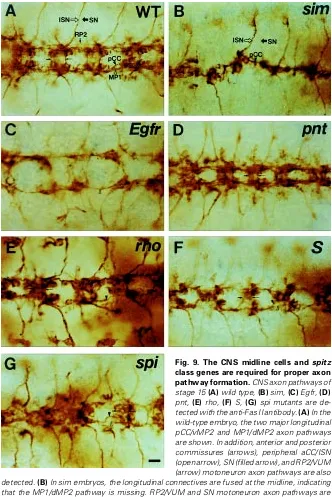
Since the CNS phenotype of the spitz class mutants shows common characteristic features of fused anterior and posterior commissures and decreased spacing between the longitudinal connectives (Mayer and Nüsslein-Volhard, 1988), the axon path-ways of the CNS were examined using the anti-Fas II antibody. The
Fig. 4. The CNS midline cells are required for the expression of ming/ lacZ S3-S5 NB marker. (A) ming/lacZ expression in a late stage 11 wild-type embryo showing a total of 15 S5 NBs including NBs 1-2, 1, 2, 2-4, 3-1, 3-2, 3-3, 3-2-4, 4-1, 5-1, 5-2, 5-3, 7-1, 7-2, and 7-4. (B) ming/lacZ expression in medial and intermediate NBs 1-2, 2-2, 2-4, 3-1, and 5-1 of a late stage 11 sim embryo is absent, although residual β-galactosidase expression remains in NBs 2-1, 4-1, and 7-1. (C) ming/lacZ expression in a stage 15 wild-type embryo is shown in dividing GMCs and neurons in the developing ventral nerve cord. (D) ming/lacZ expression in a stage 15 sim embryo shows the fused and narrow ventral nerve cord at the midline. All panels show the ventral view with the ventral midline marked with an open arrowhead, and anterior is to the left. Bar, 15.0 µm.
anti-Fas II antibody is known to be suitable for the analysis of axon pathways of the longitudinal connectives (Goodman and Doe, 1993; Lin et al., 1994; Hildago and Brand, 1997). It recognizes the first pioneering longitudinal MP1/dMP2 and pCC/vMP2 axon pathways that are the main components of longitudinal connectives. It also recognizes aCC axons that fasciculate in the intersegmental nerve
Fig. 6. The CNS midline cells are also required for the formation of the GMCs and neurons during mid-stage neurogenesis. eve expression of stage 11 (A) wild-type, (B) Egfr, (C) otd, (D) pnt, (E) rho, (F) S, (G) sim, (H) spi in the ventral side of ventral nerve cord is shown. (A) In the wild-type embryo, eve is expressed in the RP2 (open arrow) and CQ neurons (filled arrow) per segment. (G) In the sim embryo, eve expression in more than 95% of RP2 neurons is not shown. The aCC, pCC and CQ neurons become fused at the midline but the number of each neuron is almost the same as that of the wild-type embryo. (B,C,D,E,F,H) In the other spitz class mutant embryos, eve expression in the RP2 neurons is absent in some of the segments, but the number of eve-positive CQ neurons becomes much reduced compared to that of the wild-type embryo. All panels show the ventral view, and anterior is to the left. Bar, 22.5 µm.
(aCC/ISN), segmental nerve (SN), and RP2/ VUM axon pathways in wild-type embryos at stage 15 (Fig. 9A). The SN exits the CNS near the anterior commissures, whereas the ISN origi-nates from the posterior commissures and ex-tends into the adjacent posterior segment of the PNS. In the stage 14 sim mutant embryo, the pioneering MP1/dMP2 and pCC/vMP2 axon path-ways are fused at the midline, and RP2/VUM axons are absent in more than 90% of hemisegments (Fig. 9B; n=122). Considering that MP1 does not form properly and MP2 identity is changed, and aCC and pCC neurons are still present in sim mutant embryos (Figs. 6,7; per-sonal observations), the combined axon path-ways may consist of mainly pCC/vMP2 axon pathway. It is possible that defects in identity determination of GMC 4-2 and RP2 neurons observed in the sim mutant (Figs. 6,7) lead to the absence of RP2/VUM axon pathway. In the other spitz class mutants, the longitudinal MP1/dMP2 pathway is absent in rho mutant (Fig. 9E) or disrupted in pnt, S, and spi mutants, and pCC/ vMP2 axon pathway shows decreased spacing between longitudinal connectives (Fig. 9C-G). It was also shown that the spacing between ante-rior and posteante-rior commissures become narrow, and some SNs and RP2/VUM axon pathways are missing. Taken together, decreased spacing between longitudinal connectives that is observed commonly in spitz class mutants appears to be mainly due to the misrouting of pCC/vMP2 path-way close to the midline and the disruption or thinning of MP1/dMP2 pathway.
These results indicate that the CNS midline cells and spitz class genes also affect proper formation of longitudinal MP1/dMP2 and pCC/ vMP2 axon pathways, peripheral SN and RP2/ VUM axon pathways during late-stage neurogenesis. It was also demonstrated that the identity change caused by defective eve and rG335 expressions in the RP2 neurons of the sim mutants may lead to defects in the RP2/VUM axon pathway.
Discussion
The roles of CNS midline cells and spitz class genes in determination of the VNE fate
This study showed that the CNS midline cells are required for proneural gene expression essential for providing competence to generate a NB from equivalent neuroectodermal cells. Of these NBs, NB 7-1 is devoid of ac and en expression but ming/lacZ expression still remains (Figs. 2-4), and the number of aCC, pCC, and CQ neurons derived from the NB 7-1 is almost the same as that of the wild-type embryo when detected with anti-Eve antibody staining and β-gal expression of the specific enhancer trap lines in sim embryos (Figs. 6,7). These observations strongly suggest that at least in case of NB 7-1, NB formation and division is not defective but the identity of NB 7-1 has not been correctly assigned. Considering the observation that the spitz class mu-tants show a common phenotype such as the loss of ventralmost epidermis, the involvement of impaired cell division and formation cannot be completely ruled out. However, eve-positive aCC, pCC, EL, and CQ GMCs are generated, divide and differentiate into the correct number of neurons in sim embryos (Figs. 6,7), indicating that NB formation and division are essentially normal. On the other hand, the loss of specific NB marker gene expres-sions in the sim mutant may well be largely due to the NB identity change. This explanation is supported by the findings that some NB marker expressions are absent but that other marker expres-sions still remain in a subset of the identified NBs, GMCs and neurons. As described previously, the NB 7-1 shows no ac and en expression and weak ming/lacZ expression during early neurogenesis, indicating that the identity of NB 7-1 has been altered. The absence of eve and rG335 expressions in the RP2 neurons in the sim mutant may lead to the RP2/VUM axon pathway defect, as detected by the anti-Fas II antibody (Fig. 9). In addition, the loss of upg/lacZ expression and the presence of residual ming/lacZ expression in the NB 4-1 may cause alteration of NB 4-1 identity, since it is known that NB 4-1 generates the
interneurons that cross anterior and posterior commissures of the CNS (Bossing et al., 1996), and this defect may be in part responsible for the commissural defect observed in the sim mutant.
The CNS midline cells may help each NB obtain unique identity and then delaminate from the proneural clusters by sending out signal(s) throughout the VNE. It was reported that EGF receptor signaling pathway is required in the initial patterning of the entire VNE before gastrulation and later in the vicinity of the CNS midline cells during neurogenesis (Skeath, 1998; Udolph et al., 1998; Yagi et al., 1998). It was also shown that Rho and Vein expressed in the VNE activate Egfr during stage 5-6 before gastrulation. They were shown to play important roles in the specification and formation of medial and intermediate NBs by repression of proneural gene expression and promotion of NB formation in the intermediate column. In addition, they are also independently involved in the regulation of separate sets of genes, esg and msh that help medial and lateral NBs establish their unique identity, finally resulting in patterning of medial, intermediate, and lateral columns of the VNE along the D-V axis. Considering that the CNS midline cells are required for the proneural gene expression during the second stage of neurogenesis from this study, they may replace the roles of early–acting genes such as rho and vein by providing the VNE with additional ligand(s) including the secreted Spi that activates the Egfr in the second stage. This may provide the medial, intermediate and lateral NBs with additional positional information to generate diversified NB lineage from the VNE. It was reported that ac and sc expression in the VNE is necessary for the determination of NB identity as well as NB formation (Parras et al., 1996; Skeath and Doe, 1996), supporting the idea that the CNS midline cells may provide some signal(s) to generate the NBs that have unique identities along the D-V axis. In contrast to the result obtained with Egfr mutants, the CNS midline cells influence the proneural gene expression even in the
lateral NBs, indicating that there may be additional signaling pathway involved in patterning of the VNE by the CNS midline cells. Based on these observations, it remains to be resolved whether the CNS midline cells affect simply NB formation and division or NB identity determination and how the CNS midline cells influence the proper patterning of the VNE.
Fig. 8. The spitz class genes are required for the proper expression of the ming/lacZ and upg/lacZ NB markers. (A) ming/lacZ expression in a late stage 11 wild-type embryo. (B) upg/lacZ expression in a late stage 11 wild-type embryo. (C,E,G,I) ming/lacZ expression in late stage 11 spitz class mutant embryos. ming/lacZ expression is absent in the ventral midline and a number of medial and intermediate NBs. S3 NB 6-1 tends to get fused at the midline, and many NBs become disorganized. spi (I) and S (G) exhibit more severe reduction in NB number than pnt (C) and rho (E). (D,F,H,J) upg/lacZ expression in late stage 11 spitz class mutant embryos is shown. upg/lacZ expression is missing in the midline NBs and almost absent in the medial and intermediate NBs 4-1 and 5-3. All panels show the ventral view, and anterior is to the left. Bar, 22.5 µm.
The possible roles of the CNS midline cells and spitz class genes in generating NBs and VE from the VNE
It was shown that the spitz class mutants exhibit parallel defects both in the CNS and VE: the absence of ventralmost VE and the reduction in the spacing of the longitudinal connectives and anterior and posterior commissures (Mayer and Nüsslein-Volhard, 1988). Recent molecular genetic and cellular studies have demonstrated that the CNS midline cells and spitz class genes are essential for fate determination, division, and differen-tiation of VE and ventral neurons (Kim and Crews, 1993; Raz and Shilo, 1993; Menne et al., 1997). These results suggest that the spitz class genes act in the ventral neuroectodermal precursor cells during early neurogenesis, while the defects in midline glia cell determination and differentiation observed in the spitz class mutants (Klämbt et al., 1991; Sonnenfeld and Jacobs, 1994) may be due to more restricted roles of the spitz class genes at the later stage of neurogenesis. Since the spitz class genes, Egfr, and otd are known to be expressed in the VNE before and during the period of NB formation, it is possible that the spitz class genes divide the overlying NBs into similar regions and provide the NBs with positional information along the D-V axis, just as they divide VE into many specific regions and provide positional information. This would result in parallel defects in the NB and VE in the early stage of neurogenesis. On the other hand, the spitz class genes may have additional, more restricted roles in the midline glia differentiation and axon pathway formation during late neurogenesis. This would result in more specific defects in the CNS that are not simply parallel to those observed in the VE such as decreased spacing between longitudinal connectives and fused anterior and posterior commissures. The results obtained from this study appear to support the idea that the spitz class genes play homologous roles in the patterning of NBs and VE at least in the early stage of neurogenesis, since the defects in the formation of a subset of NBs and neurons in the spitz class mutants are localized mainly medial and intermediate to the CNS midline cells. These results are consistent with previous studies on fate determination of VE showing the abolished expression of VE markers in the ventralmost epidermis (Kim and Crews, 1993; Raz and Shilo, 1993). However, the absence of proneural gene ac expression in both medial and lateral NBs and of ming/lacZ expression in the lateral NB indicates that there may be distinctive roles of the CNS midline cells and spitz class genes in patterning of NBs from the VNE that are different from those in VE patterning. In conclusion, this study demonstrated that the CNS midline cells are necessary for the fate determination of a subset of NBs and formation of GMCs, neurons, and their axon pathways during Drosophila embryonic neurogenesis. The spitz class genes also play important roles in establishing NB identity and axon pathway formation. The parallel phenotypes in the CNS and VE in the spitz class mutants suggest that homologous functions are responsible for providing the VNE with unique identity along the D-V axis. This study may help understand the basic principles for genesis of neural diversity and assembly of intricate cellular networks with an outstanding degree of specificity in the CNS.
Materials and Methods
Drosophila strains and neural markers
otd9 , pnt1, rho4 , SIIN, sim2 and spi1. NB molecular markers, ming/lacZ and
unplugged (upg)/lacZ (Doe, 1992), kindly provided by Dr. C. Doe, were used to analyze the pattern of NB formation during the S3-S5 stages of neurogenesis. The ming/lacZ line contains an enhancer trap P element/ lacZ insert in the ming locus at 83°C on the third chromosome (Cui and Doe, 1992) and the upg/lacZ line contains in the locus at 44B on the second chromosome (Chiang et al., 1995). The enhancer trap lines AA31 (Grenningloh et al., 1991) and rG335 (C.S. Goodman, unpublished data) were adopted to follow the aCC/pCC and RP neurons, respectively.
Immunohistochemistry and in situ hybridization
Embryos collected from genetic crosses be-tween the spitz class mutants and neural markers were stained with monoclonal anti-β-galactosidase (β-gal) antibody (Promega) at 1:2000 dilution and detected by horseradish peroxidase immunohis-tochemistry according to Nambu et al. (1991). Ho-mozygous mutant embryos were identified by the lack of expression of a P[ry+, ftz/lacZ] or P[ry+, elav/
lacZ] from the second and third balancer chromo-somes. Mouse monoclonal engrailed (en) anti-body 4D9 and anti-Even-skipped (Eve) antianti-body 3C10 (Patel et al., 1989), kindly provided by Dr. N. Patel, were used at 1:20 dilution. Rabbit anti-Eve antibody (Frasch et al., 1987), kindly provided by Dr. M. Frasch, was diluted at 1:5000. Mouse mono-clonal anti-Fasciclin (Fas) II antibody (Grenningloh et al., 1991), kindly provided by Dr. C.S. Goodman, was used at 1:200 dilution. Whole-mount in situ hybridization using digoxigenin-labeled DNA probe was performed as described by Tautz and Pfeifle (1989). Whole-mounted embryos were viewed and photographed using an Olympus microscope with Normaski optics.
Acknowledgments
The authors are very grateful to Dr. C. Goodman for the anti-Fas II antibody and enhancer trap lines AA31 and rG335, to Dr. C. Doe for ming/lacZ and upg/ lacZ enhancer trap lines, to Dr. M. Frasch for the Eve antibody, to Dr. N. Patel for the En and anti-Eve antibodies, and to Dr. Kathy Matthews at Bloomington Drosophila Stock Center for various fly strains. This work was supported by the Basic Sci-ence Research Institute Program, Korean Ministry of Education (BSSR-95-4411) and a special grant from Konkuk University, 1993.
References
BHAT, K.M. (1996). The patched signaling pathway medi-ates repression of gooseberry allowing neuroblast speci-fication by wingless during Drosophila neurogenesis. Development 122: 2921-2939.
BHAT, K.M. (1998). Cell-cell signaling during neurogenesis: some answers and many questions. Int. J. Dev. Biol. 42: 127-139.
BIER, E., JAN, L.Y. and JAN, Y.N. (1990). rhomboid, a gene required for dorsoventral axis establishment and periph-eral nervous system development in Drosophila melanogaster. Genes Dev. 3: 190-203.
BOSSING, T., UDOLPH, G., DOE, C.Q. and TECHNAU, G.M. (1996). The embryonic nervous system lineages of Drosophila melanogaster. I. Neuroblast lineages derived from the ventral half of the neuroectoderm. Dev. Biol. 179: 41-64.
BROADUS, J., SKEATH, J.B., SPANA, E.P., BOSSING, T., TECHNAU, G.T. and DOE, C.Q. (1995). New neuroblast markers and origin of the aCC/pCC neurons in the Drosophila central nervous system. Mech. Dev. 53: 393-402.
BRUNNER, D., DUCKER, K., OEDELLERS, N., HAFEN, E., SCHOLZ, H. and KLÄMBT, C. (1994). The Ets domain protein pointed-P2 is a target of MAP kinase in the sevenless signal transduction pathway. Nature 370: 386-389.
CABRERA, C.V. (1992). The generation of cell diversity during early neurogenesis in Drosophila. Development 115: 893-901.
CAMPOS-ORTEGA, J.A. (1993). Early neurogenesis in Drosophila melanogaster.
In The Development of Drosophila melanogaster (Eds. C.M. Bate and A.M. Arias), Cold Spring Harbor Laboratory Press, New York, pp. 1091-1129.
CAMPOS-ORTEGA, J.A. and HARTENSTEIN, V. (1997). The Embryonic Develop-ment of Drosophila melanogaster. 2nd ed., Springer Verlag, New York.
CREWS, S.T. (1998). Control of cell lineage-specific development and transcription by bHLH-PAS proteins. Genes Dev. 12: 607-620.
CREWS, S.T., THOMAS, J.B. and GOODMAN, C.S. (1988). The Drosophila single-minded gene encodes a nuclear protein with sequence similarity to the per gene product. Cell 52: 143-151.
CUI, X. and DOE, C.Q. (1992). Ming is expressed in neuroblast sublineages and regulates gene expression in the Drosophila central nervous system. Develop-ment 116: 943-952.
CHIANG, C., YOUNG, K.E. and BEACH, P.A. (1995). Control of Drosophila tracheal branching by the novel homeodomain gene unplugged, a regulatory target for genes of the bithorax complex. Development 121: 3901-3912.
CHU-LAGRAFF, Q. and DOE, C.Q. (1993). Neuroblast specification and formation regulated by wingless in the Drosophila CNS. Science 261: 1594-1597.
D’ALESSIO, M. and FRASCH, M. (1996). Msh may play a conserved role in dorsoventral patterning of the neuroectoderm and mesoderm. Mech. Dev. 58: 217-231.
DOE, C.Q. (1992). Molecular markers for identified neuroblasts and ganglion mother cells in the Drosophila nervous system. Development 116: 855-864.
FINKELSTEIN, R., SMOUSE, D., CAPACI, T.M., SPRADLING, A.C. and PERRIMON, N. (1990). The orthodenticle gene encodes a novel homeo domain protein involved in the development of the Drosophila nervous system and ocellar visual structures. Genes Dev. 4: 1516-1527.
FRASCH, M., HOEY, T., RUSHLOW, C., DOYLE, H. and LEVINE, M. (1987). Characterization and localization of the even-skipped protein of Drosophila. EMBO J. 6: 749-759.
FREEMAN, M. (1997). Cell fate determination strategies in the Drosophila eye. Development 124: 261-270.
GABAY, L., SCHOLZ, H., GOLEMBO, M., KLAES, A., SHILO, B.-Z. and KLÄMBT, C. (1996). EGF receptor signaling induces pointed P1 transcription and inacti-vates Yan protein in the Drosophila embryonic ventral ectoderm. Development 122: 3355-3362.
GABAY, L., SEGER, R. and SHILO, B.-Z. (1997). In situ activation pattern of Drosophila EGF receptor pathway during development. Science 277: 1103-1106.
GOLEMBO, M., RAZ, E. and SHILO, B.-Z. (1996). The Drosophila embryonic midline is the site of Spitz processing, and induces activation of the EGF receptor in the ventral ectoderm. Development 122: 3363-3370.
GOODMAN, C.S. and DOE, C.Q. (1993). Embryonic development of the Drosophila central nervous system. In The Development of Drosophila melanogaster (Eds. M. Bate and A.M. Arias). Cold Spring Harbor Laboratory Press, New York, pp.1131-1206.
GRENNINGLOH, G., REHM, E.J. and GOODMAN, C.S. (1991). Genetic analysis of growth cone guidance in Drosophila: Fasciclin II functions as a neuronal recognition molecule. Cell 67: 45-57.
HILDAGO, A. and BRAND, A.H. (1997). Targeted neuronal ablation: the role of pioneer neurons in guidance and fasciculation in the CNS of Drosophila. Development 124: 3253-3262.
ISSHIKI, T., TAKEICHI, M. and NOSE, A. (1997). The role of msh homeobox gene during Drosophila neurogenesis: implication for the dorsoventral specification of the neuroectoderm. Development 124: 3099-3109.
JIMENEZ, F., MARTIN-MORRIS, L.E., VELASCO, L., CHU, H., SIERRA, J., ROSEN, D.R. and WHITE, K. (1995). Vnd, a gene required for early neurogenesis of Drosophila, encodes a homeodomain protein. EMBO J. 14: 3487-3459.
KIM, S.H. and CREWS, S.T. (1993). Influence of Drosophila ventral epidermal development by the CNS midline cells and spitz class genes. Development 118: 893-901.
KLÄMBT, C. (1993). The Drosophila gene pointed encodes two ets-like proteins which are involved in the development of the midline glia cells. Development 117: 163-176.
KLÄMBT, C., JACOBS, R.J. and GOODMAN, C.S. (1991). The midline of the Drosophila CNS: a model for the genetic analysis of cell fate, cell migration, and growth cone guidance. Cell 64: 801-815.
KOLODKIN, A.L., PICKUP, A.T., LIN, D.M., GOODMAN, C.S. and BANERJEE, U. (1994). Characterization of Star and its interactions with sevenless and EGF receptor during photoreceptor cell development in Drosophila. Development 120: 1731-1745
LIN, D.M., FETTER, R.D., KOPCZYNSKI, C., GRENNINGLOH, G. and GOODMAN, C.S. (1994). Genetic analysis of fasciclin II in Drosophila: defasciculation, refasciculation and altered fasciculation. Neuron 13: 1055-1069.
LÜER, K., URBAN, J., KLÄMBT, C. and TECHNAU, G.M. (1997). Induction of identified mesodermal cells by CNS midline progenitors in Drosophila. Devel-opment 124: 2681-2690.
MARTIN-BERMUDO, M.D., MARTINEZ, C., RODRIGUEZ, A. and JIMENEZ, F. (1991). Distribution and function of the lethal of scute gene product during early neurogenesis in Drosophila. Development 113: 445-454
MATSUZAKI, M. and SAIGO, K. (1996). hedgehog signaling independent of engrailed and wingless required for post-S1 neuroblast formation in Drosophila CNS. Development 122: 3567-3575.
MAYER, U. and NÜSSLEIN-VOLHARD, C. (1988). A group of genes required for pattern formation in the ventral ectoderm of the Drosophila embryo. Genes Dev. 2: 1496-1511.
MCDONALD, J. and DOE, C. (1997). Establishing neuroblast-specific gene expres-sion in the Drosophila CNS: huckebein is activated by Wingless and Hedgehog and repressed by Engrailed and Gooseberry. Development 124: 1079-1987.
MELLERICK, D.M. and NIRENBERG, M. (1995). Dorsal-ventral patterning genes restrict NK-2 homeobox gene expression to the ventral half of the central nervous system of Drosophila embryos. Dev. Biol. 171: 306-316.
MENNE, T.V., LÜER, K., TECHNAU, G.M. and KLÄMBT, C. (1997). CNS midline cells in Drosophila induce the differentiation of lateral neural cells. Development 124: 4949-4958.
MORIMOTO, A.M., JORDAN, K.C., TIETZE, K., BRITTON, O’NEILL, J.S. and RUOHOLA-BAKER, H. (1996). Pointed, an ETS domain transcription factor, negatively regulates the EGF receptor pathway in Drosophila oogenesis. Development 122: 3745-3754.
NAMBU, J.R., LEWIS, J.L., WHARTON, K.A. and CREWS, S.T. (1991). The Drosophila single-minded gene encodes a helix-loop-helix protein which acts as a master regulator of CNS midline development. Cell 67: 1157-1167.
O’NEILL, J.W. and BIER, E. (1994). Double in situ hybridization using biotin and digoxigenin tagged RNA probes. Biotechniques 17: 873-875.
PARRAS, C., GARCIA-ALONSO, L.A., RODRIGUEZ, I. and JIMENEZ, F. (1996). Control of neural precursor specification by proneural proteins in the CNS of Drosophila. EMBO J. 15: 6394-6399.
PATEL, N.H., MARTIM-BLANCO, E., COLMAN, K.G., POOLE, S.J., ELLIS, M.C., KORNBERG, J.B. and GOODMAN, C.S. (1989). Expression of engrailed proteins in arthropods, annelids and chordates. Cell 58: 955-968.
PERRIMON, N. and PERKINS, L.A. (1997). There must be 50 ways to rule the signal: the case of the Drosophila EGF receptor. Cell 89: 13-16.
RAZ, E. and SHILO, B.-Z. (1993). Establishment of ventral cell fates in the Drosophila embryonic ectoderm requires DER, the EGF receptor homolog. Genes Dev. 7: 1937-1948.
ROMANI, S., CAMPUZANO, S., MACAGNO, E.R. and MODOLLEL, J. (1989). Expression of achaete and scute genes in Drosophila imaginal discs and and their function in sensory organ development. Genes Dev. 3: 997-1007.
RUTLEDGE, B.J., ZhanG, K., BIER, E., JAN, Y.N. and PERRIMON, N. (1992). The Drosophila spitz gene encodes a putative EGF-like growth factor involved in dorsal-ventral axis formation and neurogenesis. Genes Dev. 6: 1503-1517.
SCHMIDT, H., RICKERT, C., BOSSING, T., VEF, O., URBAN, J., and TECHNAU, G.M. (1997). The embryonic central nervous system lineages of Drosophila melanogaster. 2. Neuroblast lineages derived form the dorsal part of the neuroectoderm. Dev. Biol. 189: 186-204.
SCHWEITZER, R. and SHILO, B.-Z. (1997). A thousand and one roles for the Drosophila EGF Receptor. Trends Genet. 13: 191-196.
SCHWEITZER, R., SHAHARABANY, M., SEGER, R. and SHILO, B.-Z. (1995). Secreted Spitz triggers the DER signaling pathway and is a limiting component in embryonic ventral ectoderm determination. Genes Dev. 9: 1518-1529.
SKEATH, J.B. and CARROLL, S.B. (1992). Regulation of proneural gene expres-sion and cell fate during neuroblast segregation in the Drosophila embryo. Development 114: 939-946
SKEATH, J.B. and DOE, C.Q. (1996). The achaete-scute complex proneural genes contribute to neural precusor specification in the Drosophila CNS. Curr. Biol. 6: 1146-1152
SKEATH, J.B., ZANG, Y., HOLMGREN, R., CARROLL, S.B. and DOE, C.Q. (1995). Specification of neuroblast identity in the Drosophila embryonic central nervous system bygooseberry-distal. Nature 376: 427-430.
SONNENFELD, M.J. and JACOBS, J.R. (1994). Mesectodermal cell fate analysis in Drosophila midline mutants. Mech. Dev. 46: 3-13.
STURTEVANT, M.A., ROARK, M. and BIER, E. (1993). The Drosophila rhomboid gene mediates the localized formation of wing veins and interacts genetically with components of the EGF-R signaling pathway. Genes Dev. 7: 961-973.
TAUTZ, D. and PFEIFLE, C. (1989). A nonradioactive in-situ hybridization of specific RNAs in Drosophila embryos reveals a translational control of the segmentation gene hunchback. Chromosoma 98: 81-85.
THOMAS, J.B., BASTIANI, M.J., BATE, C.M. and GOODMAN, C.S. (1984). From grasshopper to Drosophila: a common plan for neural development. Nature 310: 203-207.
THOMAS, J.B., CREWS, S.T. and GOODMAN, C.S. (1988). Molecular genetics of the single-minded locus: a gene involved in the development of the Drosophila nervous system. Cell 52: 133-141.
UDOLPH, G., URBAN, J., RUSING, G., LÜER, K. and TECHNAU, G.M. (1998). Differential effects of EGF receptor signalling on neuroblast lineages along the dorsoventral axis of the Drosophila CNS. Development 125: 3291-3300.
WANG, W., CHEN, X., XU, H. and LUFKIN, T. (1996). Msx3: a novel murine homologue of the Drosophila msh homeobox gene restricted to the dorsal embryonic central nervous system. Mech. Dev. 58: 203-215.
WIESCHAUS, E., PERRIMON, N. and FINKELSTEIN, R. (1992). orthodenticle activity is required for the development of medial structures in the larval and adult epidermis of Drosophila. Development 115: 801-808.
YAGI, Y. and HAYASHI, S. (1997). Role of the Drosophila EGF receptor in determination of the dorsoventral domains of escargot expression during primary neurogenesis. Genes to Cells 2: 41-53.
YAGI, Y., SUZUKI, T. and HAYASHI, S. (1998). Interaction between Drosophila EGF receptor and vnd determines three dorsoventral domains of the neuroec-toderm Development 125: 3625-3633.
ZAK, N.B., WIDES, R.J., SCHEJTER, E.D., RAZ, E. and SHILO, B.-Z. (1990). Localization of the DER/flb protein in embryos: Implications on the faint little ball lethal phenotype. Development 109: 865-874.
ZHANG, Y, UNGAR, A., FRESQUEZ, C. and HOLMGREN, R. (1994). Ectopic expression of either the Drosophila gooseberry-distal or proximal gene causes alterations of cell fate in the epidermis and central nervous system. Develop-ment 120: 1151-1161
ZHOU, L., XIAO, H. and NAMBU, J.R. (1997). CNS midline to mesoderm signaling in Drosophila. Mech. Dev. 67: 59-68.


