EGF, epithelium and
Ascidian maternal mRNAs segregated with the endoplasm 125
Identification and characterization of maternally expressed
genes with mRNAs that are segregated with the endoplasm
of early ascidian embryos
KAORU IMAI*, NORI SATOH and YUTAKA SATOU
Department of Zoology, Graduate School of Science, Kyoto University, Sakyo-ku, Kyoto, Japan
ABSTRACT Endoderm cells of the ascidian embryo are specified autonomously dependent on maternal cytoplasmic information or determinants that are localized in the endoplasm. In the present study, we identified three maternally expressed genes (1, 2 and CsEndo-3) by screening a cDNA library of Ciona savignyi fertilized egg mRNAs subtracted with gastrula mRNAs. CsEndo-1 encoded a protein with nuclear localization signals, CsEndo-3 predicted a protein containing both a potential transmembrane domain and the PDZ domain, and CsEndo-2 suggested a protein with no similarity to known proteins. The maternal transcripts of all of these genes were not concentrated during early stages of embryogenesis up to the 8-cell stage, but were concentrated at the endoplasmic region by the 16-cell stage and then segregated later with the endoplasm. At the 110-cell stage, the maternal transcript of CsEndo-1 was evident only in the primordial endoderm cells, while those of CsEndo-2 and CsEndo-3 were found in the primordial endoderm cells as well as the primordial notochord cells. All of the transcripts became barely detectable during gastrula-tion and neurulagastrula-tion. Later, zygotic expression of the three genes became evident again in the endoderm and notochord cells, suggesting developmental roles in the formation of these types of cell. Although we were not able to deduce their functions, this is the first report of maternal genes with mRNAs that are segregated with the endoplasm of ascidian embryos.
KEY WORDS:
ascidians, localized maternal mRNAs, endoplasm, endoderm, notochord
Int. J. Dev. Biol. 43: 125-133 (1999)
0214-6282/99/$15.00
© UBC Press Printed in Spain www.lg.ehu.es/ijdb
*Address for reprints: Department of Zoology, Graduate School of Science, Kyoto University, Sakyo-ku, Kyoto 606-8502, Japan. FAX: 81-75-705-1113. e-mail: imai@ascidian.zool.kyoto-u.ac.jp
Introduction
The endodermal tissue of an ascidian tadpole larva is com-prised of endoderm in the trunk region and an endodermal strand in the tail region (reviewed by Satoh, 1994). The lineage of the endodermal tissue is almost completely documented (Conklin, 1905; Nishida, 1987). Most of the endoderm cells, except for a small number of cells of the posterior region of the tail, are derived from the vegetal A4.1 and B4.1 blastomeres of the bilaterally symmetrical 8-cell stage embryo. The developmental fate to form endoderm is segregated into the A5.1, A5.2 and B5.1 of the 16-cell stage embryo, and then into the A6.1, A6.3 and B6.1 of the 32-cell stage embryo. As early as the 32-cell stage, the A6.1 becomes restricted to generate endoderm only. At the 64-cell stage, not only A7.1 and A7.2 (daughter cells of A6.1) but also the A7.5, B7.1, and B7.2 become endoderm-restricted. These primordial cells divide five or six times to form the endodermal tissue of about 500 cells. Reflecting such an early fate restriction, presumptive endoderm blastomeres show a high potential of autonomous differentiation
Original Article
when they are isolated from early embryos (Reverberi and Minganti, 1946; Whittaker, 1990; Nishida, 1992). This autonomy is depen-dent on maternal factors or determinants that are prelocalized in the endoplasm of eggs and early embryos (Nishida, 1993). In addition, it was shown that the endoderm cells induce the differen-tiation of notochord cells at the 32-cell stage (Nakatani and Nishida, 1994). Although recent studies identified developmental genes that are expressed zygotically in the endodermal tissues, including the LIM class homeobox gene HrLim (Wada et al., 1995), fork head/HNF-3β (Corbo et al., 1997; Olsen and Jeffery, 1997; Shimauchi et al., 1997) and alkaline phosphatase gene (Kumano and Nishida, 1998), there are no reports on maternally expressed genes with mRNAs that are segregated with the endoplasm. Therefore, the identification of maternal messages in the endo-plasm is a current research subject of ascidian embryogenesis.
subtracted with gastrula mRNAs of the ascidian Ciona savignyi. The clones were then randomly picked up from the subtracted library, and they were partially sequenced from the 3'-end to avoid analyzing the same clones any further. After partial sequencing, the localization of corresponding mRNA was examined for each clone by whole-mount in situ hybridization. cDNA clones exhibiting the localization of corresponding mRNA were selected for further analyses. Taking advantage of this strategy, we were able to isolate maternal genes with mRNAs that are localized in the posterior end of developing embryos (2, 3, 4, pem-5, and pem-6; Satou and Satoh, 1997). In the present study, we
B
A
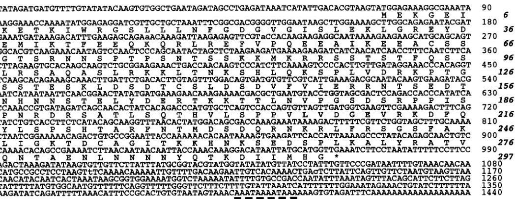
Fig. 1. Characterization of cDNA for CsEndo-1 gene. (A) Nucle-otide and deduced amino acid sequence of CsEndo-1L cDNA. The 2,676bp insert includes a
C
single open reading frame that encodes a polypeptide of 417 amino acids. The asterisk indicates the termination, and potential signal sequences for polyadenylation are dotted-underlined. Two possible nuclear localization signals (amino acid positions 74-90 and 252-255) are underlined. The nucleotide sequences that CsEndo-1S cDNA lacks are enclosed by a box. (B) A Northern blot of poly(A)+ RNA of fertilized eggs shows two distinct
EGF, epithelium and
Ascidian maternal mRNAs segregated with the endoplasm 127
extended this approach to isolate cDNA clones, the corresponding mRNAs of which exhibit localization and segregation with the endoplasm. We report here the identification and characterization of three maternally expressed genes designated CsEndo-1, CsEndo-2, and CsEndo-3. During early cleavages, these maternal messages gradually concentrated in the endoplasm, and at the 16-to 110-cell stage, CsEndo-1 mRNA was detected only in the presumptive endoderm cells, while CsEndo-2 and CsEndo-3 mRNA was detected in the endoderm cells as well as A-line notochord cells.
Results
Isolation of cDNA clones for maternally expressed genes with localized mRNAs
We constructed a cDNA library of fertilized-egg mRNAs sub-tracted with gastrula mRNAs of C. savignyi. In a previous study, we isolated five cDNA clones for maternally expressed genes with mRNAs that were localized to the posterior end of developing embryos (Satou and Satoh, 1997). The genes were named pem-2, pem-3, pem-4, pem-5, and pem-6. We extended the screening to obtain maternal mRNAs that are segregated with the endoplasm or ectoplasm. In the present study, we isolated three maternally expressed genes, the corresponding mRNAs of which were seg-regated with the endoplasm. We named these genes CsEndo-1, CsEndo-2 and CsEndo-3.
The CsEndo-1 gene
Characterization of the CsEndo-1 cDNA clone
A Northern blot analysis identified two distinct CsEndo-1 tran-scripts of about 2.3 kb (CsEndo-1S) and 2.7 kb (CsEndo-1L) in
mRNA of C. savignyi fertilized eggs (Fig. 1B). The band intensity of CsEndo-1S was stronger than that of CsEndo-1L (Fig. 1B). The cDNA clone we initially isolated was about 2.0 kb, which did not contain the entire coding sequence. Two successive 5'-RACE yielded a cDNA clone with the entire coding sequence. As shown in Figure 1, the cDNA sequence of CsEndo-1L was 2,676bp long and had a single open reading frame that encoded a polypeptide of 417 amino acids. We found that the cDNA for CsEndo-1S had the same sequence as CsEndo-1L, except that CsEndo-1S lacked the sequence between nucleotide positions 503 and 858 of CsEndo-1L (Fig. 1B,C). These transcripts are likely to be products of the alternative splicing of a single gene. The predicted CsEndo-1L and CsEndo-1S proteins contained nuclear localization signals, sug-gesting that they were nuclear proteins. However, we were not able to find any consensus motifs of transcription factors in the se-quence.
The localization and segregation of CsEndo-1 mRNA
Most of the endodermal cells of the ascidian embryo are derived from the vegetal blastomeres of the 8-cell embryo (A4.1 and B4.1 lines). The developmental fate to form most of the endoderm is further segregated into the A5.1, A5.2 and B5.1 of the 16-cell embryo (Fig. 2B), and then the A6.1, A6.3 and B6.1 of the 32-cell embryo (Fig. 2F). At the 64- and 110-cell stage, the developmental fates of A7.1, A7.2, A7.5, B7.1 and B7.2 blastomeres are restricted to the generation of only endoderm (Fig. 2H).
Although CsEndo-1 is a maternally expressed gene, whole-mount in situ hybridization failed to detect distinct signals until the 16-cell stage (data not shown). At the 16-cell stage, CsEndo-1 mRNA was detected in the endoplasm of the vegetal hemisphere (Fig. 2A,B,C). No hybridization signals were detected in
blas-Fig. 2. Expression of the CsEndo-1 gene in ascidian embryos. Localization of CsEndo-1 transcript at (A) the 16-cell, (E) 32-cell and (G) 110-cell stages, vegetal pole view. (C) The 16-cell stage embryo, posterior view, showing the maternal tran-script was restricted to the vegetal side of the embryo. (D) Hybridization of the 16-cell stage embryo with sense probe, showing no signals above background. At these stages, distinct signals were found in the presumptive endoderm cells. (B,F,H) Sche-matic representations of (A), (E), and (G). Yellow shows blastomeres with the devel-opmental fate of endoderm, and green shows the CsEndo-1-expressing cells. The blastomeres with the developmental fate of endoderm are indicated based on Conklin’s nomenclature (1905). Only the blastomeres of the right half of the embryo are shown, because the ascidian embryo is bilaterally symmetrical. (I) A gastrula, veg-etal view, showing that signals were hardly detected. (J) A tailbud embryo, lateral view, showing distinct zygotic expression of the gene in endoderm (En) and notochord (Nt).
tomeres of the animal hemisphere (Fig. 2C). The 32-cell stage embryo showed a similar and more distinct localization pattern of the transcript (Fig. 2E,F). At the 64-cell and 110-cell stages (Fig. 2G,H), CsEndo-1 mRNA was evident in all of the primordial endoderm cells.
It has been noted that, in ascidians, whole-mount in situ hybrid-ization usually detects signals of the zygotic transcripts first in nuclei of blastomeres of early embryos (e.g., Yasuo and Satoh, 1993; Satou et al., 1995). Because we were not able to detect the CsEndo-1 mRNA in nuclei of early embryos until the 110-cell stage, the signal in early embryos was thought to represent the maternal message. This was confirmed by the quantitative RT-PCR analy-sis. Because twenty-five RT-PCR cycles resulted in an appropriate level of amplification immediately below the level of saturation, we examined changes in the quantity of CsEndo-1 maternal tran-scripts by this number of amplification (Fig. 2K, left). As shown in Figure 2K (right), signal that corresponded to the CsEndo-1 mater-nal transcript was evident and constant from the stage of fertilized eggs to the 32-cell stage, although the signal became less intense at the 110-cell stage. This suggested that the zygotic expression of CsEndo-1 did not occur before the gastrulation.
As development proceeded, CsEndo-1 mRNA became barely detectable during gastrulation (Fig. 2I) and neurulation. However, zygotic transcript was evident in the endoderm cells and notochord cells of the tailbud-stage embryo (Fig. 2J).
The CsEndo-2 gene
Characterization of the CsEndo-2 cDNA clone
The cDNA sequence of CsEndo-2 is shown in Figure 3. It was 1,440 nucleotides in length. We concluded that this cDNA contained the entire coding sequence of CsEndo-2, because the 5' end of the cDNA contained stop codons in all three reading frames in front of the initiator methionine at nucleotide position 73. The decoded CsEndo-2 protein contained 297 amino acid residues. We were not able to find any similarities to other reported proteins.
The localization and the segregation of CsEndo-2 mRNA As in the case of CsEndo-1, although CsEndo-2 is also a maternally expressed gene, whole-mount in situ hybridization failed to detect a distinct pattern of signal localization until the 16-cell stage (data not shown). CsEndo-2 mRNA was detected in the endoplasmic region of the vegetal hemisphere at the 16-cell (Fig. 4A,B,C) and 32-cell stages (Fig. 4D,E). No hybridization signals were detected in blastomeres of the animal hemisphere (Fig. 4C). At the 64-cell and 110-cell stages, the signal was evident in the primordial endoderm cells; namely, A7.1, A7.2, A7.5, B7.1 and B7.2 blastomeres (Fig. 4F,G). In addition, CsEndo-2 mRNA was detected in the A-line notochord cells (A8.5, A8.6, A8.13 and A8.14 blastomeres of the 110-cell embryo; Fig. 4F,G). The quantitative RT-PCR analysis demonstrated the presence of CsEndo-2 mater-nal transcript in fertilized eggs and early embryos (Fig. 4J).
Granules within the myoplasm were sometimes lightly stained (arrowheads in Fig. 4A, D, F). However, this staining was labile and seemed to be non-specific due to longer time period of the staining reaction. A longer reaction without any probes also showed the signals (data not shown).
As in the case of CsEndo-1, CsEndo-2 mRNA was barely detectable in the gastrulae (Fig. 4H), and zygotic transcripts became detectable again in endoderm cells and notochord cells of the tailbud-stage embryos (Fig. 4I).
The CsEndo-3 gene
Characterization of the CsEndo-3 cDNA clone
The cDNA sequence of CsEndo-3 is shown in Figure 5A, and was 881 nucleotides in length. We concluded that this cDNA clone contained the entire coding sequence of CsEndo-3, because the 5' end of the cDNA contained stop codons in all three reading frames in front of the initiator methionine at nucleotide position 59. The cDNA encoded a polypeptide of 141 amino acid residues. The CsEndo-3 protein had a hydrophobic region at the carboxyl terminus, and therefore seemed to be a single-pass transmembrane protein. The CsEndo-3 protein also contained a PDZ domain (Fig. 5B). The PDZ
EGF, epithelium and
Ascidian maternal mRNAs segregated with the endoplasm 129
domains are found in diverse membrane-associated proteins including the MAGUK family of guanylate kinase homologs, several protein phosphatases and kinases, neuronal nitric oxide synthase and several dystrophin-associated proteins (reviewed by Ponting et al., 1997; Craven and Bredt, 1998). The PDZ domains of these proteins are thought to bind with other proteins. Generally, it is known that the PDZ-domain proteins of animals are cytoplasmically localized. However, the CsEndo-3 is a protein that contained both a potential transmembrane domain and a PDZ domain. This is probably the first report of a PDZ domain-containing membrane protein.
The localization and the segregation of CsEndo-3 mRNA As in the cases of CsEndo-1 and CsEndo-2, any distinct localization of the CsEndo-3 message was not detected until the 16-cell stage (data not shown), although CsEndo-3 is also a maternally expressed gene. CsEndo-3 mRNA was evident in the endoplasmic region of embryos at the 16-cell (Fig. 6A,B,C) and 32-cell stages (Fig. 6D,E). The primordial endoderm 32-cells of the 64-32-cell and 110-cell stage embryos (A7.1, A7.2, A7.5, B7.1 and B7.2) showed distinct signals (Fig. 6F,G). As in the case of CsEndo-2, CsEndo-3 mRNA was also detected in the A-line notochord cells (A8.5, A8.6, A8.13 and A8.14 blastomeres of the 110-cell stage; Fig. 6F, G). The presence of CsEndo-3 maternal mRNA in fertilized eggs and early embryos was revealed by the RT-PCR analysis (Fig. 6J). Although granules of the myoplasm were weakly stained, the staining was thought to be non-specific, as described above. CsEndo-3 mRNA also became barely detectable in the gastru-lae (Fig. 6H). In addition, the zygotic expression of CsEndo-3 became evident in endoderm cells and notochord cells of the tailbud-stage embryos (Fig. 6I).
Discussion
In the present study, we isolated and characterized three maternal genes, CsEndo-1, CsEndo-2 and CsEndo-3, the mRNAs of which are segregated with the endoplasm. The genes are also expressed zygotically in the endoderm and notochord cells of the tailbud embryo. During ascidian embryogenesis, the specification and subsequent differentiation of endoderm cells take place highly autonomously (reviewed by Satoh, 1994). This autonomy is due to endoderm determinants localized in the endoplasm (Nishida, 1993). In addition, Marikawa and Satoh (1996) suggested that maternal mRNA(s) is a component of the endoderm determinants. It is expected therefore that maternal mRNAs that are segregated together with endoplasm are associated with endoderm determi-nants. As far as we know, the present study is the first to identify maternal genes, the mRNAs of which are segregated with the endoplasm.
To deduce the possible functions of CsEndo-1, CsEndo-2 and CsEndo-3, we microinjected synthetic mRNA of each of these three genes into fertilized eggs to examine effects of the ectopic expression or overexpression of the genes. As a control, we injected pem mRNA into fertilized eggs, which induced a deficiency in the formation of the anterior and dorsal larval structures (Yoshida et al., 1996). However, the overexpression experiment failed to deduce the function of these genes, because injected embryos developed normally. Experiments with antisense-oligos to deduce the function of ascidian maternal genes have not yet succeeded. The exploration of their functions is an important subject of further research. In addition, the determination of localization of maternal proteins encoded by these genes will lead to a better understanding of their functions.
Fig. 4. Expression of the CsEndo-2 gene in ascidian embryos. Localiza-tion of CsEndo-2 transcript at (A) the 16-cell, (D) 32-cell and (F) 110-cell stages, vegetal pole view. (C) The 16-cell stage embryo, posterior view, showing the maternal transcript was restricted to the vegetal side of the embryo. At these stages, distinct sig-nals were found in the presumptive endoderm and the notochord cells. Weak background staining was de-tected in granules of the myoplasm (arrowheads). (B,E,G) Schematic rep-resentations of (A), (D), and (F). Yellow shows blastomeres with the develop-mental fate of endoderm or A-line no-tochord cells, and green shows the CsEndo-2-expressing cells. (H) A
Alkaline phosphatase (AP) activity has been used to monitor the differentiation of endoderm cells of the ascidian embryo (Minganti, 1954; Whittaker, 1990). The AP gene was recently isolated from H. roretzi (Kumano and Nishida, 1998). The AP gene is expressed both maternally and zygotically, and zygotic expression initiates around neurulation. In addition, an ascidian member of NK-2 gene family, Cititf1, was isolated from Ciona intestinalis (Dr. F. Ristorator, personal communication). The Cititf1 was expressed in all endo-dermal precursor cells at around the 110-cell stage. A microinjec-tion of Cititf1 mRNA into fertilized eggs resulted in the development of tadpole larvae with abnormalities in head-trunk development consequent to the formation of excess endoderm, suggesting that this gene plays an important role in normal endoderm differentia-tion (Dr. F. Ristorator, personal communicadifferentia-tion). In addidifferentia-tion, the LIM class homeobox gene HrLim (Wada et al., 1995) and an ascidian homolog of the fork head/HNF-3β gene are also ex-pressed in the presumptive endoderm and notochord cells (Corbo et al., 1997; Olsen and Jeffery, 1997; Shimauchi et al., 1997). Future studies should therefore explore the genetic cascade between CsEndo-1-3 and the developmental genes mentioned above.
Several recent lines of evidence suggest that the A-line precur-sors of endoderm and notochord have similar characteristics and that some common mechanisms are at work in the specification of the endoderm and notochord. First, lineages of endoderm and notochord cells are common up to the 16-cell stage; namely, A5.1 and A5.2 at the 16-cell stage have the ability to generate both endoderm and notochord, and at the 32-cell stage, A6.1 and A6.3 become endoderm-lineage, while A6.2 and A6.4 are of notochord lineage. Second, the As-T gene or an ascidian Brachyury is expressed exclusively in notochord cells (Yasuo and Satoh, 1993, 1994), and the ectopic expression of As-T in presumptive endo-derm cells altered the fate of endoendo-derm cells to notochord cells (Yasuo and Satoh, 1998). Third, the fate of A-line notochord cells was changed to that of endoderm cells by the treatment of early embryos with lithium (Yoshida et al., 1998). The present results regarding the CsEndo-1, CsEndo-2 and CsEndo-3 genes provide further support for the notion mentioned above. That is, maternal transcripts of CsEndo-2 and CsEndo-3 were seen not only in the endoderm cells but also in notochord cells of early cleavage-stage embryos. In addition, the zygotic expression of all three genes was evident in both endoderm and notochord cells of the tailbud
EGF, epithelium and
Ascidian maternal mRNAs segregated with the endoplasm 131
embryos. The effect of lithium was mimicked by the overexpression of β-catenin, suggesting that this mechanism is associated with the Wnt signaling cascade (Yoshida et al., 1998). It would be of interest to address the mechanism underlying the specification of the endoderm in relation to the Wnt signaling cascade and to investi-gate how CsEndo-1, CsEndo-2 and CsEndo-3 are involved in this cascade.
CsEndo-1, CsEndo-2 and CsEndo-3 showed a similar patterns of segregation of the maternal transcript. Namely, although the presence of the maternal transcripts was revealed by the RT-PCR analysis, we did not detect any distinct localization or segregation of the maternal transcripts during ooplasmic segregation and early cleavages up to the 8-cell stage. This was partially due to a large amount of yolk in the endoplasmic region, which sometimes prevents successful whole-mount in situ hybridization. However, from the 16-cell stage onwards, the segregation of the maternal transcript with the endoplasm was evident. Several maternal genes have recently been isolated from ascidians; they showed localization of maternal transcripts. Interestingly, most of the maternal genes including pem (Yoshida et al., 1996), 2, pem-3, pem-4, pem-5 and pem-6 (Satou and Satoh, 1997) from C. savignyi, and HrWnt-5 (Sasakura et al., 1998a) and a gene for serine-threonin kinase (Sasakura et al., 1998b) from H. roretzi showed similar patterns of localization. Namely, the maternal transcripts of these genes are segregated first with the so called myoplasm up to the 8-cell stage. Then, after the 16-cell stage, the maternal transcripts are not segregated with the myoplasm but rather are localized to the posterior end of developing embryos. There may be an anchoring mechanism for the maternal tran-scripts at the posterior end of early embryos. In contrast to these pem-like maternal genes, CsEndo-1, CsEndo-2 and CsEndo-3 are concentrated and segregated with the endoplasm. This suggests
that the machinery for the segregation of these maternal genes is different from that for pem-like maternal genes. Therefore, the segregation mechanism is also an important subject of further studies.
Materials and Methods
Ascidian eggs and embryos
Ciona savignyi adults were collected near the Otsuchi Marine Research Center, Ocean Research Institute of the University of Tokyo, Iwate, Japan, and maintained under constant light to induce oocyte maturation. Eggs and sperm were obtained surgically from the gonoduct. After insemination, eggs were reared at about 18°C in Millipore-filtered seawater (MFSW) containing 50 µg/ml streptomycin sulfate.
Isolation of cDNA clones and sequencing
The construction of a cDNA library of C. savignyi fertilized egg mRNAs subtracted with gastrula mRNAs was described in a previous report (Satou and Satoh, 1997). cDNA clones for the maternal genes of CsEndo-1, CsEndo-2 and CsEndo-3 cDNA were isolated by screening the cDNA library. Briefly, clones were randomly picked up from the subtracted library and then partially sequenced from the 3'-end to avoid analyzing the same clones any further. After partial sequencing, the localization of correspond-ing mRNA was examined for each clone by whole-mount in situ hybridiza-tion. cDNA clones exhibiting the localization of mRNA corresponding to the endoplasm were selected for further analyses.
The nucleotide sequences of the candidate clones were determined for both strands with a dye primer cycle sequencing FS ready reaction kit and ABI PRISM 377 DNA sequencer (Perkin Elmer, Norwalk, CT, USA).
Northern analysis
Total RNA was isolated from the fertilized eggs by the acid guanidinium thiocyanate-phenol-chloroform (AGPC) method (Chomczynski and Sacchi, 1987). Poly(A)+ RNA was purified using Oligotex beads (Roche Japan, Tokyo). Poly(A)+ RNA was fractionated by agarose gel electrophoresis,
and transferred to a Hybond-N+ membrane (Amersham, Buckinghamshire, UK). Blots were hybridized with a [32P]-random-labeled DNA probe in 6xSSPE, 0.5% SDS, 5xDenhardt’s solution, 100 µg/ml salmon sperm DNA, and 50% formamide. The filter was washed twice in 2xSSC/0.1% SDS, and exposed to X-ray film.
RT-PCR analysis
Quantitative RT-PCR was performed basically according to the method described by Wilson and Melton (1994) with minor modifications. Total RNA was extracted by the AGPC method (Chomczynski and Sacchi, 1987). Twenty fertilized eggs or embryos were lysed in 200 µl GTC solution and stored at -80°C. Following the phenol-chloroform extraction and iso-propanol precipitation, RNA was treated with 1U of RNase-free DNase I (GIBCO BRL) and then with 0.2 mg/ml proteinase K (Sigma). After annealing with 10 pmol oligo(dT), the total RNA was incubated with 200U of Superscript II Reverse Transcriptase (GIBCO BRL) for 50 min at 42°C in a total reaction volume of 20 µl. The reaction mixture contained 1xFirst Strand Buffer, 10 nM DTT, 1.5 nM of each dNTP and 40U of RNase inhibitor (TOYOBO). One-tenth of the RT mixture was used as templates for PCR. Each 100 µl reaction mixture contained 0.2 mM dNTPs, 1.5 mM MgCl2, 0.1
µl α-[32P]-dCTP, 100 pmol of each primer, 1xTaq Buffer and 2.5U of Taq DNA polymerases (TOYOBO). Reaction proceeded through 20 to 35 amplification cycles (30 sec at 94°C, 30 sec at 55°C, and 30 sec at 72°C). Twelve µl of the PCR mixture was resolved on 8% non-denaturing polyacry-lamide gels and subjected to autoradiography. The primers used in this study were as follows: 5', ATCGCCAAATGCTGTGCA; CsEndo1-3', ATTGAAGGTGTCAGGGGT; CsEndo2-5', TGTCCAAAACACAGCCGA; CsEndo2-3', CTTAGGAGGCGGCATGTT; CsEndo3-5', GCACAGTGAAG-CAGTTGA; and CsEndo3-3', AAGCCTATGGCTGTTGCT.
Whole-mount in situ hybridization
RNA probes were prepared with a DIG RNA labeling kit (Boehringer Mannheim, Heidelberg, Germany). Whole-mount in situ hybridization was performed as described previously (Satou et al., 1995).
Acknowledgments
We thank Dr. Yasuaki Takagi, Mr. Kouichi Morita and all of the staff of the Otsuchi Marine Research Center for their hospitality and their help. Y.S. was supported by a Predoctoral Fellowship from the Japan Society for the Promotion of Science for Japanese Junior Scientists, with Research Grant 8-6806. This research was also supported in part by a Grant-in-Aid from the Ministry of Education, Science, Sports and Culture of Japan (No. 07102012) to N.S.
References
CHO, K.O., HUNT, C.A. and KENNEDY, M.B. (1992). The rat brain postsynaptic density fraction contains a homolog of the Drosophila discs-large tumor suppressor protein. Neuron 9: 929-942.
CHOMCZYNSKI, P. and SACCHI, N. (1987). Single-step method of RNA isolation by acid guanidinium thiocyanate-phenol-chloroform extraction. Anal. Biochem. 162: 156-159.
CONKLIN, E.G. (1905). The organization and cell lineage of the ascidian egg. J. Acad. Natl. Sci. 13: 1-119.
CORBO, J.C., ERIVES, A., DI GREGORIO, A., CHANG, A. and LEVINE, M. (1997). Dorsoventral patterning of the vertebrate neural tube is conserved in a protochordate. Development 124: 2335-2344.
CRAVEN, S.E. and BREDT, D.S. (1998). PDZ proteins organize synaptic signaling pathways. Cell 93: 495-498.
KUMANO, G. and NISHIDA, H. (1998). Maternal and zygotic expression of the endoderm-specific alkaline phosphatase gene in embryos of the ascidian, Halocynthia roretzi. Dev. Biol. 198: 245-252.
MARIKAWA, Y. and SATOH, N. (1996). Ultraviolet-sensitive ooplasmic factors are responsible for the development of an endoderm-specific alkaline phosphatase in the ascidian embryo. Dev. Growth Differ. 38: 167-173.
MINGANTI, A. (1954). Fosfatasi alcaline nello sviluppo delle Ascidie. Pubbl. Stn. Zool. Napoli 25: 9-17.
NAKATANI, Y. and NISHIDA, H. (1994). Induction of notochord during ascidian embryogenesis. Dev. Biol. 166: 289-299.
NISHIDA, H. (1987). Cell lineage analysis in ascidian embryos by intracellular injection of a tracer enzyme. III. Up to the tissue restricted stage. Dev. Biol. 121: 526-541.
NISHIDA, H. (1992). Developmental potential for tissue differentiation of fully dissociated cells of the ascidian embryo. Roux’s Arch. Dev. Biol. 201: 81-87. NISHIDA, H. (1993). Localized regions egg cytoplasm that promote expression of endoderm-specific alkaline phosphatase in embryos of the ascidian Halocynthia roretzi. Development 118: 1-7.
OLSEN, C.L. and JEFFERY, W.R. (1997). A forkhead gene related to HNF-3β is required for gastrulation and axis formation in the ascidian embryo. Development 124: 3609-3619.
PONTING, C.P., PHILLIPS, C., DAVIES, K.E. and BLAKE, D.J. (1997). PDZ domains: targeting signalling molecules to sub-membranous sites. BioEssays 19: 469-479.
REVERBERI, G. and MINGANTI, A. (1946). Fenomeni di evocazione nello sviluppo dell’uovo di Ascidie. Risultati dell’indagine sperimentale sull’uovo di Ascidiella aspersa e di Ascidia malaca allo stadio di otto blastomeri. Pubbl. Stn. Zool. Napoli 20: 199-252.
SASAKURA, Y., OGASAWARA, M. and MAKABE, K.W. (1998a). HrWnt-5: a maternally expressed ascidian Wnt gene with posterior localization in early embryos. Int. J. Dev. Biol. 42: 573-579.
SASAKURA, Y., OGASAWARA, M. and MAKABE, K.W. (1998b). Maternally localized RNA encoding a serine/threonine protein kinase in the ascidian, Halocynthia roretzi. Mech. Dev. 76: 161-163.
SATOH, N. (1994). Developmental Biology of Ascidians. Cambridge University Press, New York.
SATOU, Y. and SATOH, N. (1997). Posterior end mark 2 (2), 4, pem-5, and pem-6: maternal genes with localized mRNA in the ascidian embryo. Dev. Biol. 192: 467-481.
SATOU, Y., KUSAKABE, T., ARAKI, I. and SATOH, N. (1995). Timing of initiation of muscle-specific gene expression in the ascidian embryo precedes that of developmental fate restriction in lineage cells. Dev. Growth Differ. 37: 319-327.
SHIMAUCHI, Y., YASUO, H. and SATOH, N. (1997). Autonomy of ascidian fork head/HNF-3 gene expression. Mech. Dev. 69: 143-154.
SOKOL, S.Y., KLINGENSMITH, J., PERRIMON, N. and ITOH, K. (1995). Dorsalizing and neuralizing properties of Xdsh, a maternally expressed Xenopus homolog of dishevelled. Development 121: 1637-1647.
THEISEN, H., PURCELL, J., BENNETT, M., KANSAGARA, D., SYED, A. and MARSH, J.L. (1994). dishevelled is required during wingless signaling to establish both cell polarity and cell identity. Development 120: 347-360. WADA, S., KATSUYAMA, Y., YASUGI, S. and SAIGA, H. (1995). Spatially and
temporally regulated expression of the LIM class homeobox gene Hrlim suggests multiple distinct functions in development of the ascidian, Halocynthia roretzi. Mech. Dev. 51: 115-126.
WHITTAKER, J.R. (1990). Determination of alkaline phosphatase expression in endodermal cell lineages of an ascidian embryo. Biol. Bull. 178: 222-230. WILSON, P. and MELTON, D. (1994). Mesodermal patterning by an inducer
gradient depends on secondary cell-cell communication. Curr. Biol. 4: 676-686.
WILLOTT, E., BALDA, M.S., FANNING, A.S., JAMESON, B., VAN ITALLIE, C. and ANDERSON, J.M. (1993). The tight junction protein ZO-1 is homologous to the Drosophila discs-large tumor suppressor protein of septate junctions. Proc. Natl. Acad. Sci. USA 90: 7834-7838.
WOODS, D.F. and BRYANT, P.J. (1991) The discs-large tumor suppressor gene of Drosophila encodes a guanylate kinase homolog localized at septate junctions. Cell 66: 451-464.
YASUO, H. and SATOH, N. (1993). Function of vertebrate T gene. Nature 364: 582-583.
EGF, epithelium and
Ascidian maternal mRNAs segregated with the endoplasm 133
(T) gene is expressed exclusively in notochord cells at the fate restricted stage. Dev. Growth Differ. 36: 9-18.
YASUO, H. and SATOH, N. (1998). Conservation of the developmental role of Brachyury in notochord formation in a urochordate, the ascidian Halocynthia roretzi. Dev. Biol. 200: 158-170.
YOSHIDA, S., MARIKAWA, Y. and SATOH, N. (1996). posterior end mark, a novel maternal gene encoding a localized factor in the ascidian embryo. Development 122: 2005-2012.
YOSHIDA, S., MARIKAWA, Y. and SATOH, N. (1998). Regulation of the trunk-tail patterning in the ascidian embryo: A possible interaction of cascades between lithium/β-catenin and localized maternal factor pem. Dev. Biol. 202: 264-279.