Darryl G Kitneya,b, Ph.D., Rita I Jabra,b, Ph.D., Bahareh Vahabib,c, Ph.D., Christopher H Fryb, Ph.D, DSc, FRCS.
Footnotes
a University of Surrey, Faculty of Health and Medical Sciences, Guildford, GU2 7XH.
b University of Bristol, School of Physiology, Pharmacology & Neuroscience, University Walk, Bristol, BS8 1TD.
c University of the West of England, Department of Biological, Biomedical and Analytical Sciences, Bristol, BS16 1QY.
Corresponding author.
Prof C H Fry, School of Physiology, Pharmacology & Neuroscience University of Bristol
University Walk, Bristol BS8 1TD, UK email: [email protected]
Keywords: Heating; overactive bladder; spontaneous contractions. 2
3
4
5
6
7
8
9
10
12
13
14
15
16
17
18
19
Introduction
The urinary bladder generates spontaneous contractile activity (SA) to maintain bladder wall tone but remains compliant during filling. SA is greater in isolated preparations from patients with overactive bladder (OAB) and may underlie the associated symptoms. Due to significant refractoriness and adverse side-effects of drug-based therapies, new approaches to alleviate the symptoms of OAB are desirable. One approach may be through mild heating of the bladder wall.
Heating is used as a therapeutic treatment in other organs, such as the heart. For example, raising body temperature to 42°C for 15 minutes protects against the consequences of myocardial ischaemia by inducing expression of heat shock proteins (HSPs) [1]. Several studies have shown that heating decreases contractile function in different muscle types, including smooth muscle [2,3]. However, there is little work on how heating affects the bladder, and temperature-related investigations have focused on cooling for diagnosis of say interstitial cystitis/painful bladder syndrome [4]. Bladder cooling enhances nerve-mediated contractions, attributed in part to increased ATP release from efferent nerves [5-7], but sustained cooling to 4°C down-regulates carbachol-induced contractions [8].
Some transient receptor potential (TRP) channel subtypes are activated during heating. Located on sensory nerves in the sub-urothelium and on urothelial cells they are activated during noxious stimuli, including heating. TRPV1 channels in particular respond to temperatures between 41-50°C [9,10]. Activation of TRPV
1 channels permits Ca2+ influx into cells that may modulate sensory responses to bladder filling and hence SA.
Hyperthermia affects protein structure and function and even small increases of temperature can lead to their unfolding and loss of activity [11]. However, this may be offset by 21
22
23
24
25
26
27
28
29
30
31
32
33
34
35
36
37
38
39
40
41
42
43
44
45
upregulation of protective mechanisms such as induction of chaperones including HSP70 and phosphorylated HSP27 [12]. Moreover, there is evidence that pro-apoptotic pathways mediated by activation of caspase-3 and DNA-repair pathways mediated by poly-[ADP-ribose] polymerases (PARP) are also temperature-dependent [13-15].
We hypothesised that progressive heating from 37 to 42, 46, and 50°C would continuously decrease SA, potentially through a TRPV1-dependent mechanism. We tested this hypothesis by: 1) exploring the effects of heating (42, 46, 50°C) on pig bladder wall SA, histological integrity and expression of heat-dependent signalling molecules; 2) determining the effects of TRPV1 antagonists during heating.
47
48
49
50
51
52
53
54
55
Materials and methods
In vitro preparations and solutions. Male and female pig bladders (Sus scrofa domestica, ~6 months) from a local abattoir were transported to the laboratory in gassed Tyrode’s solution (4oC). The bladder was opened sagitally through its ventral face and pinned on a Sylgard dish in Tyrode’s solution. The bladder was stored at 4oC for no longer than 24 hours when not in use. Preparations with an intact mucosa (10x20 mm, 2 mm thick) were attached to an isometric force transducer and superfused with Tyrode’s solution (12 ml.min-1). Stock solutions of various pharmacological mediators were diluted to final concentrations in Tyrode’s: the muscarinic receptor agonist carbachol (10 mM in water to 1 µM); the TRPV1 channel antagonist AMG9810 (10 mM in DMSO to 0.3 µM); the capsaicin antagonist, capsazepine (10 mM in methanol to 3 µM). Tyrode’s solution contained (mM): NaCl, 118; KCl, 4.0; MgCl2, 1.0 NaHCO3, 24, NaH2PO4, 0.4; CaCl2, 1.8; glucose, 6.1; Na pyruvate, 5.0; 95% O2/5% CO2. pH was measured at 37°C (7.49±0.04) and after heating to 42°C (7.65±0.04), 46°C (7.73±0.0.03) and 50°C (7.75±0.03).
Preparation of heating and temperature recording. A heating coil of nichrome wire (7 coils,
0.4 mm diameter; 10.5 Ω.m-1 resistance) was placed immediately above the tissue preparation to radiate heat up to 50°C onto the urothelial surface. Temperature was measured with thermistor probes (33 gauge, 0.2 mm diameter, hypodermic chromega-alomega probe, Omega) placed in the superfusate by the urothelium immediately below the coil and also at the mucosa/detrusor boundary. A temperature gradient of about 1°C between superfusate and mucosa/detrusor boundary was measured, the latter is referred to as the test temperature.
Contraction measurements. SA were initiated by exposure to 1 µM carbachol for 10 minutes
and allowed to stabilise for one-hour before any intervention. Preparations were then heated for 15 minutes and allowed to recover for 45 minutes (unless otherwise stated) before the 57
58
59
60
61
62
63
64
65
66
67
68
69
70
71
72
73
74
75
76
77
78
79
80
81
next intervention. The force integral (area-under-the-curve, AUC) of SA were measured for the final 10 minutes of each intervention and control period.
Ex vivo whole perfused pig bladders. Whole female pig bladders were excised immediately
post-mortem at the abattoir, with their associated vasculature, and stored in ice-cold gassed
Kreb’s solution, as previously described [16]. At the laboratory, the bladder was perfused with Kreb’s solution through its arterial supply at constant flow (10 ml.min-1), and the lumen filled with Kreb’s to 150 ml through a catheter via the urethra. Changes to intravesical pressure were recorded using a fluid-filled double-lumen catheter attached to a pressure transducer. Heating the bladder wall was done by altering the temperature of the perfusate and luminal fluid. Kreb’s solution was similar to Tyrode’s except (mM): KCl, 4.7, KH2PO4, 1.2; glucose, 11.7.
Histology. Intact preparations after exposure to radiant heating, or paired controls (n=6),
were immediately stored in 10% neutral-buffered formalin. Samples were wax-embedded, cut (superfrost+ slide, 5 µm) and deparaffinised. Sections were either stained with haematoxylin and eosin (H&E) or with the nuclear stain, 4′-6-diamidino-2-phenylindole (DAPI, 1:10,000 dilution, Thermofisher Scientific, UK). H&E sections were used for visualisation and DAPI-stained sections for analysis. Images (63x objective) were taken with a wide-field microscope (Leica, DM LB2) attached to a CCD camera (Leica DFC450C: 1280x960 pixels) for H&E samples. For DAPI-stained images, a z-stack was used to obtain the optimum section using different focal distances. The minimum and maximum depths of each section was obtained and the intermediate region analysed for the urothelium, sub-urothelium and detrusor smooth muscle. Images were taken by a CCD camera for 300-500 µm2 unit area. Analysis of the urothelium was; urothelial width, and nuclear diameter. A nuclear count per unit pixel was done for both the sub-urothelium and detrusor regions. 83
84
85
86
87
88
89
90
91
92
93
94
95
96
97
98
99
100
101
102
103
104
105
106
107
Western blots. Bladders were incubated with control Tyrode’s solution for 15 minutes at 37, 42, 46, or 50C, and rapidly snap-frozen in liquid N2. Whole tissue protein lysate (30 μg) from each sample was prepared using RIPA buffer, resolved by 12% polyacrylamide SDS-PAGE and transferred to polyvinylidene difluoride membranes (PVDF, Invitrogen, UK). Membranes were blocked with Odyssey blocking buffer (LI-COR Biosciences, Ltd, UK) and probed with primary antibodies (Abcam, UK, rabbit polyclonal) to caspase-3 (1:500 dilution), PARP (1:2000 dilution), phosphorylated HSP27 (HSP27-pSer82, 1:2,000 dilution) or inducible HSP70 (iHSP70, 1:500 dilution). Membranes were then washed and incubated with secondary antibodies appropriate to the source of primary antibodies (LI-COR Biosciences; 1:10,000 dilution). Resolved protein bands were imaged using an Odyssey infra-red imaging system and then quantified with Image-J software in arbitrary units. The quantified band densities were normalised to corresponding GAPDH band densities (Santa Cruz, mouse monoclonal, 1:1,000.dilution).
Data analysis and statistics. Contractile function data were normalised to the average of pre-and post-control value at 37°C unless otherwise stated. Data are medians [25,75% interquartiles], except Western blot data (means±SD). Differences between data sets were compared using ANOVA, with non-parametric or parametric post hoc comparisons using GraphPad Prism 5. The association between two variables was tested using a Spearman’s rank correlation; the null hypothesis was rejected at p<0.05.
109
110
111
112
113
114
115
116
117
118
119
120
121
122
123
124
125
126
127
128
Results
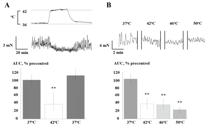
Effect of heating on bladder SA. Radiant heating to 42°C significantly and reversibly reduced the tension integral (AUC) of SA (figure 1A, n=10). A similar reduction was shown upon exposure to 46 and 50°C (figure 1B, n=10). SA recovered completely at all temperature when heating was removed. The reduction of AUC as the measure of contractile function was mirrored by reduced average amplitude and duration of individual contractions, but offset by an increased frequency (data not shown). There were no consistent changes to baseline tension during heating.
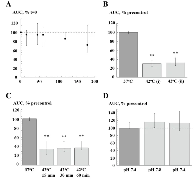
Control experiments. Because equivalent reduction of AUC occurred at the three temperatures, heating to 42°C was used for subsequent interventions. Time-control experiments, with no increases of temperature above 37°C, showed no significant changes over a 180 minute period (figure 2A, n=7). When exposure to 42°C was repeated the reduction of AUC was similar (figure 2B, n=8). Moreover the reduction of AUC was maintained between 15 and 60 minute heating (figure 2C, n=9). A confounding factor during heating was a raised superfusate pH as CO2 was driven out of solution rising to about 7.8 at 50°C. However, an increase of pH to 7.8 at 37°C had no effect on AUC and so was not a reason for the reduced values with heating (figure 2D, n=6).
Effect of TRPV1 modulators. The TRPV1 antagonist AMG9810 (0.3 µM) had no significant
effect on AUC at 37°C (105.9 [10.5, 4.8] vs 101.0 [2.0, 3.0], drug vs no drug, p>0.05, n=8), nor did it alter its reduction on heating to 42°C (34.9 [31.3, 39.1] vs 33.4 [25.4, 56.1], p>0.05,
n=8). Capsazepine, which blocks activation of TRPV1 by chemicals also had no effect on AUC at 37°C (91.8 [80.8, 107.8] vs 100.1 [2.1, 3.1], drug vs no drug, p>0.05, n=7), nor its attenuation by heating to 42°C (50.0 [35.0, 60.5] vs 43.3 [34.2, 53.8], p>0.05, n=8). Data not shown.
130
131
132
133
134
135
136
137
138
139
140
141
142
143
144
145
146
147
148
149
150
151
152
153
154
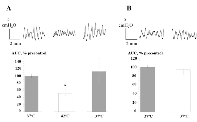
Effect of heating on the SA of the isolated whole pig bladder. Spontaneous intravesical pressure transients were also significantly reduced when perfusate temperature was raised to 42oC (figure 3B, n=5). The magnitude of reduction was similar to that from in vitro isolated preparations and also this reduction recovered to control levels on return to 37°C. Moreover, the properties of the transients changed in a similar way on heating: amplitude and duration were reduced, frequency was increased and baseline was unaffected. There were no significant changes during a time-control period at 37oC (figure 3B, n=5).
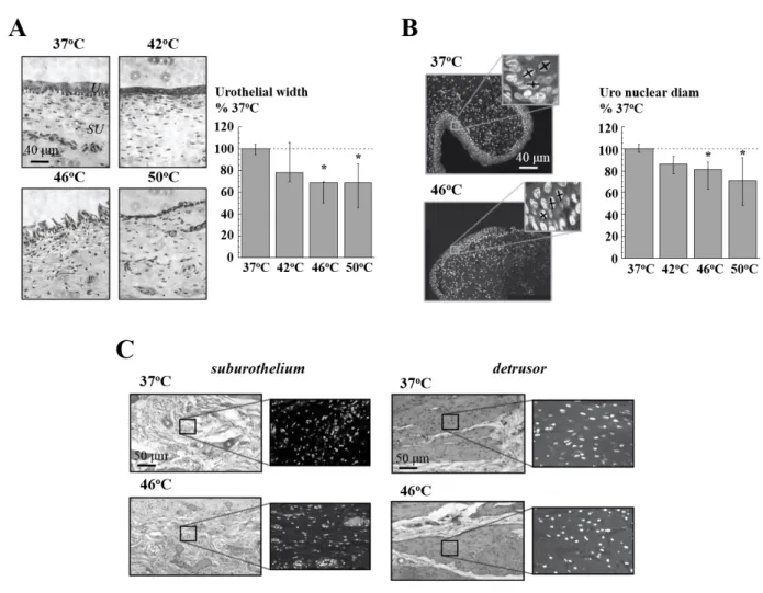
Tissue structure and protein expression changes following heating. Urothelial thickness was
not significantly altered when tissue was heated from 37 to 42oC. However, heating to 46 or 50oC significantly reduced urothelium thickness and also induced discontinuities in this layer (figure 4A, n=6). In addition, the nuclear diameter of urothelial cells was unchanged on heating to 42oC, but was significantly reduced when exposed to 46 & 50oC (figure 4B, n=6). With deeper layers of the bladder wall (figure 4C, n=6) heating to 42, 46 or 50°C induced no significant changes to the diameter or the number of nuclei in the sub-urothelium or detrusor cells at any temperature.
Normalised caspase-3 protein expression was not significantly different at 42°C when compared to that at 37C, but was greater at 46 and 50°C (figure 5A, n=3). Caspase-3 activity was measured by protein expression levels of the DNA-repair enzyme PARP, itself cleaved by caspase-3. PARP levels were similar at 37 and 42°C, but were significantly reduced at 46 and 50°C (figure 5B, n=3). Normalised protein expression of HSP27-pSer82 was unchanged on heating to 42 or 46°C when compared to 37C (figure 5C, n=3): however, at 50°C its expression was significantly lower. Similar results were also obtained for iHSP70. 156
157
158
159
160
161
162
163
164
165
166
167
168
169
170
171
172
173
174
175
176
177
178
179
Discussion
Heating to any temperature from 37°C to between 42 and 50°C reduced bladder SA and was independent of the order of heating. Moreover, heating to any temperature within this range produced a similar reduction. In addition, recovery was complete when the intervention was removed, it was reproducible when a second heat exposure was imposed and persisted for up to one hour with continuous heating. A similar observation was made in an ex vivo perfused bladder model when heated to 42°C to measure intravesical pressure variations. Thus the qualitative and quantitative observations made in isolated preparations can be carried over to the whole bladder. A previous study investigated the temperature-dependence of intravesical pressure transients between 19 and 38°C with findings that could be extrapolated to the above data (increasing temperature, decreased amplitude and increased frequency of transients) in young adult rats [17].
It was important to determine whether heat exposure to 42, 46 or 50C may have induced any structural changes to the tissue or have affected the cytoprotective HSP levels and/or pro-apoptotic pathways. Indeed, temperature-dependent effects on tissue structure were observed with the greatest effects occurring to the urothelium, with a reduction of thickness and shrinkage of nuclei within cells at 46 and 50oC. However, sub-urothelial cells and detrusor muscle cells were more resilient, with no gross changes or alterations to nuclei observed at any temperature. The lability of the urothelium to temperature-dependent damage was not due solely to the temperature gradient between this and deeper layers as no damage was evident at 50°C in detrusor or sub-urothelium, whereas changes to the urothelium were evident at 46°C. Of importance, there was no evidence of structural damage at 42°C when SA was already reduced to a maximum extent.
181
182
183
184
185
186
187
188
189
190
191
192
193
194
195
196
197
198
199
200
201
202
203
204
Expression of proteins associated with cell protection and cell death also showed no variation at 42°C, whereas changes at 46 and 50°C were evident. Thus, phosphorylated inducible HSP70 and HSP27-pSer82 protein expression levels were unaffected at 42 and 46°C, but significantly reduced at 50°C. In addition, the pro-apoptotic protein caspase-3 was also unaffected at 42°C but upregulated at 46 and 50°C. Such increase in caspase-3 protein expression levels was also associated with its enhanced apoptotic activity. This is evident from the significant cleavage and reduction in PARP protein expression levels at 46 and 50°C which in turn will impact on PARP DNA-repair capacity at these temperatures only.
Other studies have shown that heating to temperatures of 44°C or greater have significant effects on tissue damage culminating into animal mortality. Intact rat bladders, exposed to one-hour microwave treatment showed few functional changes when intravesical temperature was less than 44°C. However at higher temperatures bladder capacity decreased and animal mortality actually increased [18]. Furthermore, hyperthermia to >46°C induced DNA damage in Chinese hamster ovary cells [19,20], consistent with the data collected here concerning the reduction of PARP expression >46°C.
Overall, heating to 42°C appears to be a safe option to reduce spontaneous contractile activity in the bladder wall but without the attendant problems of tissue damage, loss of cell repair mechanisms, or induction of apoptotic pathways.
A mechanism for heat-induced reduction of SA could be related to activation of TRPV1 channels as the range of temperatures used here is similar to their activation range. However, no effect of agents that block the channel or hinder the ability of other chemicals to activate the channel has any influence on the heat-induced reduction of SA. An alternative may be through release of neuromodulators such as ATP or acetylcholine from the mucosa as this 206
207
208
209
210
211
212
213
214
215
216
217
218
219
220
221
222
223
224
225
226
227
228
229
230
influences SA [21]. However, cooling from 37 to 4°C had no effect on ATP release [22] although complementary increases of temperature were not carried out.
Heating the bladder from 37°C to any temperature in the range 42-50°C reversibly reduces SA. However, heating to 42°C offers many advantages: the effect has a rapid onset; it is reproducible and reversible; effective for up to one hour; no tissue damage is induced; proteins associated with apoptotic or DNA repair pathways are unaffected although HSPs are still induced. This approach offers an alternative to reduce SA that are associated with OAB and would be especially useful for patients refractory to conventional drug therapy.
Acknowledgements. We thank Boston Scientific for financial support 232
233
234
235
236
237
238
239
240
241
References
1 Yellon DM, Pasini E, Cargnoni A, Marber MS, Latchman DS, Ferrari R. The protective role of heat stress in the ischaemic and reperfused rabbit myocardium. J Mol Cell Cardiol 1992; 24: 895-907.
2 Everett TH, Nath S, Lynch C, 3rd, Beach JM, Whayne JG, Haines DE. Role of calcium in acute hyperthermic myocardial injury. J Cardiovasc Electrophys 2001; 12: 563-569. 3 Mustafa S, Thulesius O, Ismael HN. Hyperthermia-induced vasoconstriction of the carotid
artery, a possible causative factor of heatstroke. J Appl Physiol 2004; 96: 1875-1878. 4 Mukerji G, Waters J, Chessell IP, Bountra C, Agarwal SK, Anand P. Pain during ice
water test distinguishes clinical bladder hypersensitivity from overactivity disorders. BMC Urol 2006; 6: 31.
5 Kurihara S, Kuriyama H, Magaribuchi T. Effects of rapid cooling on the electrical properties of the smooth muscle of the guinea-pig urinary bladder. j Physiol 1974; 238: 413-426.
6 Ziganshin AU, Rychkov AV, Ziganshina LE. Effect of temperature on guinea pig urinary bladder contraction mediated via P2X-receptors. Bull Exp Physiol Med 2000; 130: 961-963.
7 Ziganshin AU, Rychkov AV, Ziganshina LE, Burnstock G. Temperature dependency of P2 receptor-mediated responses. Eur j Pharmacol 2002; 456: 107-114.
8 Demir A, Onol FF, Ercan F, Tarcan T. Effect of cold-induced stress on rat bladder tissue contractility and histomorphology. Neurourol Urodyn 2007; 26: 296-301.
9 Avelino A, Cruz C, Nagy I, Cruz F. Vanilloid receptor 1 expression in the rat urinary tract. Neuroscience. 2002; 109: 787-798.
10 Nilius B, Owsianik G, Voets T, Peters JA. Transient receptor potential cation channels in disease. Physiol Rev 2007; 87:165- 217.
243
244
245
246
247
248
249
250
251
252
253
254
255
256
257
258
259
260
261
262
263
264
265
266
11 Singh K, Shandilya M, Kundu S, Kayastha AM. Heat, acid and chemically induced unfolding pathways, conformational stability and structure-function relationship in wheat α-amylase. PLoS One 2015 8; 10: e0129203.
12 Kostenko S, Moens U. Heat shock protein 27 phosphorylation: kinases, phosphatases, functions and pathology. Cell Mol Life Sci 2009; 66: 3289-3307.
13 Wang Y, Knowlton AA, Christensen TG, Shih T, Borkan SC. Prior heat stress inhibits apoptosis in adenosine triphosphate-depleted renal tubular cells. Kidney Int 1999; 55: 2224–2235.
14 Iwashita Y, Kuwabara T, Hayata M, Kakizoe Y, Izumi Y, Iiyama J, Kitamura K, Masashi M. Mild systemic thermal therapy ameliorates renal dysfunction in a rodent model of chronic kidney disease. Am J Physiol Renal Physiol 2016; 310: F1206-F1215
15 Tramontano F, Malanga M, Farina B, Jones R, Quesada P. Heat stress reduces poly(ADPR)polymerase expression in rat testis. Mol Hum Reprod 2000; 6: 575-581. 16 Parsons BA, Drake MJ, Gammie A, Fry CH, Vahabi B. The validation of a functional,
isolated pig bladder model for physiological experimentation. Front Pharmacol 2012; 3: 52.
17 Sugaya K, de Groat WC. Influence of temperature on activity of the isolated whole bladder preparation of neonatal and adult rats. Am J Physiol Regul Integr Comp Physiol. 2000; 278: R238-246.
18 Haveman J, Smals OA, Rodermond HM. Effects of hyperthermia on the rat bladder: a pre-clinical study on thermometry and functional damage after treatment. Int J Hypertherm. 2003; 19: 45-57
19 Dewey WC, Westra A, Miller HH, Nagasawa H. Heat-induced lethality and chromosomal damage in synchronized Chinese hamster cells treated with 5-bromodeoxyuridine. Int J Rad Biol 1971; 20: 505-520.
268
269
270
271
272
273
274
275
276
277
278
279
280
281
282
283
284
285
286
287
288
289
290
291
20 Warters RL, Brizgys LM, Axtell-Bartlett J. DNA damage production in CHO cells at elevated temperatures. J Cell Physiol 1985; 124: 481-486.
21 Kushida N, Fry CH. On the origin of spontaneous activity in the bladder. BJU Int 2016; 117: 982-992.
22 Yu W. Polarized ATP distribution in urothelial mucosal and serosal space is differentially regulated by stretch and ectonucleotidases. Am J Physiol Renal Physiol 2015; 309: F864-872.
293
294
295
296
297
298
299
Figures
Figure 1. Effect of heating (42, 46, 50oC) on spontaneous contractions. A: Records:
spontaneous contractions at 37 and 42°C. (upper); output from a thermistor probe at the mucosa/detrusor boundary (lower). Bar charts: tension integral (AUC) before, during and after exposure to heating at 42°C. Data normalised to initial 37°C value (pre-control, =100%). B: Faster time-base contraction recordings at 37, 42, 46 and 50°C. Bar charts: AUC values normalised to the precontrol value. Median values (25, 25% interquartiles). ** p<0.01 vs precontrol, n=10.
301
302
303
304
305
306
307
308
309
310
311
312
313
314
315
Figure 2. Characteristics of heating to 42°C on spontaneous contractions. A: Time-control of AUC values over 180 minutes at 37°C, normalised to value at t=0; n=6. B: Repeated exposure to heating at 42°C (60 min interval); each AUC value at 42°C normalised to pre-control values, n=6. C: Effect of heating to 42°C for 15, 30 and 60 minutes; AUC values normalised to pre-control values, n=6. D: Effect of raised superfusate pH on AUC at 37°C; values normalised to pre-control values, n=6. * p<0.05, **p<0.01 vs control.
317
318
319
320
321
322
323
324
325
326
Figure 3. Effect of heating to 42°C on intravesical spontaneous pressure transients in perfused pig bladder. A: Records: examples at 37 and 42°C. Bar charts: AUC values, n=6. B: time control of changes to pressure transients over the time course of the experiment in A. AUC values normalised to the pre-control values, , n=6. * p<0.05 vs control.
328
329
330
331
332
333
334
335
336
337
338
339
340
341
342
Figure 4. The effect of heating on tissue structure. A: Sections of bladder wall (H&E stained) at 37, 42, 46 and 50°C. x63 magnification. U = urothelium; SU = suburothelium. Right: bar chart of urothelium thickness at test temperatures. B: Sections showing urothelium and underlying suburothelium after heating to 37 and 46°C, DAPI labelling. Insets show magnified regions of urothelium; crosses on the nuclei show major and minor diameters to calculate nuclear size (average of two diameters). Right: bar chart of nuclear size of urothelial cells at the test temperatures. C: H&E (left) and DAPI (right) sections showing the suburothelial layer (left) and detrusor layer (right). All data normalised to values obtained at 37°C, *p<0.05 vs control, n=6.
344
345
346
347
348
349
350
351
352
353
354
355
356
Figure 5. Expression levels of protective and apoptotic proteins at 37, 42, 46 and 50°C. A: Western blot of capase-3 (upper panel) and band densities normalised to GAPDH (lower panel) at different temperatures. B: Western blot of PARP (upper panel) and band densities normalised to GAPDH (lower panel) at different temperatures. C: Western blot of HSP27-pSer82 (upper panel) and band densities normalised to GAPDH (lower panel) at different temperatures. Data are means±SD; *p<0.05 vs control, n=3.
358
359
360
361
362
363