Cochrane
Database of Systematic Reviews
Drugs for treating Buruli ulcer (Mycobacterium ulcerans
disease) (Review)
Yotsu RR, Richardson M, Ishii N
Yotsu RR, Richardson M, Ishii N.
Drugs for treating Buruli ulcer (Mycobacterium ulcerans disease).
Cochrane Database of Systematic Reviews2018, Issue 8. Art. No.: CD012118. DOI: 10.1002/14651858.CD012118.pub2.
www.cochranelibrary.com
Drugs for treating Buruli ulcer (Mycobacterium ulceransdisease) (Review)
T A B L E O F C O N T E N T S
1 HEADER . . . .
1 ABSTRACT . . . .
2 PLAIN LANGUAGE SUMMARY . . . .
4 SUMMARY OF FINDINGS FOR THE MAIN COMPARISON . . . .
5 BACKGROUND . . . .
7 OBJECTIVES . . . .
7 METHODS . . . .
9 RESULTS . . . .
Figure 1. . . 10
Figure 2. . . 13
Figure 3. . . 14
Figure 4. . . 16
Figure 5. . . 17
Figure 6. . . 18 19 ADDITIONAL SUMMARY OF FINDINGS . . . .
21 DISCUSSION . . . .
22 AUTHORS’ CONCLUSIONS . . . .
23 ACKNOWLEDGEMENTS . . . .
23 REFERENCES . . . .
30 CHARACTERISTICS OF STUDIES . . . .
60 DATA AND ANALYSES . . . .
60 ADDITIONAL TABLES . . . .
83 CONTRIBUTIONS OF AUTHORS . . . .
83 DECLARATIONS OF INTEREST . . . .
83 SOURCES OF SUPPORT . . . .
84 DIFFERENCES BETWEEN PROTOCOL AND REVIEW . . . .
i Drugs for treating Buruli ulcer (Mycobacterium ulceransdisease) (Review)
[Intervention Review]
Drugs for treating Buruli ulcer (
Mycobacterium ulcerans
disease)
Rie R Yotsu1,2, Marty Richardson3, Norihisa Ishii4
1Department of Dermatology, National Center for Global Health and Medicine, Tokyo, Japan.2Department of Dermatology, National
Suruga Sanatorium, Shizuoka, Japan.3Cochrane Infectious Diseases Group, Liverpool School of Tropical Medicine, Liverpool, UK. 4Leprosy Research Center, National Institute of Infectious Diseases, Tokyo, Japan
Contact address: Rie R Yotsu, Department of Dermatology, National Center for Global Health and Medicine, 1-21-1 Toyama, Shinjuku-ku, Tokyo, 162-8655, Japan.yotsurie@hotmail.com.
Editorial group:Cochrane Infectious Diseases Group. Publication status and date:New, published in Issue 8, 2018.
Citation: Yotsu RR, Richardson M, Ishii N. Drugs for treating Buruli ulcer (Mycobacterium ulceransdisease).Cochrane Database of Systematic Reviews2018, Issue 8. Art. No.: CD012118. DOI: 10.1002/14651858.CD012118.pub2.
Copyright © 2018 The Authors. Cochrane Database of Systematic Reviews published by John Wiley & Sons, Ltd. on behalf of The Cochrane Collaboration. This is an open access article under the terms of theCreative Commons Attribution-Non-Commercial
Licence, which permits use, distribution and reproduction in any medium, provided the original work is properly cited and is not used for commercial purposes.
A B S T R A C T
Background
Buruli ulcer is a necrotizing cutaneous infection caused by infection withMycobacterium ulceransbacteria that occurs mainly in tropical and subtropical regions. The infection progresses from nodules under the skin to deep ulcers, often on the upper and lower limbs or on the face. If left undiagnosed and untreated, it can lead to lifelong disfigurement and disabilities. It is often treated with drugs and surgery.
Objectives
To summarize the evidence of drug treatments for treating Buruli ulcer. Search methods
We searched the Cochrane Infectious Diseases Group Specialized Register; the Cochrane Central Register of Controlled Trials (CEN-TRAL), published in the Cochrane Library; MEDLINE ( PubMed); Embase ( Ovid); and LILACS ( Latin American and Caribbean Health Sciences Literature; BIREME). We also searched the US National Institutes of Health Ongoing Trials Register (clinicaltrials.gov) and the World Health Organization ( WHO) International Clinical Trials Registry Platform ( ICTRP) (www.who.int/ictrp/search/ en/). All searches were run up to 19 December 2017. We also checked the reference lists of articles identified by the literature search, and contacted leading researchers in this topic area to identify any unpublished data.
Selection criteria
We included randomized controlled trials (RCTs) that compared antibiotic therapy to placebo or alternative therapy such as surgery, or that compared different antibiotic regimens. We also included prospective observational studies that evaluated different antibiotic regimens with or without surgery.
Data collection and analysis
Two review authors independently applied the inclusion criteria, extracted the data, and assessed methodological quality. We calculated the risk ratio (RR) for dichotomous data with 95% confidence intervals (CI). We assessed the certainty of the evidence using the GRADE approach.
1 Drugs for treating Buruli ulcer (Mycobacterium ulceransdisease) (Review)
Main results
We included a total of 18 studies: five RCTs involving a total of 319 participants, ranging from 12 participants to 151 participants, and 13 prospective observational studies, with 1665 participants. Studies evaluated various drugs usually in addition to surgery, and were carried out across eight countries in areas with high Buruli ulcer endemicity in West Africa and Australia. Only one RCT reported adequate methods to minimize bias. Regarding monotherapy, one RCT and one observational study evaluated clofazimine, and one RCT evaluated sulfamethoxazole/trimethoprim. All three studies had small sample sizes, and no treatment effect was demonstrated. The remaining studies examined combination therapy.
Rifampicin combined with streptomycin
We found one RCT and six observational studies which evaluated rifampicin combined with streptomycin for different lengths of treatment (2, 4, 8, or 12 weeks) (941 participants). The RCT did not demonstrate a difference between the drugs added to surgery compared with surgery alone for recurrence at 12 months, but was underpowered (RR 0.12, 95% CI 0.01 to 2.51; 21 participants; very low-certainty evidence).
An additional five single-arm observational studies with 828 participants using this regimen for eight weeks with surgery (given to either all participants or to a select group) reported healing rates ranging from 84.5% to 100%, assessed between six weeks and one year. Four observational studies reported healing rates for participants who received the regimen alone without surgery, reporting healing rates ranging from 48% to 95% assessed between eight weeks and one year.
Rifampicin combined with clarithromycin
Two observational studies administered combined rifampicin and clarithromycin. One study evaluated the regimen alone (no surgery) for eight weeks and reported a healing rate of 50% at 12 months (30 participants). Another study evaluated the regimen administered for various durations (as determined by the clinicians, durations unspecified) with surgery and reported a healing rate of 100% at 12 months (21 participants).
Rifampicin with streptomycin initially, changing to rifampicin with clarithromycin in consolidation phase
One RCT evaluated this regimen (four weeks in each phase) against continuing with rifampicin and streptomycin in the consolidation phase (total eight weeks). All included participants had small lesions, and healing rates were above 90% in both groups without surgery (healing rate at 12 months RR 0.94, 95% CI 0.87 to 1.03; 151 participants; low-certainty evidence). One single-arm observational study evaluating the substitution of streptomycin with clarithromycin in the consolidation phase (6 weeks, total 8 weeks) without surgery given to a select group showed a healing rate of 98% at 12 months (41 participants).
Novel combination therapy
Two large prospective studies in Australia evaluated some novel regimens. One study evaluating rifampicin combined with either ciprofloxacin, clarithromycin, or moxifloxacin without surgery reported a healing rate of 76.5% at 12 months (132 participants). Another study evaluating combinations of two to three drugs from rifampicin, ciprofloxacin, clarithromycin, ethambutol, moxifloxacin, or amikacin with surgery reported a healing rate of 100% (90 participants).
Adverse effects were reported in only three RCTs (158 participants) and eight prospective observational studies (878 participants), and were consistent with what is already known about the adverse effect profile of these drugs. Paradoxical reactions (clinical deterioration after treatment caused by enhanced immune response toM ulcerans) were evaluated in six prospective observational studies (822 participants), and the incidence of paradoxical reactions ranged from 1.9% to 26%.
Authors’ conclusions
While the antibiotic combination treatments evaluated appear to be effective, we found insufficient evidence showing that any particular drug is more effective than another. How different sizes, lesions, and stages of the disease may contribute to healing and which kind of lesions are in need of surgery are unclear based on the included studies. Guideline development needs to consider these factors in designing practical treatment regimens. Forthcoming trials using clarithromycin with rifampicin and other trials of new regimens that also address these factors will help to identify the best regimens.
P L A I N L A N G U A G E S U M M A R Y
Drugs for treating Buruli ulcer (Mycobacterium ulceransdisease)
2 Drugs for treating Buruli ulcer (Mycobacterium ulceransdisease) (Review)
What was the aim of this review?
The aim of this Cochrane Review was to summarize the evidence for drug treatments for Buruli ulcer. Key messages
Antibiotics are an important component of treatment of Buruli ulcers, but there is no evidence to suggest that any particular drug is more effective than another.
What was studied in the review?
Buruli ulcer is a disease caused by mycobacterium (tuberculosis and leprosy are other types of diseases caused by mycobacterium), which results in lumps in the skin and deep ulcers, often on the arms or the face. When diagnosed late, those affected may be left with lifelong disfigurements and disabilities. The disease is most prevalent in West Africa, but it is also found in non-tropical areas including Australia and Japan. It is often treated with drugs and surgery. This review compared different drug treatments for Buruli ulcer. What are the main results of the review?
We included 18 studies from eight countries in West Africa and Australia (1984 participants). Antibiotic combination treatments evaluated appear to be effective, but the evidence is insufficient to show that any particular drug is more effective than another. Testing treatments in Buruli ulcer is challenging as different sizes, lesions, and stages of the disease contribute to healing rates. Surgery also plays an important role in treating Buruli ulcer, and consequently the independent effect of drugs is difficult to assess. Trials of new regimens that also address these factors will help to identify the best regimens.
How up-to-date is this review?
We searched for studies published up to 19 December 2017.
3 Drugs for treating Buruli ulcer (Mycobacterium ulceransdisease) (Review)
S U M M A R Y O F F I N D I N G S F O R T H E M A I N C O M P A R I S O N [Explanation]
Rifampicin combined with streptomycin compared with surgery alone for Buruli ulcer
Patient or population:people with Buruli ulcer, non-ulcerated lesions m easuring less than 10 cm in diam eter, aged 15 years or older
Settings:Ghana
Intervention:rif am picin com bined with streptom ycin
Comparison:surgery alone
Outcomes Illustrative comparative risks* (95% CI) Relative effect (95% CI)
Number of participants (studies)
Certainty of the evi-dence
(GRADE)
Comments
Assumed risk Corresponding risk
Surgery alone Surgery plus rifampicin combined with strepto-mycin
Recurrence, 12 m onths
20 per 100 2.4 per 100
(< 1 to 50)
RR 0.12(0.01 to 2.51) 21 participants (1 trial)
⊕
VERY LOWa,b
due to risk of bias and im precision
We do not know if the treatm ent reduces re-currence.
* The basis f or theassumed risk(f or exam ple, the m edian control group risk across studies) is provided in f ootnotes. Thecorresponding risk(and its 95% CI) is based on the assum ed risk in the com parison group and therelative effectof the intervention (and its 95% CI).
Abbreviations:CI: conf idence interval; RR: risk ratio
GRADE Working Group grades of evidence
High certainty:f urther research is very unlikely to change our conf idence in the estim ate of ef f ect.
M oderate certainty:f urther research is likely to have an im portant im pact on our conf idence in the estim ate of ef f ect and m ay change the estim ate.
Low certainty:f urther research is very likely to have an im portant im pact on our conf idence in the estim ate of ef f ect and is likely to change the estim ate.
Very low certainty:we are very uncertain about the estim ate. aDowngraded by 1 f or risk of bias: study sm all and not concealed.
bDowngraded by 2 f or im precision: very f ew events and wide CIs.
B A C K G R O U N D
Buruli ulcer is a necrotizing cutaneous infection caused by infec-tion withMycobacterium ulceransbacteria, which is categorized as a non-tuberculous mycobacterium. It is an emerging disease first de-scribed byMacCallum 1948in six Australian patients. The disease was named after Buruli County in Uganda, where a large number of cases were reported in the 1960s (Clancey 1961;Uganda Buruli Group 1970). Since then, the number of Buruli ulcer cases has gradually increased (Yotsu 2015). In spite of this, the disease is still poorly understood, especially its transmission mode. Several stud-ies have demonstrated that the infection is linked to aquatic envi-ronments (Lunn 1965;Bradley 1971;Marsollier 2002;Eddyani 2004;Johnson 2005b). However, the natural reservoir and mode of transmission of the infection remain a mystery and may differ between endemic foci worldwide (Merritt 2010).
Currently, over 33 countries worldwide report cases of Buruli ul-cer, mainly in people living in tropical and subtropical regions (WHO 2013). About 2000 to 5000 new cases are reported an-nually, mostly in countries in West and Central Africa (WHO 2013). Most people who are infected in these countries are chil-dren aged under 15 years, living in remote rural areas with limited access to health facilities (Marston 1995; Asiedu 1998;Phanzu 2006; Wansbrough-Jones 2006). Other important foci include Australia (Boyd 2012;Tai 2018), French Guiana (Couppié 2015), Papua New Guinea (Igo 1988;Joseph 2003), and more recently, Japan (Yotsu 2012). In addition, a number of cases have been reported in international travellers from non-endemic areas, in-cluding North America and European countries (van Oye 1950;
Farber 1967;Bär 1998;Semret 1999;Faber 2000;Evans 2003;
Ezzedine 2009). Nevertheless, awareness and knowledge of the disease among health practitioners and the community are still lacking, hence the possibility of hidden unreported cases (WHO 2013). In endemic countries, poor health infrastructure and geo-graphical challenges also contribute to the underreporting of cases (WHO 2013). If left undiagnosed and untreated, the disease can lead to lifelong disfigurement and disabilities, which impact greatly on the lives of those affected, especially in resource-poor condi-tions where most of these people reside.
Description of the condition
The subcutaneous tissue is the primary site of infection by M ulcerans (van der Werf 1999). The bacteria produce mycolac-tone, an immunomodulatory macrolide toxin, which is the main pathogenic factor of the disease. This toxin induces tissue necrosis, particularly in subcutaneous fat (van der Werf 2003). Initially, the disease presents as a nodule, papule, plaque (firm, painless, and raised lesion, which is larger than a papule), or oedema, which when left alone eventually breaks open the skin and forms an ul-cer. A typical ulcer usually has necrotic slough, undermined edges,
and is often painless (unless complicated with a secondary infec-tion) (van der Werf 1999).M ulceransinfection often affects the upper and lower limbs and the face, as these are exposed body areas. It can progress sideways to become a larger lesion involving the joints, as well as deeper into the tissue and cause osteomyelitis in some cases. However, it is rare for the infection to disseminate systemically and cause death (Sizaire 2006). If death occurs, it is usually related to sepsis from a secondary infection or tetanus (van der Werf 1999).
The World Health Organization (WHO) has classified Burui ul-cer lesions into three groups according to important clinical fea-tures and size, with implications for their management (WHO 2012). Category I is a small, early lesion less than 5 cm in diame-ter; category II is a lesion of 5 to 15 cm in diamediame-ter; and category III is a lesion more than 15 cm in diameter, multiple lesions, or lesion(s) at a critical site (eye, breast, genitalia) and osteomyeli-tis (WHO 2012). Some people experience spontaneous healing during the course of the disease, but the mechanism for this is unclear (Johnson 2005a;Gordon 2011). In severe cases, lifelong sequelae may develop.Vincent 2014areported that among their 1043 laboratory-confirmed cases of Buruli ulcers in Benin, 229 people (22%) developed permanent functional impairment one year after their treatment.
The association between Buruli ulcer and HIV/AIDS is not yet clear; there have been some reports on the possible increased rate of infection and severity in those with HIV/AIDS (Vincent 2014b;
Tuffour 2015).
Diagnosis
Buruli ulcer possesses characteristic clinical features, and hence clinical diagnosis is possible to a certain extent in endemic ar-eas. However, for definitive diagnosis, laboratory microbiologi-cal methods are required, including Ziehl-Neelsen (ZN) staining for detecting acid-fast bacilli (AFB), in vitro culture, polymerase chain reaction (PCR) assay targeting genomic region IS2404, and histopathology. Findings from at least one of these laboratory mi-crobiological methods should be suggestive of Buruli ulcer to con-firm diagnosis (WHO 2014). Samples can be obtained by fine-needle aspiration from a non-ulcerative lesion, and purulent dis-charge fluid or swab from the undermined wound edge of an ul-cerative lesion. Skin biopsy is a reliable sample source, but this can only be performed with adequate skills, tools, and hygienic environment, which may be limited in places where Buruli ulcer is endemic. The WHO is currently promoting PCR confirmation for at least 70% of all reported cases of Buruli ulcer (WHO 2014).
Description of the intervention
Since the first description of the disease in 1948, the standard treatment for Buruli ulcers was extensive surgical debridement of affected skin and surrounding tissue, with or without subsequent 5 Drugs for treating Buruli ulcer (Mycobacterium ulceransdisease) (Review)
skin grafting (Darie 1994;van der Werf 2003). However, surgical treatment alone was insufficient to eradicate all theM ulcerans
bacteria, and recurrence was common. Although the recurrence rate varied between studies, it was reported to be from 6% to 32% (Amofah 1998;Kanga 2003;Debacker 2005;Kibadi 2006;
O’Brien 2013a). Moreover, surgery is available only to a small fraction of the population in the most affected areas of low- and middle-income countries due to limited hospital capacities, and difficulties relating to accessibility and cost (WHO 2004). Lesion site is another challenge. If the ulcer involves the face, joints, or other important body parts, which is not a rare occurrence in people with Buruli ulcer, surgical excision may cause disfiguring or disabling consequences (Sizaire 2006). For these reasons, there has been a continuous exploration for other medical approaches that can effectively cure Buruli ulcer, including topical treatments using nitrogen oxide (Phillips 2004a;Phillips 2004b), phenytoin powder (Klutse 2003), local heat treatment (Meyers 1974;Krieg 1979;Junghanss 2009;Vogel 2016), hyperbaric oxygen therapy (Krieg 1975;Krieg 1979), and antibiotic treatments (WHO 2004;
WHO 2012;WHO 2017).
Several trials of different antibiotic treatments have been con-ducted, including clofazimine and sulfamethoxazole/trimetho-prim (Revill 1973;Fehr 1994), but results of these monothera-pies were disappointing. Rifampicin, when used alone, caused the development of a rifampicin-resistantM ulceransstrain in a mice model, suggesting that it should never be used as monotherapy in people, as in people with tuberculosis (TB) or leprosy (Marsollier 2003). In 2004, based on in vitro findings and pilot clinical stud-ies, the WHO introduced a combination of rifampicin (10 mg/ kg orally once daily) and streptomycin (15 mg/kg intramuscularly once daily) for eight weeks (critical base drugs in TB) as a first-line therapy for people with Buruli ulcer (WHO 2004), which has greatly simplified the treatment and delivery of care for those affected. Nevertheless, surgical treatment adjunctive to antibiotics still plays an important role in Buruli ulcer management, espe-cially for people with severe, large ulcers. The WHO recommends surgical intervention for category III cases and some category II cases, following careful assessment of the efficacy of the antibiotic treatment. In Buruli ulcer, surgical debridement is performed ex-tensively with a wide margin, as mycolactone exists in the subcu-taneous fat tissue beyond the wound edges.
Despite antibiotic treatment being effective to an extent, some concerns remain with the current recommended regimen. Strep-tomycin requires intramuscular injection, which is invasive, there-fore patient acceptance and adherence are affected. It is also op-erationally demanding and of limited availability to people living in remote areas where Buruli ulcer is most endemic, especially ru-ral Africa. Additionally, in these areas, administration of drugs by injection carries the risk of HIV transmission. Potential adverse effects from streptomycin, including ototoxicity and nephrotoxic-ity, are another concern. There is also concern about encouraging the development of multidrug-resistant TB, as both rifampicin
and streptomycin are also effective antituberculosis drugs. Active TB would need to be confidently ruled out before treatment, and considering that this judgement may not always be completely accurate, there may be substantial consequences for the future of TB treatment. The search for a fully orally administered treatment regimen to replace rifampicin and streptomycin combination for the treatment of Buruli ulcer is thus ongoing. Several options have already been explored as replacements for the curative rifampicin and streptomycin combination, including: rifampicin and dap-sone (Espey 2002), rifampicin and clarithromycin (BURULICO Study 2010;Chauty 2011;Phillips 2014a;Friedman 2016), ri-fampicin and ciprofloxacin (O’Brien 2012;Friedman 2016), and rifampicin, levofloxacin, and clarithromycin (Sugawara 2015). To date, evaluating the efficacy of treatments for Buruli ulcer has been challenging for several clinical and biological reasons. Firstly, there have been cases in which deterioration was observed dur-ing the course of treatment, which are now defined as paradoxi-cal reactions. This phenomenon is now understood to be the re-sult of antibiotic suppression of mycolactone synthesis, leading to the reversal of host immune response toM ulcerans(Nienhuis 2012). Paradoxical reactions may occur at the same site as the ini-tial lesion, or at other sites. When it is at the same site, it is es-pecially difficult to differentiate paradoxical reactions from recur-rences; this identification largely influences the clinical decision. The WHO defines recurrences as new and culture-confirmed le-sions occurring more than three months after completion of an-tibiotic treatment (WHO 2012). However, the two conditions cannot be fully differentiated based on this definition alone. Since paradoxical reactions have only recently been documented, some past data on recurrences may have mistakenly included paradoxi-cal reactions. Secondly, microbiologiparadoxi-cal cure and cliniparadoxi-cal cure are not always the same. In other words, even thoughM ulceranswas successfully eliminated from the lesion site with antibiotic treat-ment (microbiological cure), this does not correspond to clini-cal cure if the patient has already manifested an ulcer. Moreover, in such ulcerated cases, methods used in wound care would also modify the healing process; this is another challenge in correctly evaluating antimicrobial treatment efficacy in people with Buruli ulcer. Selection of wound care methods is often dependent upon daily practice and resource availability.Velding 2014documented that there was a wide diversity in local wound care methods prac-ticed by health practitioners/healthcare givers in Ghana and Benin. Due to these atypical clinical features and medical practices re-lated to the disease, it has been difficult to develop a clear case definition for cure. Many studies evaluating treatment efficacy in Buruli ulcer disease have used complete epithelialization,Chauty 2007;Sugawara 2015, or reduction in wound size,Etuaful 2005;
BURULICO Study 2010;Sugawara 2015, as their definition of cure (clinical cure), while a few studies have also used microbio-logical cure as their case definition of cure, employing laboratory methods (Etuaful 2005;Sarfo 2010).
6 Drugs for treating Buruli ulcer (Mycobacterium ulceransdisease) (Review)
How the intervention might work
As Buruli ulcer is a mycobacterial disease and with growing ex-perience in its management, antibiotic drugs are now an essential part of its treatment (WHO 2012;Yotsu 2015). After the intro-duction of antibiotic drugs for the treatment of Buruli ulcer by the WHO in 2004, recurrence rates reportedly decreased substan-tially to 0% to 2%, and the need for surgical intervention has di-minished (Chauty 2007;BURULICO Study 2010;Sarfo 2010). With this simplified treatment and delivery of care, the quality of life of patients has increased not only during treatment, but also after treatment as use of antibiotic drugs has played a role in decreasing the number of those affected by the disease who are left with disabilities and disfigurements (Klis 2014c). In West Africa, where over 40% of those affected are children under 15 years of age, better treatment further provides better opportunity for edu-cation, and thus a better future (Agbenorku 2011;WHO 2012). The use of antibiotic drugs has also decreased the socioeconomic impact on families, as the cost of treatment of surgeries and hospi-talization is far beyond the means of those most severely affected (Asiedu 1998;Grietens 2008;Agbenorku 2011).
Why it is important to do this review
No systematic review of the literature on Buruli ulcer has previ-ously been performed. A review of the efficacy of daily adminis-tration of rifampicin and streptomycin in the treatment of early-stage Buruli ulcer including data from 2005 to 2012 was published in 2013 (Vouking 2013). In that review, evidence of diagnostic accuracy and ascertainment of cure was not clear. Also, the review did not include treatment modalities other than rifampicin and streptomycin. In this Cochrane Review, we aimed to assess the effects of antibiotic treatment with or without surgical interven-tion (debridement, skin grafting, etc.) for people with Buruli ulcer. As the search for more efficacious and/or convenient treatment modalities continues, it was an appropriate time to evaluate and summarize the evidence on current treatment options.
O B J E C T I V E S
To summarize the evidence of drug treatments for treating Buruli ulcer.
M E T H O D S
Criteria for considering studies for this review
Types of studies
Randomized controlled clinical trials (RCTs) and prospective ob-servational studies.
Types of participants
We included participants diagnosed as having Buruli ulcer due to the presence of a suggestive lesion and any one of the following:
• a culture ofM ulceransfrom the lesion;
• a positive IS2404 dry-reagent-based PCR from a swab or biopsy of the lesion;
• histopathological finding indicative ofM ulceransinfection (for example, necrotic granuloma, presence of AFB), irrespective of age.
Types of interventions
We included studies that compared:
• antibiotic therapy to placebo or alternative therapy such as surgery;
• different antibiotic regimens.
We also included prospective observational studies that evaluated different antibiotic regimens with or without surgery.
Types of outcome measures
Primary outcomes
• Cure: healing of skin lesions without recurrence at 12 months or longer.
• Probable cure: healing of skin lesions with follow-up to 12 months.
• Possible cure: healing of skin lesions at follow-up.
Secondary outcomes
• Surgery.
• Healing time needed for wound closure. • Reduction in ulcer size.
• Recurrence of skin lesion(s) after healing. • Adverse effects.
• Paradoxical reactions.
Search methods for identification of studies
We attempted to identify all potential studies regardless of lan-guage or publication status (published, unpublished, in press, and in progress).
7 Drugs for treating Buruli ulcer (Mycobacterium ulceransdisease) (Review)
Electronic searches
We searched the following databases using the search terms and strategy described in Appendix 1: the Cochrane Infectious Dis-eases Group Specialized Register; the Cochrane Central Register of Controlled Trials ( CENTRAL), published in the Cochrane Library ( Issue 11, 2017); MEDLINE ( PubMed; from 1966); Embase ( Ovid; from 1947); and LILACS ( Latin American and Caribbean Health Sciences Literature; BIREME) ( from 1982). All searches were conducted on 19 December 2017. We also searched the US National Institutes of Health Ongoing Trials Register Clin-icalTrials.gov (clinicaltrials.gov) and the World Health Organiza-tion ( WHO) InternaOrganiza-tional Clinical Trials Registry Platform ( IC-TRP) (www.who.int/ictrp/search/en/) up to 19 December 2017 using “Buruli ulcer*” as a search term.
Searching other resources
We reviewed the reference lists of all included studies. We also contacted leading researchers in this topic area to identify any unpublished data.
Data collection and analysis
Selection of studies
Vittoria Lutje, the Cochrane Infectious Diseases Group (CIDG) Information Specialist, searched the literature and retrieved studies using the search strategy outlined in Appendix 1. In the initial stage of selection, two review authors (Rie Roselyne Yotsu (RRY) and Marty Richardson (MR)) independently screened the abstracts of studies retrieved by the search to identify those that met the inclu-sion criteria. We retrieved the full-text articles of published or un-published potentially relevant study reports for further assessment. Rie Roselyne Yotsu or Marty Richardson contacted the study au-thors for further details regarding study methodology if eligibility was unclear. A third review author (Norihisa Ishii (NI)) was con-sulted when there was a difference of opinion between RRY and MR. If there was still disagreement between the review authors, we consulted one of the CIDG Co-ordinating Editors to reach a consensus. We examined study reports to ensure that we included multiple publications from the same study only once.
Data extraction and management
Two review authors (RRY and MR) extracted and summarized data from the included studies on standardized data extraction forms. Any differences of opinion were resolved through discus-sion. If important data were missing from the included studies, we contacted the study authors for further information. We extracted the number of participants randomized and the num-ber of participants followed up in each treatment arm, with a list
of each study’s inclusion and exclusion criteria, a description of the intervention(s), and primary and secondary outcome mea-sures. The data extraction form also included baseline character-istics of participants in the control group such as age, sex, stage of lesions, ulcer size, WHO category, diagnostic results, healing time, side effects, outcome, post-treatment surgery, and recurrence. Rie Roselyne Yotsu entered the data into Review Manager 5 (RevMan 2014).
For dichotomous outcomes, we extracted the number of partic-ipants experiencing the event and the number of particpartic-ipants in each treatment group. For continuous outcomes, we extracted arithmetic means, standard deviations, and the numbers of par-ticipants for each treatment group.
Assessment of risk of bias in included studies
All review authors (RRY, MR, and NI) independently assessed the risk of bias for each included study. We assessed RCTs using the Cochrane ‘Risk of bias’ assessment tool with seven domains of bias including: random sequence generation, allocation concealment, blinding of participants and personnel, blinding of outcome as-sessment, incomplete outcome data, selective reporting, and other potential sources of bias (Higgins 2011). We assessed prospective observational studies in accordance with methods adopted from ‘A Cochrane Risk of Bias Assessment Tool: for Non-Randomized Studies of Interventions’ (ACROBAT-NRSI) (Sterne 2014). We assessed five domains of bias including: selection of participants into the study, measurement of outcomes, incomplete outcome data, selective reporting, and other potential sources of bias. We assigned a judgement of either ‘high’, ‘low’, or ‘unclear’ risk of bias for each component. We chose ‘unclear’ either when the avail-able information was inadequate to judge or when it was neither ‘high’ nor ‘low’. Any discrepancies regarding ‘Risk of bias’ analysis results were resolved through discussion. We consulted one of the CIDG Co-ordinating Editors if necessary. We presented the find-ings in a ‘Risk of bias’ table, and produced figures to summarize the risk of bias across included studies. For domains that did not pertain to the study design, we assigned ‘unclear risk of bias’ for RCTs and ‘low risk of bias’ for prospective observational studies so that all studies could be handled in a single ‘Risk of bias’ graph and summary figure. We also labelled the study name and the domains with the study design in order to enable differentiation between the two study designs.
We further assessed the certainty of the evidence using the GRADE approach for any RCTs for which we could apply this method (Juni 2001). We used GRADEpro GDT software to construct a ‘Summary of findings’ table (GRADEpro GDT 2015).
Measures of treatment effect
For RCTs using dichotomous outcomes, we presented the effect of treatment within studies as the risk ratio (RR) with corresponding 95% confidence interval (CI).
8 Drugs for treating Buruli ulcer (Mycobacterium ulceransdisease) (Review)
Unit of analysis issues
Had we identified studies for inclusion that had multiple inter-vention arms, we would have included data from these studies by either combining treatment arms, or by splitting the control group so that participants would only be included in the meta-analysis once.
Dealing with missing data
In the case of missing data, we attempted to contact the study authors to request the missing information. If the study authors did not collect or assess the needed data as part of their study, or if we received no response, we analysed the available data only using a complete-case analysis.
Assessment of heterogeneity
Had we performed meta-analyses in this review, we would have inspected forest plots visually to assess whether statistical hetero-geneity was present. We would have deemed CIs that did not over-lap as indicating statistical heterogeneity.
Assessment of reporting biases
We planned to assess reporting bias by using funnel plots, however we did not create these as we did not perform any meta-analyses in this review.
Data synthesis
We compared studies in terms of combination of antibiotics and duration, whether adjunctive surgery was performed or not, and lesion size/types in order to determine whether it was possible, and appropriate, to perform meta-analyses. We consequently decided that it was not possible to perform meta-analyses due to the small number of studies with the same intervention, different inclu-sion criteria (for example, some studies only included small leinclu-sions while others included large lesions; some studies only included ul-cerated lesions while others included non-ulul-cerated lesions), and
different follow-up/assessment time points. We presented the key characteristics of included studies alongside outcome data in ta-bles, and discussed the results of the included studies narratively. We will refer to the methods described in the protocol should we need to conduct analyses in future updates.
Subgroup analysis and investigation of heterogeneity
Had we detected substantial heterogeneity in meta-analyses, we would have explored the possible causes of the heterogeneity by performing subgroup analyses. Subgroups for investigation included lesion sizes, clinical lesions (papule, nodule, plaque, oedema, and ulcer), and surgical intervention.
Sensitivity analysis
We did not perform sensitivity analyses as we did not perform any meta-analyses in this review.
R E S U L T S
Description of studies
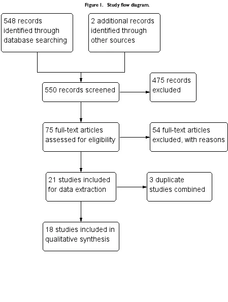
Results of the search
We conducted literature searches up to 19 December 2017 and identified 548 titles (Figure 1). While searching for additional information onArens 2015, we identified one study through its conference proceeding (Beissner 2015), and one study through ongoing trials (Barogui 2016). Two review authors (RRY and MR) closely examined 75 full-text articles. We contacted the technical team at the WHO for possible unpublished studies; there were none other than those we identified. We identified two ongoing trials on US National Institutes of Health Ongoing Trials Register ClinicalTrials.gov (NCT01432925;NCT01659437).
9 Drugs for treating Buruli ulcer (Mycobacterium ulceransdisease) (Review)
Figure 1. Study flow diagram.
10 Drugs for treating Buruli ulcer (Mycobacterium ulceransdisease) (Review)
We identified six RCTs and 15 prospective observational studies that met the inclusion criteria. Two review authors (RRY and MR) independently extracted data for these studies.
Included studies
Study design
Six articles reported a total of five RCTs. The BURULICO study was reported in two different articles with different outcomes (
Nienhuis 2010;Klis 2014; seeBURULICO Study 2010). A total of 15 articles reported prospective observational studies. Five articles were from the same Australian group using the data of Buruli ulcer patients from their registry that they had started col-lecting in January 1998 (O’Brien 2007;O’Brien 2012;Friedman 2013;O’Brien 2013b;Friedman 2016), and evaluated a number of different combinations of antibiotics. We identified two sets of articles reporting data for the same groups of participants at dif-ferent time points (Friedman 2013andFriedman 2016;O’Brien 2007andO’Brien 2012), therefore we extracted data from only the more recent papers (O’Brien 2012;Friedman 2016). Some partic-ipants inO’Brien 2012,O’Brien 2013b, andFriedman 2016may be included in more than one study, as they were from the same registry.Barogui 2016combined participants of theBURULICO Study 2010and the ongoingNCT01432925trial, therefore there is overlap of participants. However,Barogui 2016measured an outcome (paradoxical reactions) that was not an outcome measure of the original RCTs. We counted this study as an independent, prospective observational study.
We henceforth describe results of the qualitative synthesis of five RCTs and 13 prospective observational studies.
Interventions and comparisons
Randomized controlled trials
The included RCTs evaluated the following.
• Monotherapy in comparison to placebo, with surgery when indicated: clofazimine in one trial (Revill 1973), and
sulfamethoxazole/trimethoprim in a second trial (Fehr 1994). • Combination therapy:
◦ rifampicin + streptomycin before surgery with different lengths of treatment (varying from two to 12 weeks), in comparison to surgery alone (Etuaful 2005);
◦ rifampicin + streptomycin for four weeks followed by rifampicin + clarithromycin for four weeks in comparison to rifampicin + streptomycin for eight weeks, with surgery when indicated (BURULICO Study 2010);
◦ rifampicin + dapsone for eight weeks in comparison to no treatment, with no surgery in either arm (Espey 2002).
Prospective observational studies
Two studies evaluated different treatment regimens in multiple treatment arms (O’Brien 2012;Friedman 2016). All of the other prospective observational studies were single-arm studies. Prospec-tive observational studies evaluated the following.
• Monotherapy with clofazimine for one to four weeks before surgery (Lunn 1964).
• Combination therapy with rifampicin + streptomycin for: ◦ 12 weeks with surgery at week 4 (Kibadi 2010); ◦ eight weeks with surgery when indicated (Chauty 2007;Sarfo 2010;Adu 2013;Beissner 2015);
◦ eight weeks with surgery (Agbenorku 2011). • Combination therapy with rifampicin + clarithromycin:
◦ rifampicin + clarithromycin for eight weeks, with surgery when indicated (Chauty 2011);
◦ rifampicin + streptomycin for two weeks followed by rifampicin + clarithromycin for six weeks, with surgery when indicated (Phillips 2014a).
• Other combination therapy:
◦ rifampicin + either ciprofloxacin, clarithromycin, or moxifloxacin, with no surgery or with limited debridement (Friedman 2016);
◦ rifampicin + ciprofloxacin, rifampicin + clarithromycin, rifampicin + clarithromycin + ethambutol, ciprofloxacin + clarithromycin, rifampicin + moxifloxacin, clarithromycin + ethambutol, rifampicin + ethambutol + amikacin, or clarithromycin only, with surgery in all cases, in comparison to surgery alone (O’Brien 2012);
◦ single or combination administration of rifampicin, ciprofloxacin, clarithromycin, ethambutol, amikacin, and/or moxifloxacin, with surgery when indicated (O’Brien 2013b);
◦ either rifampicin + streptomycin for eight weeks or rifampicin + streptomycin for four weeks followed by rifampicin + clarithromycin for four weeks, with surgery when indicated (Barogui 2016).
Location and participants
All studies were conducted in areas with high Buruli ulcer en-demicity: of the RCTs, three were conducted in Ghana and one in Côte d’Ivoire and in Uganda; of the prospective observational studies, four were conducted in Ghana, three in Australia, two in Benin, one in Uganda, one in Democratic Republic of Congo, and one in Togo.Barogui 2016was a joint study between Ghana and Benin.
11 Drugs for treating Buruli ulcer (Mycobacterium ulceransdisease) (Review)
Some studies set inclusion criteria for age and lesion type or size given in diameter. Of the RCTs, theBURULICO Study 2010 re-cruited participants over five years with lesion size less than 10 cm;
Etuaful 2005recruited participants over 15 years with lesion size less than 10 cm; andEspey 2002recruited participants over four years with ulcers. Of the prospective observational studies,Chauty 2011recruited participants over five years with lesion size less than 10 cm;Phillips 2014arecruited participants over five years with lesion size less than 15 cm;Kibadi 2010recruited participants be-tween three and 75 years with lesion size larger than 10 cm; and theNCT01432925trial (a part ofBarogui 2016) recruited partic-ipants over three years of age. All other included studies recruited all age groups and lesion sizes.
Three RCTs,Fehr 1994;Etuaful 2005;BURULICO Study 2010, and 10 prospective observational studies,Sarfo 2010;Agbenorku 2011;Chauty 2011;O’Brien 2012;Adu 2013;O’Brien 2013b;
Phillips 2014a;Beissner 2015;Barogui 2016;Friedman 2016, had laboratory confirmation as part of their inclusion criteria. The remaining included studies did not have laboratory confirmation as an inclusion criterion.
Outcomes and length of follow-up
Outcomes in the RCTs varied. One trial measured “cure” (
BURULICO Study 2010), and one trial measured “possible cure” (Revill 1973). Both trials also measured healing time (Revill 1973;
BURULICO Study 2010). Otherwise, change in ulcer size was investigated in three trials (Fehr 1994;Espey 2002;Etuaful 2005), recurrence in three trials (Revill 1973;Etuaful 2005;BURULICO Study 2010), and adverse effects in three trials (Espey 2002;
Etuaful 2005;BURULICO Study 2010).
Of the prospective observational studies, seven studies measured “cure” (Phillips 2004;Kibadi 2010;Sarfo 2010;Agbenorku 2011;
Chauty 2011;O’Brien 2012;Friedman 2016); one study mea-sured “probable cure” (Chauty 2007); and three studies measured “possible cure” (Lunn 1964;Adu 2013;Beissner 2015). Healing time was investigated in five studies (Sarfo 2010;Chauty 2011;
Phillips 2014a;Beissner 2015;Friedman 2016), change in ulcer size in one (Sarfo 2010), recurrence in eight (Chauty 2007;Kibadi 2010;Sarfo 2010;Agbenorku 2011;Chauty 2011;O’Brien 2012;
Phillips 2014a; Beissner 2015), adverse effects in eight (Lunn 1964;Chauty 2007;Sarfo 2010;Agbenorku 2011;Chauty 2011;
O’Brien 2012; Phillips 2014a;Friedman 2016), and paradoxi-cal reactions in six studies (Sarfo 2010;O’Brien 2012;O’Brien 2013b;Phillips 2014a;Barogui 2016;Friedman 2016). Follow-up period varied in the RCTs.Etuaful 2005followed up participants until one year after completion of treatment. In the
BURULICO Study 2010, Nienhuis and colleagues first followed up participants until one year, and then Klis and colleagues re-visited participants again during four to six years after treatment. Two trials did not specify their follow-up time (Fehr 1994;Espey 2002). In the earlier study byRevill 1973, their follow-up pe-riod ranged from 17 to 40 months, with a median of 32 months. Follow-up in the prospective observational studies was one year in six studies (Chauty 2007;Sarfo 2010;O’Brien 2012;O’Brien 2013b;Phillips 2014a;Friedman 2016). Otherwise, it was seven months inBarogui 2016, 1.5 years inChauty 2011, two years inAgbenorku 2011andKibadi 2010, and not specified inLunn 1964,Adu 2013, andBeissner 2015.
Excluded studies
We excluded 475 studies after title and abstract screening. We assessed 75 full-text articles for eligibility, of which we excluded 37 on the basis of their study design (retrospective observational studies, cross-sectional surveys, case series, or qualitative studies), eight because they were either reviews or commentaries, five be-cause they were conference proceedings, and four bebe-cause they were duplicates.
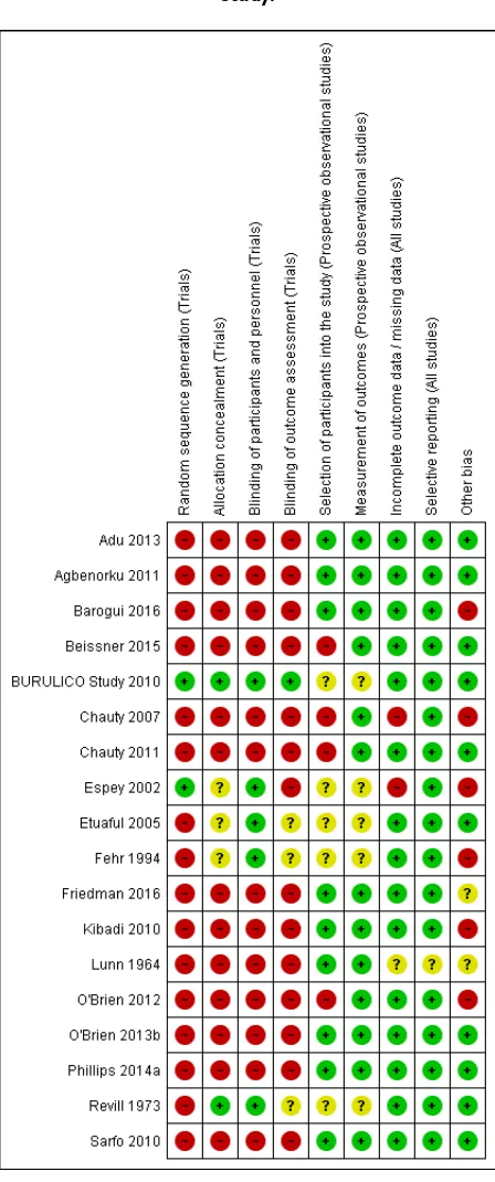
Risk of bias in included studies
We have summarized the risk of bias in included studies inFigure 2andFigure 3.
12 Drugs for treating Buruli ulcer (Mycobacterium ulceransdisease) (Review)
Figure 2. ‘Risk of bias’ graph: review authors’ judgements about each risk of bias item presented as percentages across all included studies.
13 Drugs for treating Buruli ulcer (Mycobacterium ulceransdisease) (Review)
Figure 3. ‘Risk of bias’ summary: review authors’ judgements about each risk of bias item for each included study.
14 Drugs for treating Buruli ulcer (Mycobacterium ulceransdisease) (Review)
Randomized controlled trials
Of the five included RCTs, onlyBURULICO Study 2010used adequate methods. Otherwise, either methods were either inade-quate or details were poorly reported in the remaining studies.
Prospective observational studies
Of the 13 prospective observational studies, we rated seven recent studies as at low risk of bias (Sarfo 2010;Agbenorku 2011;Adu 2013;O’Brien 2013b;Phillips 2014a;Barogui 2016;Friedman 2016). The older studies were associated with a higher risk of bias (Lunn 1964;Chauty 2007).
Allocation
Of the five RCTs, two were blinded and were rated as at low risk of bias (Revill 1973;BURULICO Study 2010). Otherwise, no information,Espey 2002;Etuaful 2005, or no clear statement,
Fehr 1994, was provided, and these studies were rated as at unclear risk of bias.
Blinding
Of the five RCTs, two were blinded and were rated as at low risk of bias (Revill 1973;Fehr 1994). Otherwise, the RCTs were open-label,BURULICO Study 2010, or no clear statement was provided,Espey 2002;Etuaful 2005, but were rated as at low risk of bias as the outcome was unlikely to be influenced by lack of blinding.
Incomplete outcome data
Of the five RCTs, we rated one as at high risk of bias, as 10 out of 30 participants (33%) were lost to follow-up (Espey 2002). The proportion of missing data was relatively large in one RCT (6/18 participants, 33%) (Fehr 1994), however reasons for exclusions/ missing data were relatively well balanced or unlikely to be related to true outcome, and the RCT was rated as at low risk of bias. Otherwise, no participants,Etuaful 2005, or a minimal number of participants,Revill 1973;BURULICO Study 2010, were lost to follow-up, and we judged these RCTs as at low risk of bias. Of the 13 prospective observational studies, we rated two studies as at high risk of bias: the assessment time point was unclear inLunn 1964, and 17 participants were lost to follow-up during the study period but were included in the final analysis inChauty 2007. Otherwise, either no participants,Kibadi 2010;Agbenorku 2011;
Chauty 2011;O’Brien 2012;Adu 2013;O’Brien 2013b;Beissner 2015;Barogui 2016;Friedman 2016, or a minimal number of participants,Sarfo 2010;Phillips 2014a, were lost to follow-up, and we considered these studies as at low risk of bias.
Selective reporting
Of the five included RCTs, we rated one as at unclear risk of bias as there were no predefined outcomes (Lunn 1964). All of the other RCTs reported all expected outcomes, and we rated these as at low risk of bias.
All 13 prospective observational studies reported all expected out-comes and were rated as at low risk of bias.
Other potential sources of bias
Five studies either did not have laboratory confirmation as their inclusion criteria or only performed laboratory exams in a por-tion of their participants, therefore non-Buruli ulcer cases may be included in their study results (Lunn 1964;Revill 1973;Espey 2002;Chauty 2007;Kibadi 2010). The standard treatment for Buruli ulcer has transitioned from surgery to drugs plus surgery as adjunctive treatment after the recommendation of drug treatment by the WHO in 2004 (WHO 2014), and this may have created some bias.
Potential comorbidities such as osteomyelitis, HIV/AIDS, dia-betes mellitus, cancer, and use of immunosuppressant drugs may have affected some results, especially on severity and healing rate and time. Two studies reported on comorbidities of their study participants: 9.5% inFriedman 2016and 16.3% inO’Brien 2012; there may be an overlap of participants in these two studies.
Effects of interventions
See:Summary of findings for the main comparisonRifampicin combined with streptomycin compared with surgery alone for Buruli ulcer; Summary of findings 2 Rifampicin with clarithromycin compared with rifampicin with streptomycin in the consolidation phase for Buruli ulcer
We first assess the effects of a variety of treatments on healing and recurrence, stratified by monotherapy and combination therapy. We then summarise adverse effects and paradoxical reactions across all comparisons.
Healing and recurrence
Monotherapy
SeeTable 1.
One RCT and one prospective observational study evaluated the efficacy of clofazimine, and one RCT evaluated the efficacy of sul-famethoxazole/trimethoprim. All three studies had small sample sizes, and no treatment effects were demonstrated.
15 Drugs for treating Buruli ulcer (Mycobacterium ulceransdisease) (Review)
Clofazimine
Revill 1973compared clofazimine to placebo, with similar recur-rence in the two arms (clofazimine 8/51 (15.7%); placebo 10/ 54 (18.5%); difference 2.8%, 95% confidence interval (CI) not given). The authors examined a subgroup of participants with non-ulcerated lesions who were withheld from immediate surgery: the number that healed was slightly higher with clofazimine, but the difference was small, and this was a post hoc subgroup analy-sis (clofazimine, 5/13 (38%); placebo, 6/21 (29%)). The median healing time was measured in this same subgroup also those with a lesion less than 5 cm in diameter (clofazimine, 8 participants; placebo, 17 participants) and was 21 weeks and 14 weeks, respec-tively.
One prospective observational study,Lunn 1964, examined the effects of clofazimine with surgery in 10 participants with ulcers. Six participants (60%) achieved complete healing in 3 to 12 weeks. The remaining four participants were still under treatment for their ulcers at the time of reporting.
Sulfamethoxazole/trimethoprim
Fehr 1994compared sulfamethoxazole/trimethoprim to placebo in 12 participants with ulcers. The mean ulcer size in the sul-famethoxazole/trimethoprim group at baseline was 73.8 cm2(9 to
247) and in the placebo group was 38.7 cm2(15 to 80). The
au-thors reported that sulfamethoxazole/trimethoprim reduced ulcer size by an average of 10.9%, while an average increase of 24.5% was observed in the placebo group (P = 0.15). The percentage ulcer area covered by granulation tissue at study end was 92% in
the sulfamethoxazole/trimethoprim group and 57% in the placebo group (P = 0.17).
Combination therapy
Rifampicin combined with streptomycin SeeTable 2.
One RCT and six prospective observational studies investigated the efficacy of rifampicin and streptomycin. Five prospective ob-servational studies evaluated this regimen administered for 8 weeks (828 participants) with surgery given to either all participants or a select group. Four studies reported healing rates for all participants, regardless of whether they had received surgery or not (84.5% to 100%, assessed at various time points). Four studies reported heal-ing rates for participants who received combination therapy alone (48% to 95%, assessed at various time points).
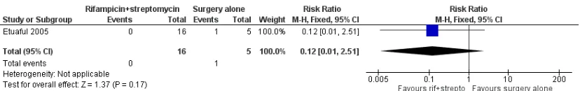
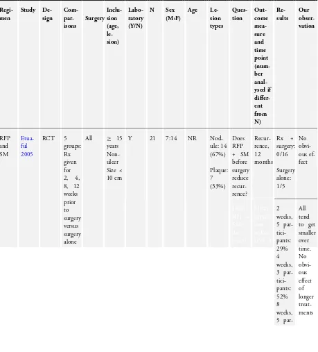
One RCT,Etuaful 2005, examined 21 participants with non-ul-cerative lesions to test the efficacy of rifampicin and streptomycin. They divided the participants into 5 groups: 4 groups were given ri-fampicin and streptomycin for 2, 4, 8, and 12 weeks before surgery respectively, and one group received only surgery. No recurrence was observed in participants in any group receiving combination therapy at 12 months, compared with one case of recurrence in a participant who received only surgery. No difference in recurrence was observed between these two groups (risk ratio (RR) 0.12, 95% CI 0.01 to 2.51;Figure 4; Analysis 1.1). Reduction in lesion sur-face area in participants who received rifampicin and streptomycin was the highest (52%) in the group that underwent four weeks of the regimen before surgery.
Figure 4. Forest plot of comparison: 1 Rifampicin plus streptomycin (experimental) versus surgery alone (control), outcome: 1.1 Recurrence.
One prospective observational study,Kibadi 2010, examined ri-fampicin and streptomycin given for 12 weeks with surgery per-formed at week 4, in 92 participants with ulcerative lesions mea-suring more than 10 cm in diameter. The study showed a high healing rate at week 12 (85/92, 92.4%) and low recurrence rate at 2 years (2/92, 2.2%).
Five prospective observational studies examined treatment with rifampicin and streptomycin for eight weeks (Chauty 2007;Sarfo 2010;Agbenorku 2011;Adu 2013;Beissner 2015). In one study all participants received surgery either during or after treatment (in this study surgery included debridement and skin grafting,
16 Drugs for treating Buruli ulcer (Mycobacterium ulceransdisease) (Review)
not just excision) (Agbenorku 2011); in one study a select group received surgery after assessment at week 4 and week 8 (Chauty 2007); and in three studies a select group of participants received after eight weeks of treatment (Sarfo 2010;Adu 2013;Beissner 2015).
• Where surgery was given to a select group participants, surgery rate differed among studies: 5% inSarfo 2010, 27% in
Beissner 2015, 52% inChauty 2007, and 52% inAdu 2013. • Four studies reported healing rates for all participants, regardless of whether they received surgery or not: 84.5% in
Beissner 2015, 96.3% inAgbenorku 2011, 99.3% inSarfo 2010
and 100% inChauty 2007.
• Four studies reported healing rates for participants who received combination therapy alone: 48% at week 8 inAdu 2013, 48% after week 8 inChauty 2007, 69.8% after minimum of 6 months follow-up inBeissner 2015, and 95% at 12 months inSarfo 2010.
• Follow-up showed recurrence was unusual: 0% inSarfo 2010andBeissner 2015, 0.5% inAgbenorku 2011, and 1.4% in
Chauty 2007.
Rifampicin combined with clarithromycin SeeTable 3.
Two prospective observational studies (51 participants) evaluated the use of rifampicin and clarithromycin. Both studies included surgery, either to all participants or a select group. All participants were healed at 12 months.
Chauty 2011evaluated rifampicin and clarithromycin for eight weeks in 30 participants with lesions measuring less than 10 cm in diameter. They reported a high healing rate at 12 months with no
recurrence at 18 months (30/30, 100%). Half of the participants (50%) healed without any form of surgery; 11 participants (37%) healed with limited surgery including curettage of the lesion or a minor excision; and 4 participants (13%) healed with extensive surgery including major excision followed by skin grafting.
O’Brien 2012 evaluated rifampicin and clarithromycin with surgery in 21 participants and reported a high healing rate (100%) and no recurrence at one year. Duration of the regimen was de-termined by the attending physician.
Rifampicin with streptomycin initially, changing to rifampicin with clarithromycin in consolidation phase SeeTable 3.
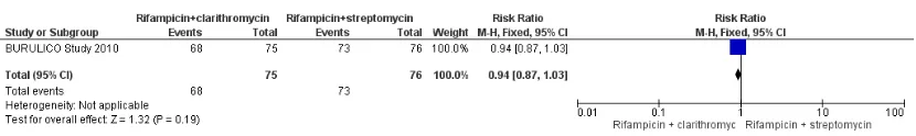
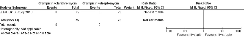
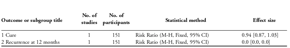
One RCT and one prospective observational study examined heal-ing rates startheal-ing with rifampicin and streptomycin, and then swapping to rifampicin and clarithromycin, with surgery as indi-cated. Both studies only included participants with small lesions, and more than 90% of participants healed without surgery. One RCT,BURULICO Study 2010, evaluated a regimen of ri-fampicin plus streptomycin for 4 weeks followed by riri-fampicin plus clarithromycin for 4 weeks in 151 participants with lesions measuring less than 10 cm in diameter. They compared this to the standard treatment at the time of eight weeks of rifampicin and streptomycin. Both groups achieved high healing rates at 12 months without surgery (a small number in each group had skin grafting): new regimen 68/75 (91%), standard regimen 73/76 (96%). There was no significant difference in healing rate or re-currence between the two groups (RR 0.94, 95% CI 0.87 to 1.03; not estimable due to 0 cases in both groups;Figure 5, Analysis 2.1;Figure 6; Analysis 2.2) or in healing time.
Figure 5. Forest plot of comparison: 2 Rifampicin combined with clarithromycin versus rifampicin combined with streptomycin in the consolidation phase, outcome: 2.1 Cure.
17 Drugs for treating Buruli ulcer (Mycobacterium ulceransdisease) (Review)
Figure 6. Forest plot of comparison: 2 Rifampicin plus clarithromycin (experimental) versus rifampicin plus streptomycin in the consolidation phase (control), outcome: 2.2 Recurrence at 12 months.
One prospective observational study,Phillips 2014a, evaluated a regimen of rifampicin plus streptomycin for 2 weeks followed by rifampicin plus clarithromycin for 6 weeks in 43 participants with lesions measuring less than 15 cm in diameter. Forty of 41 (98%) participants achieved healing by 52 weeks without surgery. Novel combination therapy
SeeTable 4.
One RCT and two prospective observational studies investigated the efficacy of combinations of one to three drugs from the fol-lowing: rifampicin, dapsone, ciprofloxacin, clarithromycin, mox-ifloxacin, ethambutol, amikacin, and azithromycin. High healing rates and low recurrence were achieved in the two prospective ob-servational studies.
One RCT,Espey 2002, examined the efficacy of rifampicin and dapsone for 8 weeks against placebo in 30 participants with ul-cerative lesions. No significant differences were observed for clin-ical improvement as judged by Buruli ulcer specialists using pho-tographs (P = 0.51). A significant change in ulcer size after two months was observed (P = 0.02), however there was a significant difference in the initial ulcer size between the two groups. Two prospective observational studies from the Australian group tested combinations of one to three oral antibiotics including ri-fampicin, ciprofloxacin, clarithromycin, moxifloxacin, ethambu-tol, amikacin, and azithromycin.Friedman 2016evaluated partic-ipants who received the regimen with no surgery or with limited surgical debridement. Among the 160 participants in their reg-istry, 28 participants (17.5%) who received extensive surgery were excluded, leaving 132 participants for their analysis. They reported that 131/132 (99%) participants healed at one year, among whom 101 (76.5%) participants healed with antibiotics alone. Median duration of antibiotic treatment was 56 days (interquartile range 24 to 96 days), and 22 participants (16.7%) needed fewer than 56 days to reach healing.O’Brien 2012compared participants who were treated with antibiotics plus surgery to surgery alone. All 90/ 90 participants (100%) who underwent combined treatment with antibiotics plus surgery healed. Fourteen (30%) participants who received only surgery had recurrence. As the participants were re-trieved from the same registry in these two studies, some partici-pants may contribute data to more than one of the studies.
Adverse effects
Three RCTs evaluated adverse effects, of which two reported none (Espey 2002;Etuaful 2005). One RCT evaluated long-term ad-verse effects of streptomycin three to six years after treatment (BURULICO Study 2010). Among those that could be retrieved from the past BURULICO study (n = 127), ototoxicity was ob-served in 23% of adults in the 4-week streptomycin group and 40% of adults in the 8-week streptomycin group (total n = 41), and in 28% of children in the 4-week streptomycin group and 26% of children in the 8-week streptomycin group (total n = 86). Nephrotoxicity during treatment was observed in 9% of adults in the 4-week streptomycin group and 20% of adults in the 8-week streptomycin group, and in 5% of children in the 4-week strep-tomycin group and 20% of children in the 8-week strepstrep-tomycin group. At long-term follow-up, one adult (2.4%) and two chil-dren (2.4%) were classified as having long-term nephrotoxicity, all from the 8-week streptomycin group.
Eight prospective observational studies evaluated adverse effects, of which two reported none (Chauty 2007(rifampicin, strepto-mycin) andAgbenorku 2011 (rifampicin, streptomycin)). One study reported no discontinuation of antibiotics (rifampicin, clar-ithromycin) due to adverse effects (Chauty 2011).Lunn 1964
reported one participant with gastrointestinal intolerance from clofazimine.Sarfo 2010reported one participant with dizziness and one with vomiting and dizziness from streptomycin, and one participant with rash probably from rifampicin.Phillips 2014a
reported one participant with ototoxicity from streptomycin.
O’Brien 2012reported that of 90 participants who received an-tibiotic treatment, 28 (31%) developed adverse effects including gastrointestinal intolerance, hepatitis, rash, hypoglycaemia, joint or tendon effects, palpitations, and hallucinations.Friedman 2016
reported that 21 of the 132 participants (16%) developed adverse effects (unspecified) that required cessation of one or more antibi-otics during treatment.
Paradoxical reactions
SeeTable 5.
Six prospective observational studies evaluated paradoxical reac-tions (Sarfo 2010;O’Brien 2012;O’Brien 2013b;Phillips 2014a;
18 Drugs for treating Buruli ulcer (Mycobacterium ulceransdisease) (Review)
Barogui 2016;Friedman 2016), of which two studies evaluated solely this outcome (O’Brien 2013b;Barogui 2016).
The incidence of paradoxical reactions ranged from 1.9% inSarfo 2010to 26% inFriedman 2016. Median onset time of paradoxical reactions ranged from 5.6 weeks (39 days) inO’Brien 2013bto 12 weeks inPhillips 2014a. As the participants were retrieved from the same registry in three studies (O’Brien 2012;O’Brien 2013b;
Friedman 2016), some participants may contribute data to more than one of the studies.
19 Drugs for treating Buruli ulcer (Mycobacterium ulceransdisease) (Review)
A D D I T I O N A L S U M M A R Y O F F I N D I N G S [Explanation]
Rifampicin with clarithromycin compared with rifampicin with streptomycin in the consolidation phase for Buruli ulcer
Patient or population:people with Buruli ulcer, early lesions m easuring less than 10 cm in diam eter, aged 5 years or older
Settings:Ghana
Intervention:rif am picin with streptom ycin, f ollowed by rif am picin with clarithrom ycin af ter 4 weeks
Comparison:rif am picin with streptom ycin continued
Outcomes Illustrative comparative risks* (95% CI) Relative effect (95% CI)
Number of participants (studies)
Certainty of the evi-dence
(GRADE)
Comments
Assumed risk Corresponding risk
Streptomycin
contin-due to im precision
We do not know if the treatm ent is superior to the control.
* The basis f or theassumed risk(f or exam ple, the m edian control group risk across studies) is provided in f ootnotes. Thecorresponding risk(and its 95% CI) is based on the assum ed risk in the com parison group and therelative effectof the intervention (and its 95% CI).
Abbreviations:CI: conf idence interval; RR: risk ratio
GRADE Working Group grades of evidence
High certainty:f urther research is very unlikely to change our conf idence in the estim ate of ef f ect.
M oderate certainty:f urther research is likely to have an im portant im pact on our conf idence in the estim ate of ef f ect and m ay change the estim ate.
Low certainty:f urther research is very likely to have an im portant im pact on our conf idence in the estim ate of ef f ect and is likely to change the estim ate.
Very low certainty:we are very uncertain about the estim ate.
aCure is def ined as ‘‘healing of skin lesions without recurrence at 12 m onths or longer.’’ There were no recurrences in this
study.
bDowngraded by 2 f or im precision: very f ew events and wide CIs.
D I S C U S S I O N
Summary of main results
SeeSummary of findings for the main comparisonandSummary of findings 2.
We included 18 studies, of which five were RCTs, in this review. Earlier studies conducted before 2000 that assessed monother-apy (clofazimine, sulfamethoxazole/trimethoprim) demonstrated no treatment effect. The remaining studies assessed combination therapy with or without surgery. The main regimens included ri-fampicin plus streptomycin, riri-fampicin plus clarithromycin, and rifampicin plus streptomycin switching to rifampicin plus clar-ithromycin during the consolidation phase.
It is evident that antimicrobials are important in treating Buruli ul-cers; this was an already established fact, but also learned from this review. Different combinations of antibiotics are given for eight weeks to treat Buruli ulcer, irrespective of the stage. However, there were insufficient studies and data to be able to determine which regimen is the most effective. In 2004, the WHO first recom-mended a combination of rifampicin and streptomycin for eight weeks (WHO 2014). However, there is no evidence from RCTs to support this treatment. Five prospective observational studies tested this regimen, which reported healing rates from 84.5% to 100% with or without surgery. Four studies reported healing rates for participants who received combination therapy alone to be from 48% to 95%. The time points assessed in the studies varied, and therefore a comparison or calculation of a combined healing rate was not possible.
There has recently been movement from the current regimen, which requires injection, to an all-oral treatment, with the goal of reducing the burden of treatment for patients. Of the stud-ies included in this review, BURULICO Study 2010 was the only RCT with adequate methods. This study tested rifampicin plus streptomycin for four weeks followed by rifampicin and clar-ithromycin for four weeks against rifampicin plus streptomycin for eight weeks, so that the patients will receive fewer injections of streptomycin. The study showed that there was no significant dif-ference in healing rate and time between the two regimens. Other studies have investigated different combinations of oral drugs, with most regimens yielding high healing rates (Chauty 2011;O’Brien 2012;Friedman 2016). The study sample sizes were small, and their study design was weak to examine the effects of these regi-mens, however these studies show the potential of all-oral treat-ments. The WHO currently lists use of rifampicin (10 mg/kg once daily) with either streptomycin (15 mg/kg once daily) or clar-ithromycin (7.5 mg/kg twice daily) for eight weeks as the treat-ment choices for Buruli ulcer, depending on the patient (WHO 2012;WHO 2017).
When assessing the efficacy of treatments for Buruli ulcers, le-sion size, lele-sion type, and whether surgery was applied or not are important factors to be considered. We attempted to perform a subanalysis, but this was not possible due to the heterogeneity of
studies. It may also be important to consider the impact of the severity of lesions (WHO category) on treatment efficacy, how-ever not all studies reported these data. It is important to note that some studies that reported high healing rates recruited only par-ticipants with small lesions, which may be important to consider when interpreting the results from these studies (Etuaful 2005;
BURULICO Study 2010;Chauty 2011;Phillips 2014a). Six prospective observational studies measured incidence of para-doxical reactions, which ranged from 1.9% to 26%. The patho-genesis of paradoxical reactions remains unclear, but recent studies report a possible association with antibiotic treatment and types of antibiotics used (O’Brien 2009;Nienhuis 2012;O’Brien 2013b).
Overall completeness and applicability of evidence
All studies included both males and females. With regards to age, participants from African countries were younger compared to those from Australia, which could have influenced the results. This is reflected by the different age distributions of the affected pop-ulation between the two areas (Asiedu 1998;Wansbrough-Jones 2006;Boyd 2012). Comorbidities (including HIV) in participants were uncommon, or those with comorbidities were excluded from the study, with the exception of the Australia group studies. Rates of comorbidities in the two Australian studies (9.5% and 16.3%) could have affected their study results.
Five studies (26%) diagnosed Buruli ulcer based only on clinical presentation, otherwise all studies had laboratory confirmation of Buruli ulcer either by Ziehl-Neelsen test for AFB, polymerase chain reaction (PCR), or histopathology. All recent studies (after 2007) had laboratory confirmation of Buruli ulcer as part of their inclusion criteria.
Treatment was often given for eight weeks, which has been the WHO recommendation since 2004, and different durations were not tested. Dosages of the drugs were the same between studies: 10 mg/kg/day for rifampicin, 15 mg/kg/day for streptomycin, and 7.5 mg/kg/day for clarithromycin. Intervention with surgery made it a challenge to compare the outcomes between studies. However, it is an important adjunctive intervention to drugs for treating Buruli ulcer, and participants who received surgery were included in the study results. The extent/definition of surgical intervention differed between studies: for example, skin grafting was not con-sidered to be surgery inBURULICO Study 2010andFriedman 2016. Furthermore, the decision of when to intervene with surgery differs among surgeons/clinicians, and this may have affected the results. It is also important to note that earlier studies tended to perform surgery more often than current studies, as it used to be the standard treatment.
Healing as defined by complete epithelialization was the primary outcome in most studies, but not in the earlier studies, where it was change in ulcer size (Fehr 1994;Espey 2002;Etuaful 2005). The only other outcome that was comparable between studies 21 Drugs for treating Buruli ulcer (Mycobacterium ulceransdisease) (Review)