IN VITRO CYTOTOXIC AND APOPTOSIS STUDY OF CHEMICAL CONSTITUENTS FROM
CLERODENDRUM PHLOMIDIS
LEAF ON MCF-7 AND A549 CANCER CELL LINES
HABEELA JAINAB N
1,2*, MOHAN MARUGA RAJA MK
11Department of Pharmacy, Centre for Research and Development, PRIST University, Vallam, Thanjavur - 613 403, Tamil Nadu, India. 2Department of Pharmaceutical Chemistry, Hillside College of Pharmacy and Research Centre, Raghuvanahalli, Bengaluru - 560 062,
Karnataka, India. Email: habi.asheesh@gmail.com Received: 14 December 2017, Revised and Accepted: 01 January 2018 ABSTRACT
Objective: Cancer is a molecularly heterogeneous diseaseand the leading cause of death worldwide. The methanol extract of Clerodendrum phlomidis leaf has been reported for cytotoxicity. Hence, the current investigation was planned to evaluate the cytotoxic activity of the chemical constituents isolated from the methanol extract of the C. phlomidis leaf against the Michigan cancer foundation-7 (MCF-7) breast cancer and adenocarcinomic human alveolar basal epithelial cells (A549) lung cancer cell lines by the apoptotic study.
Methods: Cytotoxic activity of the chemical constituents of the methanol extract of the C. phlomidis leaf was evaluated by the 3-(4,5-dimethylthiazol -2-yl)-5-diphenyltetrazolium bromide (MTT) assay against breast cancer (MCF-7) and lung cancer (A549) cell lines. The MCF-7 and A549 cell lines were tested at different concentrations to determine 50% of growth inhibition (inhibitory concentration [IC50]) by MTT assay. Apoptosis of nuclei was
detected by 4’,6-diamidino-2-phenylindole staining assay.
Results: In MCF-7 breast cancer cell line study, Compounds 6 and 9 exhibited good cytotoxic activity with an IC50 value of 83.80 and 75.16 µg/ml,
respectively. In A549 lung cancer cell line, again Compounds 6 and 9 exhibited good cytotoxic activity with an IC50 value of 84.46 and 78.60 µg/ml,
respectively. Percentage of apoptosis induced by the Compounds 6 and 9 in the MCF-7 cancer cells was found to be 74.50 and 85.48, respectively. Conclusion: The results of the current study prove that the Compounds 6 and 9 are potential agents for breast and lung cancer. In future research, these potential agents can further be evaluated by animal studies and their mechanism of action apart from the induction of apoptosis of the cancer cells can be determined.
Keywords: Methanol extract, 3-(4,5-dimethylthiazol-2-yl)-5-diphenyltetrazolium bromide assay, 4’,6-diamidino-2-phenylindole staining, DNA fragmentation, Anticancer.
INTRODUCTION
Cancer is a molecularly heterogeneous disease [1] and the leading cause of death worldwide. National Institute of Cancer Preventive and Research reported that the estimated number of people living with cancer in India is around 2.5 million, with over 7 lakh new cases being registered every year and 5,56,400 deaths. Cancers of oral cavity and lungs in males and cervix and breast in females account for over 50% of all cancer deaths in India [2]. Due to the existence of various types of tumors with different histopathology, genetic, epigenetic variations, and clinical outcomes [3], it has become difficult to understand this disease, the mechanism of action of chemotherapeutics and the creation of novel therapies. Studies of cancer rely on the use of primary tumors [1,4], paraffin-embedded samples [1], cancer cell lines [1,4,5], xenografts [2,6], tumor primary cell cultures [4,5], and/or genetically engineered mice [3]. These diverse models are used for different studies, mainly because certain types of manipulations for the genetic and DNA methylation analysis and drug testing are ethically, and in practice, difficult to perform in animals.
Cell lines emerge as a feasible alternative to overcome these issues, being at the same time easy to manipulate and molecularly characterize (e.g., genetic and/or epigenetically) [4]. Cancer cell lines have been widely used for research purposes and proved to be a useful tool in the genetic approach, and its characterization shows that they are, in fact, an excellent model for the study of the biological mechanisms involved in cancer [1]. The results of the research in cancer cell lines are
usually extrapolated to in vivo human tumors [4], and its importance as models for drug testing and translational study has been recognized by biomedical and pharmaceutical industries [7]. In comparative studies made between cancer cell lines derived from earlier stage tumors and the original tumor tissues showed good concordance in several parameters, including the state of tumor protein - TP53 (100%) and epidermal growth factor - ERBB2/EGFR2 (93%). This shows that this type of cells is more representative of an original tumor [5], reflecting more accurately the events that occur in cancer cells in vivo [6].
Plant-derived natural products have long been and will continue to be extremely important as sources of medicinal agents and models for the design, synthesis, and semi-synthesis of novel substances for treating humankind diseases. Plant-derived compounds which have recently undergone development include the anticancer agents, taxol and camptothecin, the Chinese antimalarial drug, artemisinin, and the ayurvedic drug, forskolin. These and many other examples serve to illustrate the continuing value of plant-derived secondary metabolites as viable compounds for modern drug development. However, despite these many important past contributions from the plant kingdom, a great many plant species have never been described and remain unknown to science, and relatively few have been surveyed systematically to an extent for biologically active chemical constituents [8].
Clerodendrum phlomidis is a small herb common in India and Sri Lanka. The leaves and roots are used in Ayurveda, Siddha, Chinese and Unani medicines © 2018 The Authors. Published by Innovare Academic Sciences Pvt Ltd. This is an open access article under the CC BY license (http://creativecommons. org/licenses/by/4. 0/) DOI: http://dx.doi.org/10.22159/ajpcr.2018.v11i5.24264
[9]. C. phlomidis leaf has been reported for various biological activities such as antifungal [10], antioxidant [11], antibacterial [11], analgesic [12], antiasthmatic [13], antidiarrheal [14], anti-inflammatory [15], antiplasmodial [16], and hypoglycemic [17]. The methanol extract of C. phlomidis leaf has been reported for cytotoxicity [18]. Thus, in the current investigation, we planned to evaluate the cytotoxic activity of the chemical constituents isolated from the methanol extract of the C. phlomidis leaf against breast cancer (Michigan cancer foundation-7 [MCF-7]) and lung cancer (A549) cell lines with the help of apoptosis study. The findings of the current study will be useful in identifying the potential cytotoxic agent/s against the breast and lung cancer cell line with fewer side effects.
MATERIALS AND METHODS Collection of plant material
Leaves of C. phlomidis were collected from the outskirts of Tirupati, Andhra Pradesh, India. Plant material was authenticated by Dr. K. Madhava Chetty, Taxonomist, Department of Botany, Sri Venkateswara University, Tirupati, Andhra Pradesh. A voucher specimen (CP/L/2015/1160) was also submitted.
Extraction
The leaves were dried in shade and ground to coarse powder and stored in a glass jar until use. Powdered drug was packed into Soxhlet apparatus, and extraction was carried out with methanol till exhausted. The extract was then concentrated using rotary vacuum evaporator at 50°C under vacuum. The dried residue was then stored in an opaque glass bottle for further studies.
Isolation of compounds by column chromatography
The methanol extract of C. phlomidis leaf was subjected to column chromatography for the isolation of chemical constituent/s with cytotoxic potential. The methanol extract was chromatographed on a silica gel column, and gradient elution technique was followed using chloroform: Methanol as the mobile phase in different ratios.
Spectral characterization studies of the isolated compounds The isolated compounds were characterized by infrared spectra, proton nuclear magnetic resonance spectra (1H NMR), carbon NMR (13C NMR),
and mass spectra.
3-(4,5-dimethylthiazol-2-yl)-5-diphenyltetrazolium bromide (MTT) assay chemicals
MTT, fetal bovine serum (FBS), phosphate buffered saline (PBS), Dulbecco’s Modified Eagle’s Medium (DMEM), and trypsin were obtained from Sigma-Aldrich Co, St Louis, USA. Ethylene diamine tetraacetic acid (EDTA), glucose and antibiotics were obtained from HiMedia Laboratories Ltd., Mumbai, India. 4’,6-diamidino-2-phenylindole (DAPI), dimethyl sulfoxide (DMSO), ethidium bromide, glycerin, and agarose gel were procured from E Merck Ltd., Mumbai, India. MCF-7 (human breast cancer cell line) and A549 (lung cancer cell line) cell cultures were procured from National Centre for Cell Sciences, Pune, India.
Cell treatment procedure
Stock cells were cultured in DMEM supplemented with 10% inactivated FBS, penicillin (100 IU/ml), streptomycin (100 mg/ml), and amphotericin B (5 mg/ml) in a humidified atmosphere of 5% CO2 at
37°C until confluent. The cells were dissociated with trypsin phosphate versene glucose solution (0.2% trypsin, 0.02 % EDTA, and 0.05% glucose in PBS). The stock cultures were grown in 25 cm2 culture flasks,
and all experiments were carried out in 96 µ titer plates.
Preparation of test solutions
To carry out the cytotoxicity studies, each of the Compounds (1, 2, 4, 6, 9, and 11) were separately dissolved in distilled DMSO and volume was made up with DMEM supplemented with 2% inactivated FBS to get a stock solution concentration of 1 mg/ml and sterilized by filtration. Serially two-fold dilutions of 10, 50, 100, 150, and 200 µg/ml were prepared from the stock for carrying out cytotoxic studies.
In vitro cytotoxicity MTT assay
MTT assay is based on the capacity of mitochondria succinate dehydrogenase enzymes in living cells to reduce the yellow water-soluble substrate MTT into an inwater-soluble, colored formazan product which was measured spectrophotometrically. Since only the metabolically active cells are capable of reducing MTT, the level of the activity is a measure of the cells. The MTT cell proliferation assay was used to evaluate the cytotoxicity of the compounds against human cancer cell lines, namely, MCF-7 (breast cancer) and A549 (lung cancer) at the concentration ranging from 10 to 200 µg/ml.
Determination of cell viability by MTT assay
For screening experiment, the cells were seeded into 96 well plates in 100 µl of DMEM containing 5% FBS, at a plating density of 10,000 cells/well and incubated at 37°C, 5% CO2, 95% air, and 100 %
relative humidity for 24 h before the addition of samples. 100 µl of the sample solution was added to the respective wells. The plate was then incubated for 48 h maintaining the following conditions of 37°C, 5% CO2, 95% air, and 100% relative humidity. The medium without the
sample served as control. After 48 h, 15 µl of MTT (5 mg/ml) in PBS was added to each well and incubated at 37°C for 4 h. The medium with MTT was then flicked off, and the formed formazan crystals were solubilized in 100 µl of DMSO, and then the absorbance was measured at 545 nm using microplate reader. The concentration of the samples required to inhibit the cell growth by 50% (inhibitory concentration [IC50]) value
was calculated from the dose-response curve. The percentage growth inhibition was calculated using the following formula [19,20].
%GrowthInhibition=OD control OD sample OD control
− ×
100
Apoptosis induction assay - nuclear staining with DAPI fluorescent dye Apoptosis of nuclei was detected by a DAPI staining assay. DAPI dye is a fluorescent dye that stains the nuclear DNA of a cell and is, therefore, used to determine the effect of samples on inducing morphological changes in the nuclei of cancer cells undergoing apoptosis. MCF-7 cell line at 500 µl (1×10⁶ cells/well) was seeded on 24 well plates and incubated until cell
growth at log phase for 24 h. The cells were then treated at 2×IC50 of test
samples (at the highest concentration of 75.03 and 83.80 µg/ml, 0.1% DMSO) for 24 h. After treatment, the cells were washed with PBS (1.0 ml)
and then fixed with 50 µl of methanol and water (1:1) under −20°C for
10 min. The fixed cells were washed and stained with 100 µl (1 µg/ml) of DAPI dye and then incubated at 37°C in a dark room for 30 min. The excess dye was then removed and 20 µl of PBS: Glycerin (1:1) was added to the mixed cells. The cells undergoing apoptosis, represented by the morphological changes of apoptotic nuclei, were observed and imaged at 40× magnifications under an inverted fluorescence microscope [21,22]. Percentage of apoptotic cells was calculated as follows:
% of apoptotic cells=Amount of apoptotic nuclei Amount of alll cells ×100
DNA fragmentation detection assay
DNA fragmentation was used to determine the induction of apoptosis by observing the biochemical change. After the cancer cells were treated with 2×IC50 of samples for 24 h, the cells were collected and washed
with media. Then, cell suspensions were transferred to microcentrifuge tubes (1.5 ml) and centrifuged at 300 rpm for 5 min to collect the cell pellets. The DNA in the cell pellet was extracted with DNA extraction Kit, 2 µg of DNA was electrophoresed on 2% agarose gel containing 0.1 mg/ml ethidium bromide staining. After electrophoresis, DNA fragments were analyzed with an ultraviolet illuminated camera [23].
RESULTS AND DISCUSSION
Previous studies related to the preliminary phytochemical screening of methanol leaf extract of C. phlomidis indicated the presence of alkaloids, steroids, coumarins, and flavonoids. The methanol leaf extract of C. phlomidis has been reported for significant cytotoxic and antioxidant potential [18]. Compounds (1, 2, 4, 6, 9, and 11) were isolated from the methanol extract of the C. phlomidis leaf [24]. From the spectral studies the isolated compounds were found to be 3-hexen-1-yl benzoate (Compound 1), 2,3-dihydroxypropanal (Compound 2), 1- (2,4,5-trihydroxyphenyl)-1-butanone (Compound 4), 3,6,7-trihydroxy-2-(3-methoxyphenyl)-4H-chromen-4-one (Compound 6), isopropyl linoleate (Compound 9), and oleic acid eicosyl ester (oleic acid ester) (Compound 11).
MTT cytotoxicity assay
In vitro cytotoxicity test is mainly done to screen potentially toxic compounds that affect basic cellular functions [25]. The molecular cancer cell lines profiling is essential for the development of new anticancer drugs, for understanding the mechanism of action and the patterns involved in cell resistance to chemotherapeutics already used in the treatment of cancer. Moreover, cancer cell lines can be a powerful tool for the identification of gene alterations or pathways related to cancer and for the discovery of putative drug targets [26].
Previous studies have evaluated the cytotoxic activity on mouse embryonic fibroblasts cell line (NIH 3T3) and HeLa cell lines using MTT assay on the crude extracts of petroleum ether, ethyl acetate, chloroform, and ethanol obtained from the root of the C. phlomidis plant. Ethanol extract had no cytotoxic activity, and the other extracts had moderate to weak cytotoxic activity on both the cell lines [27]. Evaluation of the in vitro antioxidant and cytotoxic activity of isolated compounds on roots of C. phlomidis has revealed that phenylacetic acid and ethyl-2-hydroxy-4-methyl benzoate from the ethanolic extract represent, a new group of cytotoxic and antioxidant agents [28]. Lakshmi and Bai have reported that C. phlomidis (L) leaves extract (CPLE) and its silver nanoparticles, plays a dual role by blocking carcinogen metabolic activation and enhancing carcinogen detoxification. They have also reported that the protective properties of the CPLE may be due to the presence of phytochemicals such as flavonoids, terpenoids, and alkaloids [29]. Although cell line studies have been reported for various solvent extracts of C. phlomidis, there was no much systematic study
performed leading to an active cytotoxic chemical constituent against breast cancer (MCF-7) and lung cancer (A549) cell lines with the help of apoptosis study.
In MCF-7 breast cancer cell line study, Compounds 6 (3,6,7-trihydroxy-2-(3-methoxyphenyl)-4H-chromen-4-one) and 9 (isopropyl linoleate) exhibited good cytotoxic activity with an IC50 value of 83.80 and
75.16 µg/ml, Compounds 1 (3-hexen-1-yl benzoate) and 4 (1-(2,4,5 trihydroxyphenyl)-1-butanone) exhibited moderate cytotoxic activity with an IC50 value of 120.08 and 152.30 µg/ml, and Compounds
2 (2,3-dihydroxypropanal) and 11 (oleic acid eicosyl ester) exhibited less cytotoxic activity with an IC50 value of 187.50 and 208.22 µg/ml (Table 1).
In A549 lung cancer cell line, Compounds 6 and 9 exhibited good cytotoxic activity with an IC50 value of 84.46 and 78.60 µg/ml, Compounds 1 and 4
exhibited moderate cytotoxic activity with the IC50 value of 107.31 µg/
ml and 139.01 µg/ml, and Compounds 2 and 11 exhibited less cytotoxic activity with the IC50 value of 168.48 and 190.50 µg/ml against A549
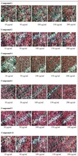
lung cancer cell lines (Table 2). The effect of isolated compounds on percentage growth of MCF-7 and A549 cell line is illustrated in Figs. 1 and 2. The results clearly indicate that the Compounds 6 and 9 decreases cell viability and induces the cytotoxic effect in MCF-7 cells and A549 cells more efficiently compared to the other isolated compounds.
Apoptotic study
Apoptosis is a process of programmed cell death. Biochemical events lead to characteristic cell changes (morphology) and death. Morphological changes in the cell include blebbing, cell shrinkage, nuclear fragmentation, chromatin condensation, chromosomal DNA fragmentation, and mRNA decay.
Compound 6 (3,6,7-trihydroxy-2-(3-methoxyphenyl)-4H-chromen-4-one), a flavonoid and Compound 9 (isopropyl linoleate) showed good cytotoxic activity against the breast and lung cancer cell lines. Flavonoids are part of the polyphenol class of phytonutrients. Research on flavonoids had shown major developments in anticancer drug discoveries with the potential to destroy cancer cells through apoptotic induction [30]. Flavonoids exert protective effects against various types of tumors including oral and pharyngeal, gastric, pancreatic, colorectal, hepatic, prostate, ovarian, endometrial, breast, and lung cancers [31,32].
Table 1: Effect of the isolated compounds from methanol extract of C. phlomidis leaf on MCF-7 cell lines
Concentration (mg/ml) % Growth inhibition
CPD-1 CPD-2 CPD-4 CPD-6 CPD-9 CPD-11
10
50 19.55±0.6034.21±0.25 24.61±0.3928.97±0.70 15.88±0.2627.23±0.49 33.86±0.6042.58±0.50 34.90±0.6642.76±0.87 17.80±0.8827.57±0.61 100
150 47.47±0.1453.23±0.31 37.00±0.1843.63±1.14 39.97±0.7348.87±0.87 51.31±0.5960.21±0.27 55.67±0.8667.54±0.60 30.54±0.3943.11±0.63 200
IC50
72.60±0.19
120.08 52.88±0.86187.50 60.56±0.68152.30 84.82±0.6483.80 83.42±1.1175.16 47.99±1.08208.22
All the values are expressed as mean±standard deviation, n=3. CPD-1, CPD-2, CPD-4, CPD-6, CPD-9, CPD-11 represents the isolated Compounds 1, 2, 4, 6, 9, and 11.
C. phlomidis: Clerodendrum phlomidis, MCF-7: Michigan cancer foundation-7
Table 2: Effect of the isolated compounds from methanol extract of C. phlomidis leaf on A549 cell lines
Concentration (µg/ml) % Growth inhibition
CPD-1 CPD-2 CPD-4 CPD-6 CPD-9 CPD-11
10
50 20.37±0.8038.68±0.54 18.72±0.4524.69±0.63 16.87±0.6429.84±0.56 27.78±0.4135.80±0.37 23.46±0.2747.94±0.75 22.63±0.8822.51±0.70 100
150 46.50±0.7564.81±0.73 37.86±0.0842.59±0.71 41.77±0.6052.67±0.40 54.53±0.2576.54±0.67 59.05±0.4174.07±0.79 36.01±0.8741.56±0.85 200
IC50
72.84±0.47
107.31 58.85±0.78168.48 63.99±0.52139.01 83.13±0.4084.46 78.40±0.3578.60 53.09±0.70190.50
All the values are expressed as mean±standard deviation, n=3. CPD-1, CPD-2, CPD-4, CPD-6, CPD-9, CPD-11 represents the isolated Compounds 1, 2, 4, 6, 9, and 11.
The molecular mechanism of flavonoids in cancer prevention remains unclear; some studies have proposed that flavonoids interact with different types of genes and enzymes. Some mechanisms of action of flavonoids have been identified, such as inactivation of carcinogens, antiproliferation, cell cycle inhibition, apoptotic induction, inhibition of angiogenesis, antioxidants, or the combination of such mechanisms [33].
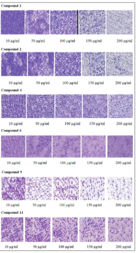
To find out whether the cytotoxic activity of the isolated chemical constituents was due to apoptosis, MCF-7 cells were treated with the Compounds 6 and 9 at 2× IC50 for 24 h. There were no prominent
morphological or nuclear changes in the control or untreated MCF-7 cells; the stained nuclei were rounded and evenly stained with DAPI. The cancer cells treated with the sample Compounds 6 and 9 showed the presence of condensed chromatin and apoptotic bodies that are the characteristic of the early and late stages of apoptosis. Compounds
6 and 9 induced 74.50 and 85.48% apoptosis in MCF-7 cells (Table 3 and Fig. 3). Determination of apoptosis was further carried out by evaluating the DNA laddering as a result of DNA fragmentation, which was typical of the late stage of apoptosis. DNA laddering was observed in the MCF-7 cells treated with the Compounds 6 and 9.
CONCLUSION
CONFLICT OF INTEREST
The authors declared that there is no conflict of interest.
AUTHOR’S CONTRIBUTION
MKMM Raja conceived the original idea and supervised the project. NH Jainab carried out the experiment and wrote the manuscript with support from MKMM Raja.
Fig. 2: Cytotoxic activity of the isolated compounds on A549 cell lines at different concentrations
Fig. 3: Nuclear morphological changes in Michigan cancer foundation -7 cell lines after treatment with the Compound 6 and
Compound 9
Table 3: Percentage of apoptotic cells of the Compounds 6 and 9 in apoptosis study of MCF-7 cell lines
S. no Isolated compounds Percentage of apoptotic cells
1 CPD-6 74.50±6.03
2 CPD-9 85.48±2.95
REFERENCES
1. Louzada S, Adega F, Chaves R. Defining the sister rat mammary tumor cell lines HH-16cl.2/1 and HH-16.cl.4 as an in vitro cell model for Erbb2. PLoS One 2012;7:e29923.
2. NICPR. Cancer Statistics; 2017. Available from: http://www. cancerindia.org.in/statistics. [Last cited on 2017 Nov 10].
3. Gogola TV, Rosen JM. Modelling breast cancer: One size does not fit all. Nat Rev Cancer 2007;7:659-72.
4. Staveren WC, Solis DY, Hebrant A, Detours V, Dumont JE, Maenhaut C. Human cancer cell lines: Experimental models for cancer cells in situ? For cancer stem cells. Biochim Biophys Acta 2009;1795:92-103. 5. Burdall SE, Hanby AM, Lansdown MR, Speirs V. Breast cancer cell
lines: Friend or foe. Breast Cancer Res 2003;5:89-95.
6. Leonetti C, Scarsella M, Zupi G, Zoli W, Amadori D, Medri L, et al. Efficacy of a nitric oxide-releasing nonsteroidal anti-inflammatory drug and cytotoxic drugs in human colon cancer cell lines in vitro and xenografts. Mol Cancer Ther 2006;5:919-26.
7. Gazdar AF, Girard L, Lockwood WW, Lam WL, Minna JD. Lung cancer cell lines as tools for biomedical discovery and research. J Natl Cancer Inst 2010;102:1310-21.
8. Balandrin MF, Kinghorn AD, Fransworth NR. Plant-derived natural products in drug discovery and development an overview. In: Kinghorn AD, Balandrin MF, editors. Human Medicinal Agents from Plants. Washington, DC: American Chemical Society; 1993. p. 2-12. 9. Prakash R. Medicinal values of Clerodendrum phlomidis: A review. Int
J Pharmacol Bio Sci 2013;7:1-7.
10. Rajasekaran A, Ponnusamy K. Antifungal activity of Clerodendrum inerme (L) and Clerodendrum phlomidis (L). Turk J Biol 2006;30:139-42. 11. Prasad MP, Sushant S, Chikkaswamy BK. Phytochemical
analysis, antioxidant potential, antibacterial activity and molecular characterization of Clerodendrum species. Int J Mol Biol 2012;3:71-6. 12. Srinivasa U, Rao JV, Krupanidhi AM, Babu PR. Analgesic activity of
leaves of Clerodendrum phlomidis. J Res Edu Indian Med 2007;13:23-5. 13. Vadnere GP, Somani RS, Singhai AK. Studies on antiasthmatic activity
of aqueous extract of Clerodendrum phlomidis. Pharmacol Online 2007;1:487-94.
14. Rani S, Ahamed N, Rajaram S, Saluja R, Thenmozhi S, Murugesan T. Anti-diarrhoeal evaluation of Clerodendrum phlomidis Linn. Leaf extracts in rats. J Ethnopharmacol 1999;68:315-9.
15. Prakash BN, Pandikumar P, Ignacimuthu S. Lysosomal membrane stabilization and anti-inflammatory activity of Clerodendrum phlomidis L.F., a traditional medicinal plant. J Ethnopharmacol 2011;135:779-85.
16. Simonsen HT, Nordskjold JB, Smitt UW, Nyman U, Palpu P, Joshi P,
et al. In vitro screening of Indian medicinal plants for antiplasmodial activity. J Ethnopharmacol 2001;74:195-204.
17. Chaturvedi GN, Subramaniyam PR, Tiwari SK, Singh KP. Experimental and clinical studies on diabetic mellitus evaluating the efficacy of an indigenous oral hypoglycaemic drug-arani (Clerodendrum phlomidis). Ancient Sci Life 1983;3:216-24.
18. Jainab NH, Raja MK. In vitro cytotoxic, antioxidant and GC-MS study of leaf extracts of Clerodendrum phlomidis. Int J Pharm Sci Res 2017;8:4433-40.
19. Denizot F, Lang R. Rapid colorometric assay for cell growth and survival modifications to the tetrazolium dye procedure giving improved sensitivity and reliability. J Immunol Methods 1986;89:271-7. 20. Wilson AP. Cytotoxicity and viability assays. In: Masters JR, editor.
Animal Cell Culture. 3rd ed. New York: Oxford University Press; 2000.
p. 175-219.
21. Escarate CG, Borrego JA, Brand EV, Dupre E, Portilla MA. Relationship between DAPI-fluorescence fading and nuclear DNA content: An alternative method to DNA quantification. Biol Res 2007;40:29-40. 22. Elmore S. Apoptosis: A review of programmed cell death. Toxicol
Pathol 2007;35:495-516.
23. Kalinina TS, Bannova AV, Dygalo NN. Quantitative evaluation of DNA fragmentation. Bull Exp Biol Med 2002;134:554-6.
24. Jainab NH, Raja MK. Antioxidant study of isolated chemical constituents from methanol extract of the Clerodendrum phlomidis leaf. World J Pharm Res 2017;6:1122-33.
25. Fotakis G, Timbrell JA. In vitro cytotoxicity assays: Comparison of LDH, neutral red, MTT and protein assay in hepatoma cell lines following exposure to cadmium chloride. Toxicol Lett 2006;160:171-7. 26. Camarillo CL, Ocampo EA. Oncogenomics and cancer
proteomics - Novel approaches in biomarkers discovery and therapeutic targets in cancer. Available from: http://www.intechopen. com/books/oncogenomics-and-cancerproteomics-novelapproaches-in-biomarkers-discovery-and-therapeutic-targets-incancer. [Last cited on 2017 Nov 20].
27. Sathish M, Tharani CB, Niraimathi V, Satheesh KD. In-vitro cytotoxic activity on roots of Clerodendrum phlomidis against NIH 3T3 cell line and Hela cell line. Pharmacol Online 2011;3:1112-8.
28. Sathish M, Niraimathi V, Noorulla KM, Palanimuthu A. In vitro
antioxidant and cytotoxic activity of isolated compounds on roots of
Clerodendrum phlomidis Linn. Asian J Pharm Clin Res 2015;8:196-201. 29. Lakshmi V, Bai GV. In vitro anticancer activity of Clerodendrum
phlomidis leaves and its silver nanoparticles on human breast cancer cell line (MCF-7). Asian J Innov Res 2016;1:1-5.
30. Monasterio A, Urdaci MC, Pinchuk IV, Moratalla NL, Irujo JJ. Flavonoids induce apoptosis in human leukemia U937 cells through caspase and caspase-calpain-dependent pathways. Nutr Cancer 2004;50:90-100.
31. Romagnolo DF, Selmin OI. Flavonoids and cancer prevention: A review of the evidence. J Nutr Gerontol Geriatr 2012;31:206-38.
32. Haris M, Mahmood R, Rahman H, Rahman N. In vitro cytotoxic activity of Clerodendrum infortunatum L. against T47D, PC3, A549 and HCT-116 human cancer cell lines and its phytochemical screening. Int J Pharm Pharm Sci 2016;8:439-44.
33. Bashari MH, Hidayat S, Ruswandi YA, Putri T, Qomarilla N, Dwiwina RG, et al. The n-hexane fraction of Myrmecodia pendans