Sox17-dependent gene expression and early heart and
gut development in Sox17-deficient mouse embryos
SABINE PFISTER
1, VANESSA J. JONES
1, MELINDA POWER
1, GERMAINE L. TRUISI
1,
POH-LYNN KHOO
1, KIRSTEN A. STEINER
1, MASAMI KANAI-AZUMA
2, YOSHIAKIRA KANAI
3,
PATRICK P. L. TAM
1,4and DAVID A. F. LOEBEL*
,1,4,51Embryology Unit, Children’s Medical Research Institute, New South Wales, Australia, 2Center for Experimental Animals, Tokyo Medical and Dental University, Tokyo, Japan,
3Department of Veterinary Medicine, University of Tokyo, Tokyo, Japan and 4Sydney Medical School, University of Sydney, New South Wales, Australia
ABSTRACT Sox17 is a transcription factor that is required for maintenance of the definitive endoderm in mouse embryos. By expression profiling of wild-type and mutant embryos and Sox17-overexpressing hepatoma cells, we identified genes with Sox17-dependent expression. Among the genes that were up-regulated in null embryos and down-regulated by Sox17-expressing HepG2 cells is a set of genes that are expressed in the developing liver, suggesting that one function of Sox17 is the repression of liver gene expression, which is compatible with a role for Sox17 in maintaining the definitive endoderm in a progenitor state. Consistent with these findings, Sox17-/- cells display a diminished capacity to contribute to the definitive endoderm
when transplanted into wild-type hosts. Analysis of gene ontology further revealed that many genes related to heart development were downregulated in Sox17-null embryos. This is associ-ated with the defective development of the heart in the mutant embryos, which is accompanied by localised loss of Myocd-expressing cardiogenic progenitors and the malformation of the anterior intestinal portal.
KEY WORDS:
Sox17, downstream gene, endoderm, heart morphogenesis
Introduction
Endoderm specification and differentiation during embryogen-esis in frogs and fish involves transcriptional activation of zinc finger GATA factors, Mix-homeodomain proteins and High Mobil-ity Group SOX proteins by TGF/Nodal signalling and T-box and POU-domain transcription factors (Kiefer, 2003; Lunde et al., 2004; Reim et al., 2004; Shivdasani, 2002; Stainier, 2002; Wood-land and Zorn, 2008; Zorn and Wells, 2007; Zorn and Wells, 2009). A similar set of genes including Mix/Mixer, Sox17 and Foxa
is associated with endoderm formation in mouse, frogs and fish, but their function and regulation differ in mouse development.
The mouse Mixl1 gene is expressed in the primitive streak where the endoderm progenitors reside but not in the definitive endoderm (Pearce and Evans, 1999), unlike in Xenopus where
Mix/Mixer expression is endoderm-specific. Mixl1 function is critical for the generation of definitive endoderm in the mouse.
BIOLOGY
www.intjdevbiol.com*Address correspondence to: David Loebel. Embryology Unit, Children’s Medical Research Institute, Locked Bag 23 Wentworthville, NSW Australia 2145. Fax: +61-2-9687-2120. e-mail: [email protected]
Supplementary Material (7 tables) for this paper is available at: http://dx.doi.org/10.1387/ijdb.103158sp
Accepted: 11 January 2011. Final author corrected PDF published online: 1 February 2011.
ISSN: Online 1696-3547, Print 0214-6282 © 2011 UBC Press
Printed in Spain
Abbreviations used in this paper: APS, anterior primitive streak; GO, gene ontology.
Mixl1-null embryos contain a reduced population of endoderm that lacks the expression of molecular markers of definitive endoderm (Hart et al., 2002) and Mixl1-null embryonic stem cells and primitive streak cells are less efficient in populating the definitive endoderm (Hart et al., 2002; Tam et al., 2007). Whereas
Sox17 is essential for endoderm formation in Xenopus and Zebrafish embryos, loss of Sox17 in the mouse does not appear to affect lineage allocation but impairs the viability of the endo-derm of the foregut and the proliferation of endoendo-derm in the posterior gut (Kanai-Azuma et al., 2002; Tam et al., 2003). In
Xenopus, the endodermal genes HNF1, Foxa1, Foxa2 and
Endodermin are direct transcriptional targets of Sox17 (Clements
Foxa2 is severely reduced in Sox17-/- mouse embryos
(Kanai-Azuma et al., 2002), but it is not clear whether they are direct transcriptional targets of Sox17.
At present, the transcriptional targets of Sox17 in the mouse embryo at early-organogenesis when the null-mutant phenotype first manifests have not been identi-fied. Previous studies dealing with Sox17-dependent gene expression in mammalian cells have used mouse or human embryonic stem (ES) cells, in which Sox17 was over-expressed. Results conflicted between the two stem cell types, with Sox17 inducing molecular characteristics of definitive endoderm in human ES cells (Seguin et al., 2008) but those of extraembryonic endoderm in mouse ES cells (Niakan et al., 2010). However, in mouse ES cells, Sox17 can induce both definitive and visceral endoderm differentiation under some circumstances (Qu
et al., 2008). The variation in results between in vitro
models suggests that Sox17-dependent changes in the transcriptome and the identification of potential Sox17 transcriptional targets is influenced by the inherent
prop-identify possible Sox17 target genes, we compared the changes in gene expression profiles of mutant embryos with those result-ing from Sox17-overexpression in HepG2 cells. The findings show that changes in Sox17 activity impact on the genetic activity associated with endoderm development. Embryological analysis
0
Cyp51 Idi1 Insig1 Ldlr Sc4mol
relative expression
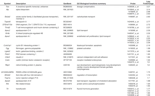
Fig. 1. Validation of the down-regulation of genes associated with choles-terol metabolism in Sox17-/- embryos. Real-time quantitative RTPCR analysis was performed on cDNA generated from RNA samples of E8.25 Sox17+/+ and
Sox17-/- embryos. Expression levels are shown relative to wild-type. Gapd was the
reference gene. Error bars indicate standard error of the mean. N=3 for each assay. **Significant difference at p<0.01 by two-tailed t-test from Sox17+/+ embryos.
erties of the cell model used.
In this study, we examined gene expression profiles in the mouse embryo, in which Sox17 has been shown to be required for development of the definitive but not visceral endoderm, to identify Sox17-dependent changes in gene transcription. To
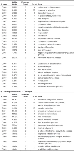
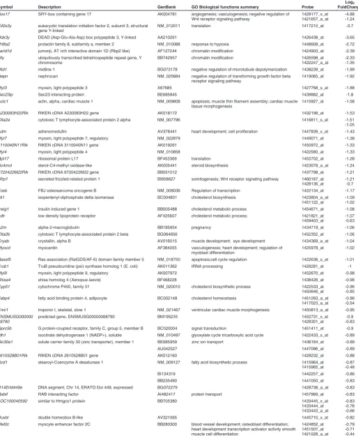
Symbol Description GenBank GO Biological functions summary Probe
Log2
FoldChange
Tsix X (inactive)-specific transcript, antisense BG806300 dosage compensation; 1436936_s_at 3.27
Afp alpha fetoprotein NM_007423 1416645_a_at
1416646_at 1436879_x_at
3.14 2.96 2.63 Slc2a2 solute carrier family 2 (facilitated glucose transporter),
member 2
NM_031197 carbohydrate transport 1449067_at 2.83
Tspan8 tetraspanin 8 BC025461 1424649_a_at 2.77
D7Ertd715e DNA segment, Chr 7, ERATO Doi 715, expressed BB314814 1436964_at 2.72
Timd2 T-cell immunoglobulin and mucin domain containing 2 BC028829 1418766_s_at 2.37
Apoc2 apolipoprotein C-II NM_009695 lipid transport 1418069_at 2.35
Xlr4b X-linked lymphocyte-regulated 4B NM_021365 1449347_a_at 2.35
Apoa1 apolipoprotein A-I NM_009692 endothelial cell proliferation; lipid transport 1438840_x_at 1419232_a_at Ccnb1ip1 cyclin B1 interacting protein 1 BG066504 blastocyst formation; apoptosis 1435998_at 2.19
Fgg fibrinogen, gamma polypeptide NM_133862 platelet activation 1416025_at 1.99
Lgals2 lectin, galactose-binding, soluble 2 NM_025622 1417079_s_at 1.89
Pdzk1 PDZ domain containing 1 AK006269 1431701_a_at 1.85
Cldn2 claudin 2 NM_016675 calcium-independent cell-cell adhesion 1417231_at 1.82 Cubn cubilin (intrinsic factor-cobalamin receptor) AF197159 receptor-mediated endocytosis 1426990_at
1452270_s_at
1.94 1.8 Rbp4 retinol binding protein 4, plasma U63146 eye development; spermatogenesis; lung development;
cardiac muscle development female genitalia morphogenesis
1426225_at 1.8
2410003J06Rik RIKEN cDNA 2410003J06 gene AK010362 1429701_at 1.75
Sfmbt2 Scm-like with four mbt domains 2 BM200222 regulation of transcription 1434353_at 1.72
Trap1a tumor rejection antigen P1A NM_011635 1460226_at 1.7
Apoa4 apolipoprotein A-IV BC010769 lipid transport; regulation of cholesterol absorption 1417761_at 1.68
Nrk Nik related kinase AK012873 protein amino acid phosphorylation 1450078_at
1450079_at
1.94 1.65
Ttr transthyretin BG141874 thyroid hormone generation 1455913_x_at
1459737_s_at
further shows that Sox17-null cells are less efficient in contribut-ing to the definitive endoderm. Loss of Sox17 in the endoderm also disrupts form-shaping activity in the anterior intestinal portal and the morphogenesis of the heart tube. Sox17-null mutant embryos also showed defective differentiation of the cardiogenic mesoderm. These findings are consistent with the definitive endoderm being a source of morphogenetic cues that patterns the tissues associated with the gut (Lewis and Tam, 2006),
Results
Identification of Sox17-dependent changes in gene expres-sion
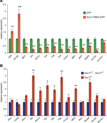
To identify Sox17-dependent gene expression we compared the expression profile of Sox17-null mutant and wild type embryos at the 4-5 somite stage, in order to capture the changes in the transcriptome before the Sox17-null phenotype becomes mor-phologically evident (Kanai-Azuma et al., 2002). This was ex-pected to minimize the confounding effects caused by secondary changes in gene expression due to aberrant development. Among the up-regulated transcripts in the mutant embryos, there is a preponderance of those encoding apolipoproteins and solute transporters (Table 1, Supplementary Table S1). This is reflected in the over-representation of genes defined by Gene Ontology (GO) biological function terms relating to homeostasis, absorp-tion, transport as well as cholesterol, steroid and lipid metabolism, catabolism and biosynthesis (Table 2A). These genes include
Apoa1, Apob, Apoc2, Apom and Slc2a2 that are known to be
expressed primarily in the extraembryonic visceral endoderm during post-implantation development. Other genes that are expressed in the visceral endoderm were also upregulated in mutant embryos (including Afp,Cubn,Dab,Rbp4, Rhox5 and Ttr;
Table 1, Supplementary Table S1). Some of these putative visceral endoderm genes (e.g.: Afp, Ttr and Apo family members) are also expressed in the liver later in development but they are normally only expressed at low levels in the foregut endoderm of wild-type early-somite stage embryos. The profiling results are consistent with the observation that definitive endoderm is re-placed by cells displaying phenotypes characteristic of the ex-traembryonic visceral endoderm in the embryonic gut region of the Sox17-null mutant (Kanai-Azuma et al., 2002). Alternatively, it may be that one function of Sox17 is to maintain liver-specific gene expression at a low level in the early definitive endoderm. Among the genes that were down-regulated in the mutant embryos (Table 3, Supplementary Table S1), there is an over-representation of genes involved in pathways for biosynthesis, metabolism and transport of lipids and cholesterol (Table 2B). Transcripts of genes involved in cholesterol biosynthesis (Cyp51, Idi1, Insig1 and Sc4mol) and transport (Ldlr) showed reduced expression in the absence of Sox17, and downregulation was validated by qRT-PCR (Fig. 1). Cholesterol is a key component of the Hedgehog signalling pathway that is essential for patterning the primitive gut tube (Harmon et al., 2002). Notably, the con-certed changes of the cholesterol pathway genes are consistent with the concept that cholesterogenic enzymes constitute a synexpression group in embryos and cultured fibroblasts (Iyer et
Amn amnionless NM_033603 excretion 1417920_at 1.6
Fgb fibrinogen, B beta polypeptide AK011118 platelet activation 1428079_at 1.53
Spp2 secreted phosphoprotein 2 NM_029269 bone remodeling 1418916_a_at 1.53
Xlr3a X-linked lymphocyte-regulated 3A NM_011726 1420357_s_at 1.5
Paip1 polyadenylate binding protein-interacting protein 1 BB381990 regulation of translation 1425521_att 1441955_s_a
1.49 1.61
Vil1 villin 1 NM_009509 cytoskeleton organization 1448837_at 1.41
Mfap3l microfibrillar-associated protein 3-like AV262974 1428804_at
1441481_at
1.46 0.92 Rab11fip5 RAB11 family interacting protein 5 (class I) BF682225 protein transport 1434314_s_at
1427405_s_at
1.4 0.62
Ctsh cathepsin H NM_007801 proteolysis 1443814_x_at
1418365_at
1.39 1.46
2610528J11Rik RIKEN cDNA 2610528J11 gene AK012175 1450947_at 1.39
Mbl2 mannose-binding lectin (protein C) 2 NM_010776 innate immune response 1418787_at 1.38 Npl N-acetylneuraminate pyruvate lyase BC022734 carbohydrate metabolic process 1424265_at 1.37 Slc7a9 solute carrier family 7 (cationic amino acid transporter, y+
system), member 9
NM_021291 amino acid transport 1448783_at 1.36
Tdh L-threonine dehydrogenase NM_021480 cellular metabolic process 1449064_at 1.36
Car7 carbonic anhydrase 7 BB193643 1443824_s_at 1.35
Soat2 sterol O-acyltransferase 2 BC025931 lipid; steroid; cholesterol metabolic process 1460722_at 1.35
Trf transferrin AF440692 iron ion transport 1425546_a_at 1.33
Fga fibrinogen, alpha polypeptide BC005467 blood coagulation 1424279_at 1.31
Rhox5 reproductive homeobox 5 BM210473 sperm motility; germ cell programmed cell death 1423429_at 1.3 Slco4c1 solute carrier organic anion transporter family, member
4C1
AV024403 spermatogenesis; organic anion transport c 1437870_at 1460616_at
1.27 1.29
Reep6 receptor accessory protein 6 AK002562 1430128_a_at 1.24
Aass aminoadipate-semialdehyde synthase BF687395 generation of precursor metabolites 1423523_at 1.22
Morc4 microrchidia 4 AV036158 1434436_at 1.18
Lgmn legumain NM_011175 negative regulation of growth 1448883_at 1.16
Adora2b adenosine A2b receptor BB709140 mast cell degranulation; relaxation of vascular smooth muscle
1434430_s_att 1434431_x_a
1.16 0.78 Cfi complement component factor i NM_007686 proteolysis immune response complement activation;
classical pathway
1418724_at 1.15
Gpr155 G protein-coupled receptor 155 BB762731 1452353_at 1.14
al., 1999; Laubner et al., 2003; Marijanovic et al., 2003). In the
Sox17-null embryo, expression of Ihh in the gut endoderm and
Ptch1 in the lateral plate mesoderm is down-regulated, indicating that loss of Sox17 function is associated with reduced Hedgehog signalling activity (Kanai-Azuma et al., 2002). Also of note is the reduced expression of Nepn (Table 3, Supplementary Table S1), which is normally expressed in the lateral midgut endoderm at this stage (Hou et al., 2007). Its down-regulation is consistent with the depletion of definitive endoderm in the mutant embryos.
Analysis of networks of transcriptional targets using Metacore (www.genego.com) revealed that 75 of the differentially ex-pressed genes were also potentially regulated by Hnf4, with either a known effect on transcription or evidence for binding of Hnf4 to
putative regulatory regions (Supplementary Fig. S1). Hnf4 is expressed in liver and visceral endoderm suggesting a change in cell composition of the gut or dys-regulation of genes associated with liver development. Sox17 may also be regulated by Hnf4 via a cross-acting Hnf4 and Sox17 transcriptional networks.
To identify changes in Sox17-dependent gene expression without the complicating factor of altered tissue contents due to the loss of gene function, we over-expressed Sox17 in HepG2 hepatoma cells, an endoderm cell line of epithelial morphology which originates from the liver tissue. HepG2 cells were trans-fected with either a GFP expressing construct or a bi-cistronic construct expressing Sox17 along with a GFP reporter. Trans-fected cells were sorted for GFP expression, and expression
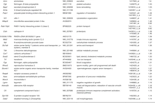
Fig. 2. Validation of the microarray results of differential gene expression in HepG2 cells over-expressing Sox17-IRES-GFP and in Sox17-/- embryos. Real-time quantitative RTPCR analysis was performed on cDNA generated from: (A) RNA samples of HepG2 cells transfected with GFP only or Sox17-IRES-GFP expression vector; and (B) RNA samples of 6-8 somite Sox17+/+ and
Sox17+/- or Sox17-/- embryos. Expression levels are shown relative to GFP only(A) or Sox17+/-/
Sox17+/-embryos (B). POLR2A was the reference gene for(A) and Gapd was the reference gene
for (B). Error bars indicate standard error of the mean. N=4 for Sox17+/+, Sox17+/-and N=3 for each
all others. **Significant difference at p<0.01 or * at p<0.05 by two-tailed t-test from GFP-only expressing cells or Sox17+/+/Sox17+/- embryos.
profiles analysed using Illumina human Sentrix-6 chips. Results were filtered for significantly different expression levels between GFP-only and Sox17-IRES-GFP transfected cells (Supplementary Table S2). The list of differentially expressed genes was compared to the list of genes that were differentially expressed between wild-type and Sox17-mutant embryos, and we identified 32 genes that were differentially expressed in both sets of data (Table 4). Of these, 21 showed concordance between the two data sets; that is, these genes were up-regulated in the HepG2 over-expressing Sox17 but down-regulated in Sox17-null embryos, and vice versa. These 21 genes are there-fore potential candidates for positive or negative regulation by Sox17. Results of qRT-PCR have validated in the majority of cases the differential expression of these genes in transfected HepG2 cells (Fig. 2A) and in embryos (Fig. 2B).
Two previous studies have addressed the question of Sox17-dependent gene expression either in mouse (Niakan et al., 2010) or human (Seguin et al., 2008) ES cells in which Sox17 was over-expressed. To gain additional insights into the genes that potentially act downstream of Sox17 we compared our embryo and HepG2 microarray data to these studies. Com-paring the list of genes that were up- or down-regulated in Sox17-null embryos with those that were changed in the op-posite direction in Sox17-over-express-ing mouse (Niakan et al., 2010) and hu-man (GEO accession GSE10809) ES cells revealed an overlap of 6 genes each in the mouse ES and embryo-human ES sets. These genes are in-volved in diverse processes including metabolism (folic acid, TCA, sorbitol, car-bohydrate), cytoskeletal arrangement,
CRYAB ABCC AFP APOC2 FGA FGB FGG FOXA3 MBL2 NRP1 REEP6 SLC2A2 SLCO4C1
Adm Cryab Abcc Afp Apoc2 Fga Fgb Fgg Foxa3 Mbl2 Nrp1
invagination) that forms the upper digestive tract and associated organs (Tremblay and Zaret, 2005). In wild type and heterozy-gous mutant embryos, formation of the anterior intestinal portal proceeded first by invagination, followed by anterior extension and narrowing of the foregut pocket (Fig. 4 A, B, 0, 5 and 23 hrs)
(A) Upregulated in Sox17 embryos
0.001 68.800 0 2 regulation of cholesterol absorption
0.001 68.800 0 2 cholesterol efflux
0.002 45.863 0 2 negative regulation of blood coagulation
0.002 45.863 0 2 lipid digestion
0.002 13.826 0 3 regeneration
0.002 13.826 0 3 vasodilation
0.003 34.394 0 2 lipoprotein catabolic process
0.003 11.519 0 3 cholesterol homeostasis
0.003 5.358 1 5 cholesterol metabolic process
0.004 10.912 0 3 blastocyst formation
0.004 10.912 0 3 zinc ion transport
0.005 4.796 1 5 negative regulation of multicellular organismal
process
0.006 3.879 2 6 di-, tri-valent inorganic cation homeostasis
0.007 3.808 2 6 cellular cation homeostasis
0.007 2.235 7 14 reproduction
0.007 19.649 0 2 axon regeneration
0.007 5.660 1 4 lipoprotein biosynthetic process
(B) Downregulated in Sox17 embryos
0.000 38.762 0 6 cholesterol biosynthetic process
0.000 6.713 2 14 cellular alcohol metabolic process
0.000 12.039 1 7 steroid biosynthetic process
0.000 3.184 5 15 oxidation reduction
0.001 11.490 0 4 cholesterol metabolic process
0.001 18.822 0 3 cholesterol homeostasis
0.002 14.724 0 3 lipid homeostasis
0.004 6.822 1 4 steroid metabolic process
0.004 4.325 1 6 lipid biosynthetic process
0.006 20.544 0 2 isoprenoid biosynthetic process
0.009 3.580 2 6 heart development
0.009 Infinite 0 1 S-adenosylmethionine biosynthetic process
0.009 Infinite 0 1 isoprenoid catabolic process
0.009 Infinite 0 1 low-density lipoprotein receptor metabolic
process
0.009 Infinite 0 1 regulation of low-density lipoprotein receptor
catabolic process
0.009 Infinite 0 1 muscle cell fate specification
0.009 Infinite 0 1 arachidonic acid secretion
were no genes common to both sets. Most likely, this reflects the different effects of Sox17 over-expression in the two cell models. In mouse ES cells, Sox17 appears to direct extraembryonic endoderm differentiation, whereas in human ES cells, Sox17 overexpression results in up-regulation of genes that are charac-teristic of definitive endoderm. Among the genes of the embryo-mouse ES and embryo-human ES sets, Adm, Fgb, Folr1 were also detected by ChIP from mouse ES or extraembryonic endo-derm stem (XEN) cells (Niakan et al., 2010) suggesting that Sox17 may act directly on these genes.
Comparison of genes with changed expression levels in Sox17 -mutant embryos and Sox17-overexpressing human ES cells (Seguin et al., 2008) revealed a set of genes that encode proteins that affect diverse processes including actin cytoskeleton organi-zation (CDC42, RDX), proliferation and differentiation (FST, GNG4, SERPINE2) and responses to signalling (ACSL4, ADM, FST, GNG4, STC2; Supplementary Table S4). The best candidates Sox17-regulated genes in the definitive endoderm are those that are differentially expressed in Sox17-null embryos and in the over-expression HepG2 cells and to which Sox17 binds. Com-parison with published data (Niakan et al., 2010), (Supplementary Table S5) revealed a set of six genes that fulfilled this criterion amongst those identified by ChIP of ES cells only (Fgb, Slco4c1), XEN cells only (Mbl2) or both ES and XEN cells (Adm, Nrp1,
Rbm4b).
Sox17-deficient cells contribute less efficiently to gut endo-derm
Our microarray studies revealed a higher representation of transcripts of genes that are characteristic of extraembryonic visceral endoderm in the Sox17-null embryo, suggesting a pre-ponderance of this type of endoderm in the early-somite stage
Sox17-null embryo. This is consistent with findings that Sox17
-/-embryo is depleted of definitive endoderm, which is partly re-placed by cells that display properties of extraembryonic visceral endoderm (Kanai-Azuma et al., 2002). To test directly whether
Sox17-deficient progenitor cells are compromised in their ability to populate the gut endoderm, the fates of cells of the anterior segment of the primitive streak (APS) of gastrula-stage embryos, where the progenitors of definitive endoderm are localised (Kinder
et al., 2000), were analysed by orthotopic transplantation to wild type recipient embryos (Fig. 3 A, H). Fluorescence imaging of the recipient embryos cultured for 29 hours revealed a widespread distribution of the EGFP and lacZ-expressing APS-derived cells from Sox17+/+ and Sox17+/- donor in the host embryos (Fig. 3 B,
B’, E, E’) and to tissues derived from different germ layers (Fig. 3 C, D, F, G). Although a similar average number of cells were generated by grafts of Sox17-/- APS cells, a smaller proportion of
graft-derived cells were found in the gut endoderm and the presomitic mesoderm (Fig. 3 I-N, Table 5). Instead, Sox17-/- cells
were found more frequently in the ectoderm of the posterior neural tube and the extraembryonic mesoderm (Table 5). Sox17-/- APS
cells therefore are impaired in their ability to contribute to gut endoderm.
Morphogenesis of the anterior intestinal portal is disrupted in Sox17-/- embryos
Cells of the anterior definitive endoderm cells are fated to become the endoderm of the anterior intestinal portal (the foregut
TABLE 2
Symbol Description GenBank GO Biological functions summary Probe
Log2
FoldChange
Sox17 SRY-box containing gene 17 AK004781 angiogenesis; vasculogenesis; negative regulation of Wnt receptor signaling pathway
1429177_x_at 1421657_a_at
-4.88 -1.24 Eif2s3y eukaryotic translation initiation factor 2, subunit 3, structural
gene Y-linked
NM_012011 translation 1417210_at -3.7
Ddx3y DEAD (Asp-Glu-Ala-Asp) box polypeptide 3, Y-linked AA210261 1426438_at -3.65
Prl8a2 prolactin family 8, subfamily a, member 2 NM_010088 response to hypoxia 1448608_at -2.72 Jarid1d jumonji, AT rich interactive domain 1D (Rbp2 like) AF127244 chromatin modification 1424903_at -2.39 Uty ubiquitously transcribed tetratricopeptide repeat gene, Y
chromosome
BB742957 chromatin modification 1426598_at
1422247_a_at
-2.33 -1.35 Mid1 midline 1 BG073178 negative regulation of microtubule depolymerization 1438239_at -1.99 Nepn nephrocan NM_025684 negative regulation of transforming growth factor beta
receptor signaling pathway
1419065_at -1.92
Myl3 myosin, light polypeptide 3 X67685 1427768_s_at -1.88
Sec23ip Sec23 interacting protein BE685845 1439882_at -1.8
Actc1 actin, alpha, cardiac muscle 1 NM_009608 apoptosis; muscle thin filament assembly; cardiac muscle tissue morphogenesis
1415927_at -1.58
A230083H22Rik RIKEN cDNA A230083H22 gene AK018172 1432198_at -1.53
Ctla2a cytotoxic T lymphocyte-associated protein 2 alpha NM_007796 1416811_s_at -1.51 -1.05
Adm adrenomedullin AV378441 heart development; cell proliferation 1447839_x_at -1.43
Myl7 myosin, light polypeptide 7, regulatory NM_022879 1449071_at -1.38
3110040N11Rik RIKEN cDNA 3110040N11 gene AK019261 1450972_at -1.33
Myl4 myosin, light polypeptide 4 NM_010858 1422580_at -1.33
Rpl17 ribosomal protein L17 BF453369 translation 1453752_at -1.28
Sc4mol sterol-C4-methyl oxidase-like AK005441 steroid biosynthesis 1423078_a_at -1.24
6720422M22Rik RIKEN cDNA 6720422M22 gene BB051012 1437798_at -1.21
Sfrp1 secreted frizzled-related protein 1 BI658627 somitogenesis; Wnt receptor signaling pathway 1460187_at 1428136_at
-1.21 -0.7 Fosb FBJ osteosarcoma oncogene B NM_008036 Regulation of transcription 1422134_at -1.17 Idi1 isopentenyl-diphosphate delta isomerase BC004801 cholesterol biosynthesis 1423804_a_at
1451122_at
-1.09 -1.02 Insig1 insulin induced gene 1 BB005488 cholesterol metabolic process 1454671_at -1.08 Ldlr low density lipoprotein receptor AF425607 cholesterol metabolic process; 1421821_at
1459403_at
-1.07 -0.63
A2m alpha-2-macroglobulin BB185854 pregnancy 1434719_at -1.06
Ctla2b cytotoxic T lymphocyte-associated protein 2 beta BG064656 1452352_at -1.06
Cryab crystallin, alpha B AV016515 muscle development; eye development 1434369_a_at -1.04 Myocd myocardin AF384055 vasculogenesis; heart development; regulation of
myoblast differentiation
1425978_at -1.02
Rassf5 Ras association (RalGDS/AF-6) domain family member 5 NM_018750 apoptosis;cell cycle regulation 1422638_s_at -1.01 Trub1 TruB pseudouridine (psi) synthase homolog 1 (E. coli) AK011362 tRNA processing 1428281_at -1
Myl9 myosin, light polypeptide 9, regulatory AK007972 1452670_at -0.98
Shisa4 shisa homolog 4 (Xenopus laevis) BF468228 1438426_at -0.98
Cyp51 cytochrome P450, family 51 NM_020010 cholesterol biosynthetic process 1422533_at 1450646_at
-0.96 -0.85 Fabp4 fatty acid binding protein 4, adipocyte BC002148 cholesterol homeostasis 1451263_a_at
1417023_a_at
-0.96 -0.54 Tnni1 troponin I, skeletal, slow 1 NM_021467 ventricular cardiac muscle morphogenesis 1450813_a_at -0.95 ENSMUSG000000
68790
predicted gene, ENSMUSG00000068790 BM195235 1452731_x_at
1428301_at
-0.9 -0.83 Gprc5b G protein-coupled receptor, family C, group 5, member B BC020004 signal transduction 1451411_at -0.9 Idh1 isocitrate dehydrogenase 1 (NADP+), soluble NM_010497 glyoxylate cycle tricarboxylic acid cycle 1422433_s_at -0.89 Slc30a1 solute carrier family 30 (zinc transporter), member 1 BE685959 zinc ion transport 1436164_at -0.89
AU042527 1447096_at -0.89
2610528B01Rik RIKEN cDNA 2610528B01 gene AK012160 1429232_at -0.88
Scd1 stearoyl-Coenzyme A desaturase 1 NM_009127 fatty acid biosynthetic process 1415964_at 1415965_at
-0.87 -0.48
BI134319 1442257_at -0.86
BB235490 1441050_at -0.83
D14Ertd449e DNA segment, Chr 14, ERATO Doi 449, expressed BG072279 1428738_a_at -0.83
Rabif RAB interacting factor AI482417 protein transport 1457969_at -0.83
LOC100040592 similar to Hmgcs1 protein BB705380 1433445_x_at
1433444_at 1433443_a_at
-0.83 -0.78 -0.66
Duxbl double homeobox B-like AV321065 1445710_x_at -0.82
Mef2c myocyte enhancer factor 2C BB280300 blood vessel development; osteoblast differentiation; heart development transcription activator activity smooth muscle cell differentiation
Symbol (mouse/human) Name Affymetrix probe ID
Sox17-/- mouse embryos
(Log2 fold change) Illumina target ID
Sox17-IRES-GFP HepG2 cells
(Log2 fold change)
Abcc2/ABCC2* ATP-binding cassette, sub-family C (CFTR/MRP), member 2
1450109_s_at 1 ILMN_9691 -0.6043
Adm/ADM* adrenomedullin 1447839_x_at -1.43 ILMN_29514 1.009719
Afp/AFP* alpha fetoprotein 1416646_at
1436879_x_at 1416645_a_at
3.14 2.96 2.63
ILMN_19039 -0.20155
Agpat5/AGPAT5 1-acylglycerol-3-phosphate O-acyltransferase 5 (lysophosphatidic acid acyltransferase, epsilon)
1453257_at -0.44 ILMN_9737 -0.72299
Apoc2/APOC2* apolipoprotein C-II 1418069_at 2.35 ILMN_26723 -0.35034
Bmp4/BMP4 bone morphogenetic protein 4 1422912_at 0.59 ILMN_27187 -0.46856
Cryab/CRYAB* crystallin, alpha B 1434369_a_at -1.04 ILMN_6827 1.910507
Eno3/ENO3 enolase 3, beta muscle 1417951_at -0.46 ILMN_16651 -0.36165
Eps8/EPS8 epidermal growth factor receptor pathway substrate 8 1422823_at 1422824_s_at
0.52 0.79
ILMN_17717 0.556275
F10/F10* coagulation factor X 1449305_at 0.65 ILMN_138620 -0.71965
Fga/FGA* fibrinogen, alpha polypeptide 1424279_at 1.31 ILMN_11182 -0.47527
Fgb/FGB* fibrinogen, B beta polypeptide 1428079_at 1.53 ILMN_13882 -0.32784
Fgg/FGG* fibrinogen, gamma polypeptide 1416025_at 1.99 ILMN_26176 -0.20352
Foxa3/FOXA3* forkhead box A3 1431900_a_at 0.42 ILMN_22171 -1.193
Ldlr/LDLR* low density lipoprotein receptor 1421821_at 1459403_at
-1.07 -0.63
ILMN_10126 0.282144
Lgmn/LGMN legumain 1448883_at 1.16 ILMN_20242 0.620039
Lmna/LMNA* lamin A 1421654_a_at 0.39 ILMN_12442 -0.38805
Lrp8/LRP8 low density lipoprotein receptor-related protein 8, apolipoprotein e receptor
1440882_at 1442347_at
-0.54 -0.46
ILMN_2030 -0.42656
Mbl2/MBL2* mannose-binding lectin (protein C) 2 1418787_at 1.38 ILMN_6942 -1.53863
Nrp1/NRP1* neuropilin 1 1457198_at
1448944_at
0.53 0.54
ILMN_17483 -1.10432
Nsdhl/NSDHL NAD(P) dependent steroid dehydrogenase-like 1416222_at -0.63 ILMN_13529 -0.42729
Rbm4b/RBM4B* RNA binding motif protein 4B 1430032_at 0.41 ILMN_29996 -0.28508
Reep6/REEP6* receptor accessory protein 6 1430128_a_at 1.24 ILMN_15192 -0.64812
Sc4mol/SC4MOL sterol-C4-methyl oxidase-like 1423078_a_at -1.24 ILMN_2770 -0.41272
Sec23ip/SEC23IP Sec23 interacting protein 1439882_at -1.8 ILMN_7522 -0.4024
Slc16a10/SLC16A10 solute carrier family 16 (monocarboxylic acid transporters), member 10
1434592_at 0.31 ILMN_10556 0.425748
Slc2a2/SLC2A2* solute carrier family 2 (facilitated glucose transporter), member 2
1449067_at 2.83 ILMN_28285 -1.20611
Slc30a1/SLC30A1* solute carrier family 30 (zinc transporter), member 1 1436164_at -0.89 ILMN_5933 0.439786 Slc39a5/SLC39A5* solute carrier family 39 (metal ion transporter), member 5 1429523_a_at 0.78 ILMN_14803 -0.64595 Slco4c1/SLCO4C1* solute carrier organic anion transporter family, member 4C1 1460616_at
1437870_at
1.29 1.27
ILMN_3183 -0.37455
Slu7/SLU7 SLU7 splicing factor homolog (S. cerevisiae) 1425488_at 0.46 ILMN_12938 0.927178 Zcchc14/ZCCHC14* zinc finger, CCHC domain containing 14 1418170_a_at 0.6 ILMN_138708 -0.63098
TABLE 4
GENES THAT WERE SIGNIFICANTLY DIFFERENTIALLY EXPRESSED IN SOX17-/- EMBRYOS COMPARED TO WILD TYPE EMBRYOS AND IN SOX17-IRES-GFP TRANSFECTED HEPG2 CELLS COMPARED WITH GFP TRANSFECTED HEPG2 CELLS
Asterisk indicates genes that were up-regulated when Sox17 was over-expressed and down-regulated in its absence, and vice versa
which led to the medial convergence and longitudinal extension of the lateral endoderm cell populations. Subsequently, these cells were found primarily in the lateral wall of the anterior intestinal portal (Fig 4 A, B left and right). A unique morphogenetic feature of the anterior intestinal portal is the asymmetric displacement of the endoderm cells in the plane of the epithelium (Franklin et al., 2008). This was observed in the wild type and Sox17+/- mutant
embryos in which cells from the left side of the portal were found on the floor (ventral wall) whereas those from the right side were found in the roof (dorsal wall) (Fig. 4 A, B left and right; Table 6). The Sox17-/- embryos characteristically showed flattened neural
folds and a shallow depression in the prospective foregut area at the initial phase of portal formation (Fig. 4C, 0hr). The lateral regions of the portal were wider apart, resulting in a short portal and with a broad entrance (Fig. 4C, 23hr). In addition, the lateral
cell populations were mostly confined to the lateral walls and infrequently found asymmetrically on the dorsal or ventral wall of the portal (Fig. 4C, left and right; Table 6). Morphogenesis of the anterior intestinal portal in Sox17-/- embryos is therefore defective
with less convergence-extension and planar rotation of the ante-rior definitive endoderm.
Loss of Sox17 leads to down-regulation of cardiac genes and abnormal morphogenesis of the heart tube
Among the genes that were down-regulated in Sox17-null embryos, there was an over-representation of genes associated with heart development (Table 2). In particular, Myocd, a tran-scription factor expressed in the heart tube (Wang et al., 2001), is down-regulated significantly in early-somite stage Sox17-/-
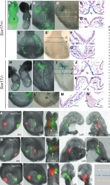
Fig. 4. Formation of the anterior intestinal portal. Distribution of contra-lateral cell population of the anterior definitive endoderm in the anterior intestinal portal at different time points (0, 5 and 23 hours) dur-ing its formation in (A)Sox17+/+, (B)
Sox17+/- and (C)Sox17-/-embryos.
Cells were labeled with DiO (green) and DiI (red) carbocyanine dye. Embryos were imaged intact
dur-ing in vitro culture and were imaged
after bisected into left and right halves at the end of culture to re-veal the localization of the labelled cells. Arrows (see legend for colour code) point to the ventral lip, ventral wall and dorsal wall of the anterior intestinal portal.
Fig. 3. Contribution of graft-derived cells in the recipient embryos. Trans-plantation of cells from the
EGFP-express-ing (A) Sox17+/- donor (d) embryo and (H)
Sox17-/- donor embryos to the anterior
region of the primitive streak of the wild type mid-streak stage recipient (r) em-bryo followed by visualization of the graft-derived cells by fluorescence imaging (for EGFP: B, E, I, L) and X-gal staining (for lacZ: B’ E’, I’, L’) in (B,B’, I, I’) the anterior and (E, E’, L, L’) posterior regions of the recipient embryo after 24 hours of in vitro development. Histology was performed on recipient embryos (in the same series of the transplantation but not necessarily of the specimens shown in B’, E’, I’ and L’). For orientation, planes of the histo-logical sections are shown in B’, E’, I’ and L’. Graft-derived ( lacZ-expressing, visu-alized by blue X-gal staining reaction) (C, D, F, G) Sox17+/- cells and (J, K, M, N)
Sox17+/- cells populate the endoderm (en)
of the foregut portal (fg), and the cranial mesoderm (cm) underneath the neural plate (np) in the anterior region; and the presomitic mesoderm (pm) and lateral plate mesoderm (lm) in the posterior re-gion of the embryo. Scale bars, 100 m.
B
Sox17+/-0hr 5hr 23hr Left Right
0hr 5hr 23hr Sox17+/+
Left Right
0hr 5hr 23hr Left Right
Sox17-/-ventral lip
ventral wall
dorsal wall
A
B
C
Sox17
+/-Sox17
-/-d
r
F
fg encm np
en
cm
pm
pm
en fg
fg np
cm
pm
C
D
G
F
M
N
J
K
en
lm
A
B
B'
C
E
E'
F
H
I
I'
J
L
L'
M
N
K
G
Examination the heart morphology of E8.5 Sox17-null em-bryos revealed abnormal heart tube phenotypes ranging from single heart tube that had not initiated looping (1/5) (Fig. 6B), incomplete union of the heart tube (1/5) (Fig. 6C) and the forma-tion of two completely separated heart tubes (cardia bifida, 3/5) (Fig. 6D). The heart of Sox17 +/- embryos (not shown) was
indistinguishable from that of the wild type (Fig. 6A). In the Sox17
mutant embryos the anterior intestinal portal was irregularly shaped and the floor crinkled extensively (Fig. 6 F, G, J), in contrast to the uniform contour in the wild type embryo (Fig. 6E). In Sox17-/- mutant embryo with incomplete fusion or separated
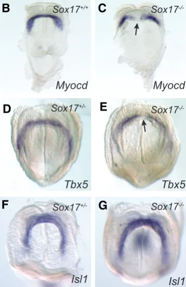
markers of the primary heart field (Myocd and Tbx5) and the secondary heart field (Isl1) are expressed in the cardiogenic mesoderm of Sox17-/- embryos at the early head-fold stage (Fig.
5 B, D, F), suggesting that the heart precursors were present in the embryo and that the initial distribution of the cardiogenic mesoderm to the heart field is not affected by the loss of Sox17
in the endoderm. However, Myocd and Tbx5 expression was absent from the midline tissues in the primary heart field of the
Sox17-/- embryos (3/4), resulting in a hiatus in the cardiac
cres-cent where the lateral population of the heart progenitors failed to unite medially (Fig. 5 C-G).
B
C
A
Fig. 5. Expression of heart genes in Sox17
-/-early head fold stage (pre-somite, E8.25) em-bryos revealed by whole mount in situ hybrid-ization.(A) Down-regulation of Myocd in Sox17
-/-embryos revealed by real time quantitative RT-PCR. Error bars indicate standard error of the mean. N=3 for each assay (* P<0.05 by t test). Whole mount in situ hybridization of embryos showing the expression pattern of (B,C)Myocd and (D,E)Tbx5 in the cardiac crescent of (B)Sox17+/+, (D)Sox17+/ - and (C,E) Sox17-/- embryos. In Sox17-null
em-bryos, expression of Tbx5 and Myocd is absent in the midline region of the cardiac crescent (arrows). (F, G) Expression of Isl1 is intact in the tissues in the secondary heart field of the Sox17-/- embryo.
heart tubes, the more caudal seg-ment of the portal was open, re-sulting a wide ventral gap (Fig. 6 K, L), which in the wild type em-bryo was closed by this stage (Fig. 6I). The defects of heart tube for-mation in the Sox17-null mutant embryos are therefore accompa-nied by the abnormal morphogen-esis of the anterior intestinal por-tal in addition to the defects in cardiac mesoderm differentiation.
Discussion
Sox17 downstream genes and molecular pathways
Microarray analysis of wild-type and Sox17-/- embryos revealed changes in expression of genes that are associated primarily with the definitive endoderm (Nepn,
Foxa3, and Hnf4a), as well as those that are expressed in both the extraembryonic visceral en-doderm and the definitive endo-derm (e.g.: Apolipoprotein genes,
Ttr, Afp). Furthermore, there are Donor genotypes
(no. of donor embryos)
Sox17+/+or Sox17
+/-Extraembryonic mesoderm* 716 20.8% 1207 30.4%
TOTAL 3587 3965
Average 239.1 264.3
TABLE 5
CONTRIBUTION OF THE ANTERIOR PRIMITIVE STREAK CELLS TO THE GERM LAYER DERIVATIVES DURING GASTRULATION
AND EARLY ORGANOGENESIS
*Significant difference between genotypes by Chi-squared test at P<0.05.
Sox17 Genotype of embryo
Distribution of labeled cells in the anterior intestinal portal+ No of
embryos
Left side Right side Ventral Lateral Dorsal Ventral Lateral Dorsal
+/+ or +/- 2
PATTERNS OF DISTRIBUTION OF CELLS ORIGINATING FROM THE LATERAL REGIONS OF THE ANTERIOR DEFINITIVE
ENDODERM IN THE ANTERIOR INTESTINAL PORTAL
DiO labeled cells; DiI labeled cells. + Three out of five null-mutant embryos showed
localization of the labeled cells only to the lateral wall of the anterior intestinal portal. Only 1/5 null-mutant as contrasted to 9/10 wild type or heterozygous embryos showed proper asymmetrical distribution of labeled cells: significant difference at P<0.01 by «2 test. * One each of null-mutant and wild type/heterozygous showed atypical localization of labeled cells from right side to the ventral wall (floor) instead of dorsal wall (roof).
Fig. 6. Heart phenotype of E8.5 (7-8 somites) Sox17-/- embryos. Wild type embryo shows a looping heart tube (A), whereas Sox17-/- embryos display (B) un-looped heart tube, (C) partially united and (D)
separate heart tube. Heart tissues are highlighted by Myocd expression by whole mount in situ hybridization (A-D) and in histological preparations (E-L, planes of sectioning indicated by arrowheads in A-D). The foregut of Sox17-/- embryo has an abnormal shape (F, arrow) and an expanded corrugated
floor (J, arrow) and opens ventrally (K, L: asterisk) in the posterior region of the foregut of embryos with bifid heart tube. ht: heart tube, fg: foregut.
L
changes in the expression of genes (e.g. heart development genes, cholesterol pathway genes) that are not associated with the endoderm.
To identify transcripts that are candidates for direct depen-dence on Sox17 expression, we over-expressed Sox17 in HepG2 cells. As the liver is derived from the definitive endoderm, we reasoned that HepG2 cells would provide a cellular environment comparable to the gut endoderm of the mouse embryo, where
Sox17 is normally expressed. Comparison of the results of the two array screens revealed a set of 21 genes whose changes in expression are consistent with either negative or positive regula-tion by Sox17. Analysis of publicly available gene expression data (http://biogps.gnf.org) revealed that 9 of these genes were specifi-cally or predominantly expressed in adult and/or fetal liver (Afp,
Apoc2, F10, Fga, Fgb, Fgg, Mbl2, Reep6 and Slc2a2). All of these genes were found in our micrarray study to be downregulated in the
Sox17-overexpressing HepG2 cells and upregulated in Sox17
-/-embryos. These data, along with the significant overlap with Hnf4a regulated genes, suggest the possibility that Sox17 plays a role in repression of liver-specific gene expression. This is consistent with a role for Sox17 in maintaining definitive endo-derm in the progenitor state, and the reported requirement for Sox17 in the delineation of specific foregut endoderm-derived lineages, including the gallbladder and bile duct (Spence et al., 2009; Uemura et al., 2010).
By comparing our mouse embryo and HepG2 micrarray ex-pression data with ChIP data from mouse ES or XEN cells, we have identified a shortlist of six genes (Adm, Fgb, Mbl2, Nrp1,
Rbm4b, Slco4c1) for which Sox17 influences their expression
and there is evidence for binding of Sox17 protein to regulatory regions. These genes are diverse in function and expression and represent strong candidates for Sox17 transcriptional target genes.
Loss of Sox17 function impairs the allocation of endoderm cells
Although the Sox17-mutant phenotype indicates a role in the development of the definitive endoderm, several lines of evi-dence also point to a requirement of Sox17 in the differentiation of extraembryonic endoderm. In mouse ESC, enforced expres-sion ofnSox17 promotes the differentiation of extraembryonic endoderm, whereas Sox17-null ES cells maintain the expression of pluripotency genes and fail to differentiate into extraembryonic endoderm. Stem cells with extraembryonic endoderm properties (XEN cells) cannot be generated from Sox17-null embryos (Niakan
et al., 2010). In contrast, in human ESC, progenitors with stable characteristics of definitive endoderm can be generated by con-stitutive expression of Sox17, whereas expression of another Group F Sox factor, Sox7, produces cells with more restricted potential reminiscent of the extraembryonic endoderm (Seguin et al., 2008). Transcriptional profiling of SOX17 and SOX7
overexpressing human ESCs revealed that the sets of genes that were specifically upregulated by either SOX17 or SOX7 were far greater than the set of genes that was upregulated by both, indicating unique, tissue-specific roles for the two transcription factors. In agreement with these findings, our microarray data do not support a role for Sox17 in promoting extraembryonic endo-derm differentiation in mouse embryos. Instead, our data show up-regulation of extraembryonic gene expression in Sox17-null embryos, consistent with apparent replacement of definitive endoderm with visceral endoderm-like cells (Kanai-Azuma
et al., 2002).
Sox17-null ES cells are unable to contribute significantly to the defini-tive endoderm of the gut in the pres-ence of wild type cells in the chimera (Kanai-Azuma et al., 2002). Cell transplantation experiments per-formed in the present study further show that cells of the Sox17-/-
the place of the gut endoderm cells. This may be the result of not only the prevalence of apoptosis and reduced proliferative activity but also of the compromised ability of the mutant cells to contrib-ute to the definitive endoderm.
Morphogenesis of the embryonic foregut and the heart tube is dependent on Sox17 function
Development of the embryonic foregut is initiated by the formation of the anterior intestinal portal beginning with the invagination of the anterior definitive endoderm underneath the head folds, accompanied by the longitudinal extension of the medial regions and the convergence of the lateral parts of the endoderm layer (Franklin et al., 2008). An intriguing feature of the morphogenesis of the portal is the pattern of movement of the contra-lateral endoderm populations of the prospective foregut. While these populations are undergoing longitudinal extension, they are also displaced asymmetrically along the wall of the portal: cells on the right populate the right and dorsal (roof) sides while cells on the left populate the left and ventral (floor) of the portal. The asymmetrical distribution of contra-lateral endoderm cells is accomplished by the directional relocation of cells within the epithelium that lines the portal and not due to the “rotation” of the foregut. However, the asymmetry of cell displacement corre-sponds with the laterality of subsequent rotation (left to ventral and right to dorsal) of the lower foregut. Sox17-null embryos form an anterior intestinal portal which is shorter and wider than the wild type counterpart, with excess folding of the floor and a split posterior ventral lip of the portal. Tracking the morphogenetic movement of anterior definitive endoderm in the null mutant embryo further revealed a much reduced longitudinal extension and medial convergence of the lateral endoderm, culminating in the distribution of these cells to wide lateral domain in the portal and very restricted rotational displacement.
Heart defects were not reported in the initial analysis of the
Sox17-null phenotype (Kanai-Azuma et al., 2002). A subsequent study revealed that the heart of the mutant embryo displayed defects in the looping of the heart tube (Sakamoto et al., 2007). Our study further shows that Sox17-null mutant embryos display additional and more severe defects of the heart tube: lack of union of the cardiac precursors from the contra-lateral part of the cardiac crescent and bifid heart tubes. One possible cause of the differ-ence in the manifestation of heart defects in our study and that of Sakamoto et al. (2007) is the difference in background. Whereas in the latter study, the mice were maintained on a mixed 129/Sv X C57BL/6 background, our mice have been maintained on a predominantly 129/Sv background. A precedent for the effect of genetic background on the heart defects is seen in Fibronectin 1
mutants which display less severe heart phenotype with an increasing contribution of C57/BL6 over the 129/Sv background (George et al., 1997).
The formation of a wide portal with a ventral hiatus and the lack of asymmetrical displacement of the cells in the portal could be the morphogenetic factors underlying the incomplete fusion and lack of looping of the heart tube. Similar cardia bifida phenotypes found in other mouse and Zebrafish mutants are also accompa-nied by defects in foregut development, such as absence or reduction of foregut endoderm and incomplete closure of the foregut pocket (Alexander et al., 1999; Kuo et al., 1997; Li et al., 2004; Molkentin et al., 1997; Reiter et al., 1999; Roebroek et al.,
1998). It is not known whether the heart tube defects are due to the abnormal morphogenesis of the anterior intestinal portal and whether, in a broader context, the laterality in tissue movement in the anterior intestinal portal may predispose the direction of heart looping and the left-right asymmetry of the heart and the digestive tract.
Sox17 activity in the endoderm influences cardiac cell differ-entiation
In the mouse embryo, the cardiac cells are derived from two sources of progenitor cells: the primary and the secondary heart field. The primary heart field, consisting of Tbx5- and Myocd -expressing cells contributes to the left ventricle and atria. The secondary heart field, populating by Isl1-expressing cells, contrib-utes to the outflow tract, right ventricle and atria and the pharyngeal mesoderm (Buckingham et al., 2005). Expression profiling analy-sis reveals that loss of Sox17 is associated with the down-regulation of Myocd expression. The heart-specific transcription factors Myocd, Tbx5 and Isl1 are expressed in the cardiac cres-cent. However, Myocd and Tbx5 are specifically down-regulated in the tissues at the vertex of the crescent. The domain where Myocd
and Tbx5 expression is lacking corresponds to the region of defective union of the lateral cardiac progenitors in the midline. The cardia bifida phenotype in the absence of Sox17 is therefore most likely to be initiated by a failure in the fusion of the cardiac crescent at the midline leading to the formation of two separate lateral heart tubes. Our findings therefore suggest that Sox17 activity in the definitive endoderm is required for the induction Myocd-expressing cardiogenic mesoderm in a specific population of cells in the primary heart field that form the atria and part of the ventricle.
Sox17 has been shown to act in a non-cell autonomous manner in eliciting cardiac differentiation in mouse ES cells. Inhibition of
Sox17 in mouse ES cells leads to a suppression of cardiac differentiation (Liu et al., 2007). The expression of Sox17 in the foregut endoderm could therefore be required for the induction of heart-specific genes in the embryo. This may occur either by direct regulation of genes encoding signalling molecules or antagonists by Sox17, or indirectly with Sox17 playing a role in the maintenance or differentiation of the definitive endoderm, which then expresses the morphogenetic signals for heart development. Consistent with the non-cell autonomous role of Sox17 in heart development, over-expressed Sox17 in the pluripotent fibroblast cell line C3H10T1/2 does not induce Myocd expression (our unpublished data). On the other hand, co-culture of C3H10T1/2 with HepG2 cells does induce
Myocd expression in the C3H10T1/2 cells, suggesting that the endoderm derived HepG2 cells are secreting a cardiac-inducing factor into the media. This is consistent with Sox17 playing an indirect role in heart induction. However, transfection of a Sox17
expression construct into the HepG2 cells does not result in increased Myocd expression in co-cultured C3H10T1/2 cells (F. Kruiswijk, DAFL and PPLT unpublished data). In this case, the HepG2 cells are already primed to express the cardiac-inducing factor independently of Sox17 expression.
Materials and Methods
Mouse strains and genotyping
generate wild type embryos, and heterozygous and homozygous mutant embryos for phenotypic analysis, expression studies and embryological experimentation. For testing the lineage potential of Sox17-/- embryonic
cells in the cell grafting experiment, Sox17+/- mice were crossed with
transgenic mice (H253) that co-express two transgenes (Hmgcr-nls-lacZ and pCAGG-EGFP) widely in embryonic tissues (Hadjantonakis et al., 1998; Kinder et al., 2001; Tam and Tan, 1992) to derive a line of Sox17+/-;H253 mice
(SGX). The SGX mice were inter-crossed to generate embryos of different
Sox17 genotypes that express the transgenic reporters, so that the cells harvested from these embryos can be tracked during the course of the cell grafting experiment.
Microarray analyses
Total RNA from embryonic specimens was isolated using the RNeasy Micro Kit (QIAGEN) according to the manufacturer’s instructions. RNA was isolated from two pairs of somite-number matched (4-5 somites) Sox17
-/-mutant and wild type embryos. The quality and quantity of the isolated RNAs were assessed on an Agilent Bioanalyser. RNA probes were amplified and labelled using the Affymetrix two-cycle process. Each RNA probe was hybridised to two or three Affymetrix GeneChip mouse genome 430 2.0 arrays, comprising all the known and predicted genes in the mouse genome. The arrays were read with an Affymetrix GS3000 scanner and raw data imported into Chipster v 1.4.4 (http://chipster.csc.fi) for normalization (RMA) and statistical analysis by linear modelling using LIMMA (signifi-cance at P<0.05), taking into account biological and technical replicates. Analysis of gene ontology was carried out within Chipster using a hypergeo-metric test for over-representation of terms.
HepG2 (Human hepatocellular carcinoma) cells were maintained in DMEM + 10 % FCS. Prior to transfection, approximately 2 x 10⁶ cells were
seeded into T75-flasks. One day later, the HepG2 cultures were transiently transfected with 8g of pCMV-Sox17-EGFP or CMV-EGFP control and 24
l FuGENE6 (Roche). After 48 hours the EGFP expressing cells were isolated using the flow cytometry (FACSVantage cell sorter). Total RNA was extracted from the cell pellets using the RNeasy Mini Kit (QIAGEN) according to the manufacturer’s protocol. RNA samples from three inde-pendent groups of transfected cells were amplified, labelled and hybridised to Illumina Sentrix® Human-6 v2 Expression BeadChips that cover the human genome. The arrays were analysed with the Illumina BeadStation 500 reader. Beadstudio was used to normalize the data (cubic spline) and identify transcripts whose expression level had significantly changed (Illumina Custom test, p<0.01).
Quantitative real-time RT-PCR
RNA samples from Sox17 mutant and wild type embryos (4-5 somites) were reverse transcribed into cDNA using the SuperScript III First-Strand Synthesis System (Invitrogen). Real-time quantitative RT-PCR was per-formed on Rotor-Gene thermocyclers (Corbett Research) using QuantiTect SYBR Green (QIAGEN) or Platinum Taq (Invitrogen). Details of PCR primers are given in Supplementary Tables S6 and S7.
Generation of riboprobes
To generate a plasmid from which to make a Myocd whole mount in situ
hybridisation probe, a fragment was amplified from mouse E8.5 cDNA using Pfu DNA polymerase (Roche). Primer sequences for generating the 750bp probe were forward: 5’-TGGGCTAGACTCTGAGAAGGAC -3’ and reverse: 5’- TGGGTGATATCTGAAACTGCTG-3’. The amplified fragments were subsequently cloned into pGEM-T (Promega) downstream of the T7 promoter. The Tbx5 antisense probe has been described previously (Chapman et al., 1996). The Isl1 probe was kindly provided by Gerhard Przemeck (Helmholtz Zentrum München). For the generation of antisense RNA probes from linearized cDNA clones the DIG RNA labelling kit (Roche) has been used according to the manufacturer’s instructions.
Whole mount in situ hybridisation
For whole mount in situ hybridisations embryos were fixed in 4% PFA
in PBS over night, dehydrated in methanol and stored at -20ºC until usage. Whole mount in situ hybridisations were performed as described previously (Chapman et al., 1996; Davidson et al., 1999). In brief, embryos were rehydrated in PBT (PBS plus 0.1% Tween-20), bleached with 6% hydrogen peroxide in PBT for 30min, washed in PBT, refixed in 4% PFA in PBS containing 0.2% glutaraldehyde for 20min and washed in PBT. Hybridisations with DIG-labelled antisense probes (1:200) were performed in hybridisation buffer (50% formamide, 5 x SSC pH4.5, 1% SDS, 50g/ml Heparin) at 70ºC over night. After two washes each in 2 x SSC, 0.1% SDS and 0.2 x SSC, 0.1% SDS at 70ºC, embryos were washed 3 times in MABT (100mM maleic acid pH 7.5, 150mM NaCl, 1% Tween-20, 2mM Levamisole). Embryos were blocked in 2% blocking reagent (Roche) in MABT for 1h, in 2% blocking reagent with 20% FCS in MABT for 1-3 h and incubated with alkaline phosphatase coupled anti-DIG antibody (Roche) (1:2000) in 2% blocking reagent with 20% FCS in MABT at 4ºC over night. After extensive washing in MABT for 1-3 days the embryos were washed twice in 2mM Levamisole, 0.1% Tween-20. BM purple AP substrate (Roche) was used for subsequent alkaline phosphatase staining. The embryos were kept in 4% PFA. After whole mount in situ hybridisations the embryos were dehydrated, embedded in paraffin wax and sectioned at 7m. The sections were counterstained with nuclear fast red and mounted using Canada balsam.
X-Gal staining of embryos
Embryos were washed twice in PBS, fixed in 4% PFA in PBS for 5min and washed again twice in PBS. In order to detect lacZ the embryos were stained in X-gal [5-bromo-5-chloro-3-indoyl--D-galactopyranoside] stain-ing solution (1mg/ml X-gal, 5mM K3Fe(CN)6, 5mM K4Fe(CN)6, 2mM MgCl2, 0.01% Tween-20, 0.2% PFA in PBS) for 40 min at 37ºC. After-wards the embryos were rinsed twice in PBS and fixed in 4% PFA in PBS over night. The stained embryos were processed for wax histology and the relative contribution of the graft-derived cells to various types of germ layer derivatives evaluated by scoring the number of X-gal stained cells in serial sections of the recipient embryos.
Cell transplantation
Embryos from SGX mating were collected at the E7.0 mid-streak stage. GFP positive embryos were selected as donors for transplantation. Tissue fragments from the anterior primitive streak of these embryos were isolated and further dissociated into small clumps of cells. The cell clumps were then transplanted to the anterior primitive streak of stage-matched wild type (ARC/s) embryos using a Leica micromanipulator. The remain-ing part of the embryo was collected for PCR genotypremain-ing. After graftremain-ing, the recipient embryos were checked by fluorescence microscopy for the correct placement of the transplanted cells. Embryos were cultured for 24-28h in a medium made up of 75% heat-inactivated rat serum and 25% Dulbecco’s modified Eagle medium at 37C in glass bottles rotating at 30 RPM with a continuously replenished gas phase of 5% CO2, 20% O2 and 75%N2 (Sturm and Tam, 1993). At the end of culture, recipient embryos were imaged by fluorescence photomicroscopy to visualise and record the distribution of the expressing cells. Embryos containing EGFP-expressing graft-derived cells were then fixed in 4 % PFA and stained in X-gal staining solution to detect lacZ expression.
Dye labelling of the foregut endoderm
E7.0 mid-streak-stage embryos were harvested from pregnant mice generated by inter-crossing Sox17+/- mice. Embryos were dissected from
the decidua and the Reichert’s membrane was removed. Embryos were selected by somite numbers to ensure stage-matching (1-3 somites) between groups of different Sox17 genotypes. They were kept in 100% rat serum in a 5%CO2 incubator at 37C prior to micro-manipulation. For
relative spatial distribution of different cell populations and to track their morphogenetic movement during foregut morphogenesis. Following dye labelling, embryos were cultured for 12 hours under the same conditions as the cell grafting studies. Labelled embryos were imaged by fluores-cence microscopy within 1 hour after labelling to ascertain the site of labelling. The embryos were re-imaged at the end of a 12-hour culture period to visualise the distribution of labelled cells in the embryonic gut. Photographs were taken using a Leica MZ16 microscope with a SPOT Advanced digital camera and fluorescent and bright field images were digitally edited and merged with the SPOT 4.0 software and Adobe Photoshop 7.0. The yolk sac of the embryo was collected to prepare the DNA for genotyping.
Acknowledgements
We acknowledge the support of a Deutscher Akademischer Austauschdienst Fellowship (SP) and studentship from University of Applied Sciences, Mannheim (GT). DL is a Kimberly-Clark Research Fellow and PT is a NMHRC Senior Principal Research Fellow. This work is supported by NHMRC project grant 321704. Our thanks also to Mr. James Fairfax.
References
ALEXANDER, J., ROTHENBERG, M., HENRY, G.L. and STAINIER, D.Y. (1999). casanova plays an early and essential role in endoderm formation in zebrafish.
Dev Biol 215: 343-357.
BILDSOE, H., FRANKLIN, V. and TAM, P.P.L. (2007). Fate-Mapping Technique: Using Carbocyanine Dyes for Vital Labeling of Cells in Gastrula-Stage Mouse Embryos Cultured In vitro. Cold Spring Harb Protoc 2007: db.
BUCKINGHAM, M., MEILHAC, S. and ZAFFRAN, S. (2005). Building the mamma-lian heart from two sources of myocardial cells. Nat Rev Genet 6: 826-835. CHAPMAN, D.L., GARVEY, N., HANCOCK, S., ALEXIOU, M., AGULNIK, S.I.,
GIBSON-BROWN, J.J., CEBRA-THOMAS, J., BOLLAG, R.J., SILVER, L.M. and PAPAIOANNOU, V.E. (1996). Expression of the T-box family genes, Tbx1-Tbx5, during early mouse development. Dev Dyn 206: 379-390.
CLEMENTS, D., CAMELEYRE, I. and WOODLAND, H.R. (2003). Redundant early and overlapping larval roles of Xsox17 subgroup genes in Xenopus endoderm development. Mech Dev 120: 337-348.
DAVIDSON, B.P., KINDER, S.J., STEINER, K., SCHOENWOLF, G.C. and TAM, P.P. (1999). Impact of node ablation on the morphogenesis of the body axis and the lateral asymmetry of the mouse embryo during early organogenesis. Dev Biol 211: 11-26.
FRANKLIN, V., KHOO, P.L., BILDSOE, H., WONG, N., LEWIS, S. and TAM, P.P. (2008). Regionalisation of the endoderm progenitors and morphogenesis of the gut portals of the mouse embryo. Mech Dev 125: 587-600.
GEORGE, E.L., BALDWIN, H.S. and HYNES, R.O. (1997). Fibronectins are essential for heart and blood vessel morphogenesis but are dispensable for initial specification of precursor cells. Blood 90: 3073-3081.
HADJANTONAKIS, A.K., GERTSENSTEIN, M., IKAWA, M., OKABE, M. and NAGY, A. (1998). Generating green fluorescent mice by germline transmission of green fluorescent ES cells. Mech Dev 76: 79-90.
HARMON, E.B., KO, A.H. and KIM, S.K. (2002). Hedgehog signaling in gastrointes-tinal development and disease. Curr Mol Med 2: 67-82.
HART, A.H., HARTLEY, L., SOURRIS, K., STADLER, E.S., LI, R., STANLEY, E.G., TAM, P.P., ELEFANTY, A.G. and ROBB, L. (2002). Mixl1 is required for axial mesendoderm morphogenesis and patterning in the murine embryo. Develop-ment 129: 3597-3608.
HOU, J., CHARTERS, A.M., LEE, S.C., ZHAO, Y., WU, M.K., JONES, S.J., MARRA, M.A. and HOODLESS, P.A. (2007). A systematic screen for genes expressed in definitive endoderm by Serial Analysis of Gene Expression (SAGE). BMC Dev Biol 7: 92.
IYER, V.R., EISEN, M.B., ROSS, D.T., SCHULER, G., MOORE, T., LEE, J.C.F., TRENT, J.M., STAUDT, L.M., HUDSON, J., BOGUSKI, M.S., LASHKARI, D., SHALON, D., BOTSTEIN, D. and BROWN, P.O. (1999). The Transcriptional Program in the Response of Human Fibroblasts to Serum. Science 283: 83-87.
KANAI-AZUMA, M., KANAI, Y., GAD, J.M., TAJIMA, Y., TAYA, C., KUROHMARU, M., SANAI, Y., YONEKAWA, H., YAZAKI, K., TAM, P.P. and HAYASHI, Y. (2002). Depletion of definitive gut endoderm in Sox17-null mutant mice. Devel-opment 129: 2367-2379.
KIEFER, J.C. (2003). Molecular mechanisms of early gut organogenesis: a primer on development of the digestive tract. Dev Dyn, 228: 287-291.
KINDER, S.J., TAN, S.S. and TAM, P.P. (2000). Cell grafting and fate mapping of the early-somite-stage mouse embryo. Methods Mol Biol 135: 425-437. KINDER, S.J., TSANG, T.E., WAKAMIYA, M., SASAKI, H., BEHRINGER, R.R.,
NAGY, A. and TAM, P.P. (2001). The organizer of the mouse gastrula is composed of a dynamic population of progenitor cells for the axial mesoderm.
Development 128: 3623-3634.
KUO, C.T., MORRISEY, E.E., ANANDAPPA, R., SIGRIST, K., LU, M.M., PARMACEK, M.S., SOUDAIS, C. and LEIDEN, J.M. (1997). GATA4 transcrip-tion factor is required for ventral morphogenesis and heart tube formatranscrip-tion.
Genes Dev 11: 1048-1060.
LAUBNER, D., BREITLING, R. and ADAMSKI, J. (2003). Embryonic expression of cholesterogenic genes is restricted to distinct domains and colocalizes with apoptotic regions in mice. Brain Res Mol Brain Res 115: 87-92.
LAWSON, K.A., MENESES, J.J. and PEDERSEN, R.A. (1991). Clonal analysis of epiblast fate during germ layer formation in the mouse embryo. Development
113: 891-911.
LEWIS, S.L., TAM, P.P. (2006). Definitive endoderm of the mouse embryo: formation, cell fates, and morphogenetic function. Dev Dyn 235: 2315-2329. LI, S., ZHOU, D., LU, M.M. and MORRISEY, E.E. (2004). Advanced cardiac
morphogenesis does not require heart tube fusion. Science 305: 1619-1622. LIU, Y., ASAKURA, M., INOUE, H., NAKAMURA, T., SANO, M., NIU, Z., CHEN, M.,
SCHWARTZ, R.J. and SCHNEIDER, M.D. (2007). Sox17 is essential for the specification of cardiac mesoderm in embryonic stem cells. Proc Natl Acad Sci USA 104: 3859-3864.
LUNDE, K., BELTING, H.G. and DRIEVER, W. (2004). Zebrafish pou5f1/pou2, homolog of mammalian Oct4, functions in the endoderm specification cascade.
Curr Biol 14: 48-55.
MARIJANOVIC, Z., LAUBNER, D., MOLLER, G., GEGE, C., HUSEN, B., ADAMSKI, J. and BREITLING, R. (2003). Closing the gap: identification of human 3-ketosteroid reductase, the last unknown enzyme of mammalian cholesterol biosynthesis. Mol Endocrinol 17: 1715-1725.
MOLKENTIN, J.D., LIN, Q., DUNCAN, S.A. and OLSON, E.N. (1997). Requirement of the transcription factor GATA4 for heart tube formation and ventral morpho-genesis. Genes Dev 11: 1061-1072.
NIAKAN, K.K., JI, H., MAEHR, R., VOKES, S.A., RODOLFA, K.T., SHERWOOD, R.I., YAMAKI, M., DIMOS, J.T., CHEN, A.E., MELTON, D.A., MCMAHON, A.P. and EGGAN, K. (2010). Sox17 promotes differentiation in mouse embryonic stem cells by directly regulating extraembryonic gene expression and indirectly antagonizing self-renewal. Genes Dev 24: 312-326.
PEARCE, J.J.H., EVANS, M.J. (1999). Mml, a mouse Mix-like gene expressed in the primitive streak. Mech. Dev. 87: 189-192.
QU, X.B., PAN, J., ZHANG, C. and HUANG, S.Y. (2008). Sox17 facilitates the differentiation of mouse embryonic stem cells into primitive and definitive endoderm in vitro. Dev Growth Differ 50: 585-593.
REIM, G., MIZOGUCHI, T., STAINIER, D.Y., KIKUCHI, Y. and BRAND, M. (2004). The POU domain protein spg (pou2/Oct4) is essential for endoderm formation in cooperation with the HMG domain protein casanova. Dev Cell 6: 91-101. REITER, J.F., ALEXANDER, J., RODAWAY, A., YELON, D., PATIENT, R., HOLDER,
N. and STAINIER, D.Y. (1999). Gata5 is required for the development of the heart and endoderm in zebrafish. Genes Dev 13: 2983-2995.
ROEBROEK, A.J., UMANS, L., PAULI, I.G., ROBERTSON, E.J., VAN LEUVEN, F., VAN DE VEN, W.J. and CONSTAM, D.B. (1998). Failure of ventral closure and axial rotation in embryos lacking the proprotein convertase Furin. Development
125: 4863-4876.
SEGUIN, C.A., DRAPER, J.S., NAGY, A. and ROSSANT, J. (2008). Establishment of endoderm progenitors by SOX transcription factor expression in human embryonic stem cells. Cell Stem Cell 3: 182-195.
SHIVDASANI, R.A. (2002). Molecular regulation of vertebrate early endoderm development. Dev Biol 249: 191-203.
SINNER, D., RANKIN, S., LEE, M. and ZORN, A.M. (2004). Sox17 and beta-catenin cooperate to regulate the transcription of endodermal genes. Development 131:
3069-3080.
SPENCE, J.R., LANGE, A.W., LIN, S.C., KAESTNER, K.H., LOWY, A.M., KIM, I., WHITSETT, J.A. and WELLS, J.M. (2009). Sox17 regulates organ lineage segregation of ventral foregut progenitor cells. Dev Cell 17: 62-74.
STAINIER, D.Y. (2002). A glimpse into the molecular entrails of endoderm forma-tion. Genes Dev 16: 893-907.
STURM, K., TAM, P.P. (1993). Isolation and culture of whole postimplantation embryos and germ layer derivatives. Methods Enzymol 225: 164-190. TAM, P.P., KANAI-AZUMA, M. and KANAI, Y. (2003). Early endoderm
develop-ment in vertebrates: lineage differentiation and morphogenetic function. Curr Opin Genet Dev 13: 393-400.
TAM, P.P., KHOO, P.L., LEWIS, S.L., BILDSOE, H., WONG, N., TSANG, T.E., GAD, J.M. and ROBB, L