Sim1 and Sim2 expression during chick and mouse
limb development
PASCAL COUMAILLEAU
#and DELPHINE DUPREZ*
CNRS, UMR7622, Biologie Moléculaire et Cellulaire du Développement, Université Pierre et Marie Curie, Paris, France
ABSTRACT The Drosophila Single minded (Sim) transcription factor is a master regulator of cell fate during midline development. The homolog mouse Sim1 and Sim2 genes are important for central nervous system development. Loss of mSim1 activity leads to an absence of specific neuroendocrine lineages within the hypothalamus, while overexpression of mSim2 leads to behavioural defects. We now provide evidence that vertebrate Sim genes might be important for limb muscle formation. We have examined by in situ hybridisation the expression of the Sim1 and Sim2 genes during limb development in chick and mouse embryos. The expression of both Sim genes is mainly associated with limb muscle formation. We found that each Sim gene has a similar temporal and spatial expression pattern in chick and mouse embryonic limbs, although with some differences for the Sim2 gene between species. In chick or mouse embryonic limbs, Sim1 and Sim2 display non-overlapping expression domains, suggesting an involvement for Sim1 and Sim2 proteins at different steps of limb muscle formation. Sim1 gene expression is associated with the early step of muscle progenitor cell migration in chick and mouse, while the Sim2 gene is expressed just after the migration process. In addition, chick and mouse Sim2 gene expression is enhanced in limb ventral muscle masses versus dorsal ventral muscle masses. Our results provide a basis for further functional analysis of the Sim genes in limb muscle formation.
KEY WORDS:
single-minded, chick, mouse, limb, muscle
Introduction
The mammalian Sim1 and Sim2 genes encode proteins of the basic helix-loop-helix and Period-Arnt-Sim (bHLH-PAS) tran-scription factor family homologous to the Drosophila single minded (Sim) gene. The Drosophila Sim gene is key regulator of the development of the midline cells of the central nervous system (CNS) (Crews, 1998; Crews and Fan, 1999). Null mutations of murine Sim1 and Sim2 genes in mice have provided evidence that both genes are important for embryonic survival, since both Sim1 and Sim2 mutant mice die shortly after birth (Michaud et al., 1998; Goshu et al., 2002, Shamblott et al., 2002). The disruption of the Sim1 gene in mice has shown that mSim1 is required for the development of two hypothalamic nuclei, the paraventricular nucleus (PVN) and supraoptic nucleus (Michaud et al., 1998). In mouse, Sim1 haploinsufficiency induces obesity by increasing food intake (Michaud et al., 2001; Holder et al., 2004). The absence of one mSim1 copy leads to a hypocellular PVN, a region of the hypothalamus that controls food intake (Michaud et al.,
BIOLOGY
www.intjdevbiol.com*Address correspondence to: Delphine Duprez. CNRS, UMR7622, Biologie Moléculaire et Cellulaire du Développement, Université Pierre et Marie Curie, 9 quai St Bernard, 75252 Paris Cedex 05, France. e-mail: [email protected]
#Present address: CNRS UMR6026, Université de Rennes 1, Campus de Beaulieu, Bat. 13, Avenue du Général Leclerc, 35042 Rennes, France.
Accepted: 14 August 2008. Published online: 6th November 2008.
ISSN: Online 1696-3547, Print 0214-6282 © 2008 UBC Press
Printed in Spain
Abbreviations used in this paper: bHLH, basic helix-loop-helix; PAS, Period-Arnt-Sim; Sim, single minded transcription factor.
and Zebrafish (Pourquié et al., 1996; Coumailleau et al., 2003, Eaton and Glasgow, 2006). cSim1 is used as a maker of lateral somitic compartments, although its function in this region is not known (Pourquié et al., 1996, Cheng et al., 2004, Ahmed et al., 2006). xSim has been identified and characterized but its function is not known (Coumailleau et al., 2000, 2003). In zebrafish, zSim1 has been recently shown to be required for isotocin cell develop-ment, indicating an evolutionary conservation of neuroendocrine cell development between mammals and fish (Eaton and Glasgow, 2006). At a molecular level, the murine Sim1 and Sim2 proteins have been shown to form heterodimers with the Arnt (Arylhydrocarbon receptor nuclear translocator) and Arnt-2 pro-teins (Probst et al., 1997; Moffet et al., 1997; Woods and Whitelaw, 2002). In vitro biochemical analyses have shown that the murine Sim1 and Sim2 proteins have different transcriptional properties. mSim1 can strongly activate transcription via the transactivation domain of Arnt, while mSim2 inhibits transcription by active repression (Moffet et al., 1997; Moffet and Pelletier, 2000).
Although most mammalian Sim studies focus on CNS, the endogenous expression of the mouse Sim2 gene has frequently been observed to be enhanced in embryonic and adult skeletal muscle tissues (Fan et al., 1996; Moffett et al., 1996; Ema et al., 1996). However, the Sim2 transcript distributions related to the different steps of limb myogenesis have not been described. Limb skeletal muscle cells originate from somites. Cells from the ventro-lateral lips of the dermomyotomes of the limb somites will undergo an epithelial to mesenchyme transition, delaminate and then migrate to limb mesenchyme. Once they have reached the limb, somitic muscle cells aggregate into dorsal and ventral limb regions. The dorsal and ventral muscle progenitors will activate the skeletal muscle program upon the expression of the myogenic regulatory factors (MRFs), a family of four bHLH transcription factors, Myf5, MyoD, Myogenin and Mrf4. Formation of limb
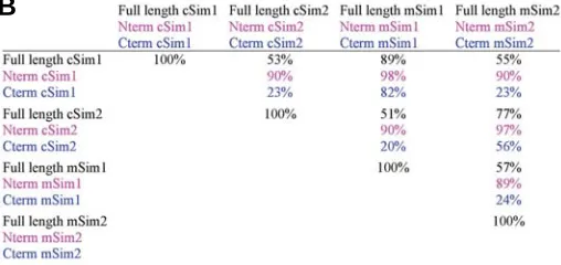
Fig. 1. Comparison of chick and mouse Sim proteins.(A) Comparison of the primary structures of chicken and mouse full length Sim proteins, cSim1, cSim2, mSim1 and mSim2. Identical amino acids (*) and conser-vative changes (: or.) are indicated according to Blast program. Dashes indicate deletions to maximize the sequence similarity. The classical domains of the bHLH/PAS transcription factors are indicated: basic, HLH, PAS-A, PAS-B and HST. (B) Table showing the percentage of homologies between the chick and mouse Sim proteins. The percentages for the N-terminal halves (Nterm) were calculated using the sequences from AAN°1 (M) to AAN°334 (D), including all the classical domains of bHLH/ PAS proteins. The percentages for the Carboxyl-terminal regions (Cterm) were calculated using the sequences from the AAN° 335(I) just after the HST domain to the stop codon.
proteins between chick and mouse showed that each chick Sim protein has a strong homology with its mouse counterpart: chick and mouse Sim1, 89%, chick and mouse Sim2, 77%, (Fig. 1B). The Sim homologies between species are higher that the homolo-gies observed between the Sim1 and Sim2 within the same species (Fig. 1B). The three classical domains of bHLH/PAS transcriptions factors (basic, Helix-Loop-Helix and PAS domains) are highly conserved between chick and mouse. The bHLH domains of the cSim1 and cSim2 proteins are 100% identical with that of the mSim1 and mSim2, respectively. In addition, bHLH domains between Sim1 and Sim2 proteins share 94% identical amino acids both in chick and mouse. Strong similarities in the PAS domains were also apparent between murine and chick sequences, since PAS domains of the Sim proteins share 98% (Sim1) and 97% (Sim2) identical amino acids. These high ho-mologies within these domains, which are included in the N-term region of the Sim proteins (Fig. 1B), strongly suggest that Sim1 and Sim2 proteins have similar DNA binding and dimerisation properties across species, but also between them. The carboxy-terminal regions of the predicted amino acid sequences, which contain the trans-regulation domains, showed relatively high conservation of sequences between chick and mouse Sims (Fig. 1B). However, there is no significant amino acid identity of the carboxyl-terminal halves between the two chick proteins (23%) and between the two mouse proteins (24%), (Fig. 1B). The low identity of the carboxyl-terminal sequences between Sim1 and
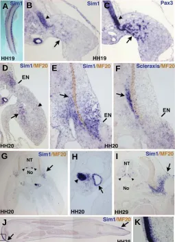
Fig. 2. cSim1 expression during chick limb development.(A) In situ
hybridisation to HH19 embryo with the cSim1 probe. (B,C) Consecutive transverse sections of HH19 embryos at the forelimb level hybridized with the cSim1 (B) and Pax3 (C) probes. Arrows point to cSim1- and Pax3 -migrating cells. Arrowheads show the lateral parts of the dermomyotomes expressing the cSim1 and Pax3 genes. Transverse sections of HH20 (D-H) and HH29 (I) embryos at the forelimb levels were hybridised with the
cSim1 (D,E,G-I) or Scleraxis (F) probes and then incubated with the MF20 antibody (brown) that recognises myosins. (D) The arrow points to the
cSim1-expressing cells in the ventral muscle mass, while the arrowhead shows the faint cSim1 expression in the dorsal muscle regions. (E,F)
Arrows point to the regions expressing the cSim1 and Scleraxis genes. (G,I) The arrows point to the lateral somitic regions expressing cSim1 of HH20 (G) and HH29 (I) embryos. The two arrowheads point to the cSim1
expression in the ventral parts of the neural tube of HH20 and HH29 embryos. (H) Arrowhead points to the mesonephros and the arrow to the Wolffian duct. (J) Longitudinal sections of forelimbs from HH35 em-bryos. (J) Arrows point the cSim1 expression sites. (K) Higher magnifica-tion of the inset drawn in (J) shows that cSim1 expression is restricted to the dermis and does not cover the ectoderm. For all the sections (B-K) dorsal is to the top. NT, neural tube; No, notochord; EN, ectodermal notch.
skeletal muscles during vertebrate embryogenesis also involves other steps such as proliferation, growth arrest and skeletal muscle differentiation (reviewed in Duprez, 2002). Null mutations in mice have revealed hierarchical relationships and apparent functional overlap among the MRFs (reviewed in Buckingham, 2006). Besides the recognized master role of the MRFs in triggering myogenesis in vertebrates, there is emerging evidence that other transcription factors are important for myogenesis. In addition, distinct genetic hierarchies have been identified control-ling the formation of each category of muscles, axial, limbs and head (Buckingham, 2006). However, the function of each of the components of the genetic network involved in limb myogenesis is not fully characterised.
In this paper, we investigated the expression pattern of the Sim1 and Sim2 genes during chick and mouse limb development. Due to the fact that the experiments of in situ hybridisation to wholemount embryos have some limitations, such as insufficient probe penetration, we focused our expression analysis using in situ hybridisation to tissue sections. Our results show that Sim1 and Sim2 gene expression are related to different steps of limb myogenesis during chick and mouse embryonic development.
Results
Comparison between chick and mouse Sim proteins To search for chick homologs of the mouse Sim genes, we analyzed the recently sequenced genome of chick Gallus Gallus (International Chicken Genome Sequencing Consortium, 2004). Our search analysis in the database concluded that the chick genome only contains two genes that are highly related to the mouse and Drosophila Sim genes, cSim1 (accession number XP_419817) and cSim2 (accession number XP_416724). No other Sim sequence could be found from our blast search in the chicken genome. The chick full length cDNA sequences for the Sim1 and Sim2 genes encode a 766-amino acid protein (pre-dicted molecular mass of 86 kDa) and a 764-amino acid protein (predicted molecular mass of 86.4 kDa), respectively (Fig. 1A). The comparison of the deduced amino acid sequences of Sim
G
B
C
D
E
F
H
I
J
A
Sim2 proteins is consistent with distinct transcriptional activities observed in cultured systems (Moffet et al., 1997; Moffet and Pelletier, 2000).
cSim1 gene expression during chick limb bud development
In situ hybridisation to whole mount HH19 embryos showed that cSim1 transcripts were located in the lateral parts of somites, all along the antero-posterior axis, as already described (Fig. 2A, Pourquié et al., 1996). At the limb level, in situ hybridisation to sections showed enhanced cSim1 expression in the lateral parts of the dermomyotomes, visualised by Pax3 expression (Fig. 2B,C, arrowheads). cSim1 transcripts were also observed in a subset of migrating muscle precursors, visualised by Pax3 expression in forelimbs (Fig. 2B,C, arrows). cSim1 transcripts appeared to be located in a ventral subpopulation of Pax3-positive migrating cells. At stage HH20, cSim1 expression was still observed in ventral muscle cells, while a faint expression was consistently observed in dorsal muscle limb regions (Figure 2D, arrow and arrowhead). Similar cSim1 expression (lateral parts of dermomyotomes and migrating cells) was also observed in HH21 hindlimbs (data not shown). From stage HH23 onwards, cSim1 expression was no longer detected in the limb muscle cells (data not shown). In addition to cSim1 expression in lateral dermomyotomes, cSim1 transcripts were also observed in lateral regions of sclerotome and in dermomyotome derivatives such as the dermatome and myo-tome at various stages of development (Fig. 2B,D,E,G,I), in line with previous studies (Cheng et al., 2004). At the limb level, at HH20, the ventral/lateral boundary of the dermal cSim1 expression domain corresponds exactly to the ectodermal notch (Fig. 2D,E,G), which is a thickening of the ectoderm demarcating the somite- and lateral plate-derived dermis (Christ et al., 1983). Comparison of cSim1 expression with that of the tendon marker Scleraxis did not highlight any obvious correlation, although the cSim1 expression domain did encompass the Scleraxis domain (Fig. 2E,F, arrows). In addition, cSim1 transcripts were never detected in differentiated skeletal muscle cells, visualised by sarcomeric myosin expression (Fig. 2I and data not shown). At the axial level, the cSim1 gene displayed the known sites of expression: the ventral regions of the neural tube (Fig. 2G,I, arrowheads), the mesonephros and the Wolffian duct (Fig. 2H). In HH35 limbs, only discrete sub-regions of the limb dermis displayed cSim1 expression (Fig. 2J,K, arrows). In summary, the cSim1 expression in the lateral parts of the dermomyotomes of the limb somites and the faint and transient
cSim1 expression in limb migrating cells suggest a role for the cSim1 protein in early steps of migration of muscle progenitor cells into the limb buds.
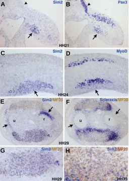
cSim2 expression during chick limb development
In contrast to cSim1, the cSim2 gene was not expressed during the migration step of the muscle precursors into the chick limbs (data not shown). The first limb cSim2 expression was observed at stage HH20 in the forelimbs (data not shown) and at stage HH21 in the hindlimbs (Fig. 3A). cSim2 transcripts were not observed in limb somites; the dermomyotomes were visualised with Pax3 expression (Fig. 3A,B, arrowheads). cSim2 transcripts were spe-cifically observed in the ventral muscle masses; the dorsal and ventral muscle masses were also visualised by Pax3 expression (Fig. 3A,B). In the ventral muscle mass, the cSim2 and Pax3 expression domains were similar (Fig. 3A,B, arrows). At stage HH24, cSim2 transcripts are still exclusively observed in the ventral muscle cells of the limbs, in a domain larger than that of MyoD (Fig. 3C,D). At stage HH29, cSim2 expression was still observed in the limb ventral muscle masses and not in the dorsal muscle masses, both masses were visualized with myosin expression (Fig. 3E). Detailed examination of the ventral muscle mass at HH29 (Fig. 3G) and on a ventral muscle at HH35 (Fig. 3H) shows that cSim2 transcripts were not observed in MF20-positive cells, indicating that cSim2 is not expressed by differentiated muscle cells. At stage
Fig. 3. cSim2 expression during chick limb development. (A,B)
Consecutive transverse sections at the hindlimb level from HH21 em-bryos were hybridised with the cSim2 (A) and Pax3 (B) probes. (C,D)
Consecutive transverse sections at the forelimb level from HH24 em-bryos were hybridized with the cSim2 (C) and MyoD (D) probes. (E,F)
Consecutive transverse sections of forelimbs from HH29 embryos were hybridized with the cSim2 (E) and Scleraxis (F) probes and then incubated with the MF20 antibody (brown). Arrows indicate non-myogenic cSim2
expression surrounding the cartilage elements. (G) High magnification of the ventral muscle mass of (E). (H) Transverse sections of a ventral muscle from HH35 forelimbs hybridized with the cSim2 probe and then incubated with the MF20 antibody (brown) shows that cSim2 transcripts are located outside the MF20-positive cells. (A-D) For transverse sec-tions of embryos (leading to longitudinal limb secsec-tions), dorsal is to the top and proximal to the left. (E-G) For transverse limb sections, posterior is to the left and dorsal to the top. r, radius, u, ulna.
G
B
C
D
E
F
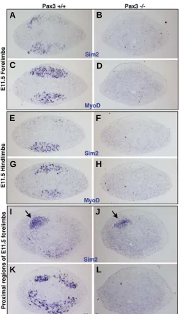
absence of muscle cells but reflects a normal expression re-stricted to myogenic cells. The absence of mSim2 expression in distal fore and hindlimbs of Pax3 mutant mice confirmed that mSim2 expression was exclusively in myogenic cells (Fig. 6A-H). However, in proximal and posterior limb regions close to the body axis, we were able to observe mSim2 expression domains, which were present in Pax3 mutant limbs (Fig. 6I-L). These non-myogenic mSim2 expression domains did not specifically corre-spond to Scleraxis expression domains (data not shown).
Discussion
In this paper we have addressed the precise tissue distribution of Sim transcripts during limb development in the chick and mouse embryos. We have established a link between the expres-Fig. 4. cSim2 expression in chick HH35 forelimbs. Consecutive longi-tudinal sections of HH35 forelimbs were hybridized with the cSim2 (A,C,E) and Scleraxis(B,D,F) probes and then incubated with the MF20 antibody (brown). (A,B) These longitudinal sections are in anterior re-gions. (A,B) Arrows show the ventral tendons expressing Scleraxis and no Sim2. Arrowheads point to dorsal tendons expressing cSim2 and Scleraxis. (C,D) Focus on dorsal and posterior forelimb muscles of HH35 embryos showing the unique dorsal muscle expressing cSim2, the ANC (Anconeus). Arrowheads in (C) point to the tendons expressing the cSim2
(C) and Scleraxis (D) genes. (E,F) The ventral and posterior muscle, FCU (Flexor carpi ulnaris) expresses the cSim2 genes; however, the associ-ated tendon does not (arrows).
HH29, the cSim2 gene was expressed around the cartilage elements (Fig. 3E, arrows). Comparison with Scleraxis ex-pression on adjacent sections showed that these cSim2 expression domains were Scleraxis-negative (Fig. 3E,F, ar-rows). At stage HH35, when the final muscle pattern is organised, cSim2 transcripts were still observed in ventral, individualised muscles (Fig. 4A). No cSim2 expression was observed in dorsal muscles, with the exception of one dorsal and posterior muscle, the Anconeus (Fig. 4A,C). This cSim2 expression in the Anconeus muscle must be late since there was no obvious sign of dorsal cSim2 expression at earlier stages. cSim2 transcripts were also expressed in some ten-dons, which appeared to be more dorsal (Fig. 4A,C, arrow-heads), while most of the ventral tendons did not display any cSim2 expression (Fig. 4A,E, arrows). The tendons were
B
C
D
E
F
A
visualised with Scleraxis expression (Fig. 4B,D,F). In summary, cSim2 is a specific marker of chick limb ventral muscle masses.
mSim1 and mSim2 gene expression during mouse limb
development
sion of the Sim1 and Sim2 genes and different steps of limb muscle formation, in chick and mouse.
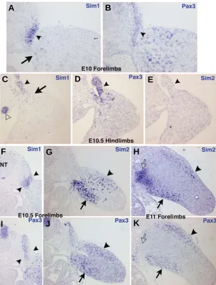
Limb Sim1 expression in chick and mouse embryos Sim1 transcripts displayed a similar expression patterns in chick and mouse limbs. In mouse embryos, mSim1 has previously been described as being restricted to the central dermomyotome at E10.5 at the interlimb region (Sporle, 2001). Our observation of
enhanced ventral expression is not clear and could reflect an involvement of the Sim2 gene in patterning the ventral limb muscles. However, embryological data have established that the positional information for limb muscle patterning is not located in myogenic cells but within limb lateral plate-derived mesenchyme cells (reviewed in Duprez, 2002). The only muscle defect de-scribed in the Sim2 mutant mice is a thinner diaphragm, contrib-uting to the pulmonary atelectasis (Goshu et al., 2002). Further Fig. 5. mSim1 and mSim2 expression during mouse limb development. Consecutive
transverse sections at the forelimb level from E10 (A,B), E10.5 (F,G,I,J) and E11 (H,K) embryos or at the hindlimb level from E10.5 embryos (C-E) were hybridized with the
mSim1 (A,C,F), mSim2 (E,G,H) and mPax3 (B,D,I-K) probes. Arrowheads in (A-E) indicate the lateral regions of the dermomyotomes at the limb levels, expressing mSim1
(A,C), Pax3 (B,D) and no mSim2 (E). Arrows in (A,C) point to the faint mSim1 expression in a subset of the migrating muscle progenitor cells. (C) The open arrowhead shows the mesonephros expressing mSim1. (F,I) Arrowheads point to the lateral somitic mSim1
expression domain (F), which include the lateral Pax3 expression (I).Arrows in (G,H,J,K) show the ventral muscle masses expressing mSim2 (G,H) and Pax3 genes (J,K). Arrowheads in (G,H,J,K) point to the dorsal muscle masses visualised with Pax3
expression, which display faint mSim2 expression. The open arrows in (H,K) indicates a
mSim2-positive domain (H), which is not Pax3-positive (K). NT, neural tube.
mSim1 expression in lateral limb dermomyotomes could reflect a difference of axial level, although it is consistent with previous observations that described mSim1 expression in the lateral compartment of the dermomyotomes at the interlimb region (Ikeya and Takada, 1998). It is also consistent with the cSim1 expression in chick embryos (Fig. 2, Pourquié et al., 1996; Cheng et al., 2004). The enhanced expres-sion of cSim1 and mSim1 in lateral parts of the dermomyotomes of limb somites and the faint, transient expression in migrating somitic cells sug-gest an involvement in the migration step of limb muscle progenitors in chick and mouse embryos. However, no limb muscle phenotype has been described in the Sim1 mutant mice. Interestingly, using a cell aggregation assay it has been shown that the cSim1-expressing cells in the lateral dermomyotomes do not mix with the medial Engrailed1-expressing cells, suggesting different properties of Sim1-expressing cells in the dermomyotomes versus the medial dermomyotomal cell population (Cheng et al., 2004). Sim1 is not exclusive to the lateral parts of chick and mouse dermomyotomes and is also expressed in lateral regions of the sclerotome, myotome and dermatome, defining a lateral somitic-derived region, for both species (Olivera-martinez et al., 2000, 2002, Ben-Yair et al., 2003)
Limb Sim2 expression in chick and mouse em-bryos
One major feature of the Sim2 limb expression was its enhancement in ventral muscle cells (ver-sus dorsal muscle cells). cSim2 expression was almost exclusive to chick ventral muscle masses. In mouse, although mSim2 expression was clearly enhanced in ventral muscle masses of fore- and hindlimbs, we could detect mSim2 expression in the dorsal muscle masses, specifically in the fore-limbs. This restricted/enhanced expression pattern in ventral limb muscles was observed until E9 chick limbs and until E12.5 mouse limbs. The transient mSim2 expression in mouse limb muscle masses and the absence of cSim2 expression in chick muscle fibres indicate that Sim2 labels limb myo-blast progenitors. To our knowledge, Sim2 repre-sents of the first example of a gene displaying this enhanced expression in ventral muscle masses, since all the known muscle genes (associated with any steps of myogenesis) are located in both dorsal and ventral muscle masses. The reason for this
G
B
C
D
E
F
H
I
J
K
work is necessary to determine the function of the Sim2 gene in the ventral muscle masses in chick and mouse embryos. cSim2 was also expressed in a subset of chick limb tendons, but we did not observe similar mSim2 expression in mouse limbs, highlight-ing a difference between Sim2 expression in chick and mouse limbs.
Sim1 and Sim2 genes display distinct expression profiles
during embryonic limb development
Sim1 and Sim2 expression domains did not overlap during chick and mouse limb myogenesis, suggesting an absence of functional redundancy in muscle formation. The only overlapping expression between the Sim1 and Sim2 genes was observed in HH20 chick forelimbs in ventral muscle masses. In addition to being expressed in different steps of muscle formation, the homologies between the Sim1 and Sim2 genes (in chick or mouse) are very low compared to those between cSim1 and
Fig. 6. Limb mSim2 expression in the absence of muscles. Transverse sections of forelimbs (A-D, I-L) or hindlimbs (E-H) from E11.5 wild type embryos (A,C,E,G,I,K) or E11.5 Pax3 mutant embryos (B,D,F,H,J,L) were hybridised with the mSim2 (A,B,E,F,I,J) or MyoD(C,D,G,H,K,L) probes. The sections hybridized with the mSim2 probe are adjacent of the sections hybridized with the MyoD probe respectively. Residual mSim2
expression in muscleless limbs of Pax3 mutant mice shows that non-myogenic expression of the mSim2 gene is located in the proximal and posterior limb region (arrows in I,J). For all the sections, dorsal is to the top and posterior to the left.
mSim1 and between cSim2 and mSim2 (Fig. 1B). Sim1 expres-sion suggests an involvement in early steps of limb muscle formation (specification or/and migration of lateral muscle precur-sors), while Sim2 expression indicates an involvement after the migration step. This is reminiscent of the mSim1 and mSim2 expression in CNS, where the murine Sim1 and Sim2 genes display different expression profiles that overlap in certain regions of the anterior hypothalamus (Fan et al., 1996). Sim mutant analysis showed that Sim1 acts upstream of Sim2 and partially compensates for the loss of Sim2 in PVN embryonic development (Michaud et al., 1998, Goshu et al., 2002, Goshu et al., 2004). However, mSim1 and mSim2 act along compensatory pathways in mammillary body axonal development (Marion et al., 2005).
In summary, the Sim1 gene is expressed mainly in early limb muscle precursor cells and Sim2 expression is enhanced in ventral limb myoblasts in chick and mouse embryonic limbs. Sim expression analysis provides a basis for analysing the function of the Sim genes within the gene network involved in limb muscle formation.
Materials and Methods
Chick and mouse embryos
Fertilized chick eggs from commercial sources (JA 57 strain, Intitut de Sélection Animale (ISA), Lyon, France) were incubated at 37°C. Embryos were staged according to Hamburger and Hamilton (1992). Embryos from wild type and Pax3-/- mutant mice were collected after natural overnight
matings (Relaix et al., 2003). For staging fertilization was considered to
take place at 6 am.
In situ hybridisation to tissue sections or to wholemount embryos
Chick or mouse embryos were fixed overnight at 4% (v/v) formalde-hyde and processed for in situ hybridisation to whole mounts and to
paraffin sections as previously described (Delfini et al., 2000). Antisense
RNA probes were labelled with digoxigenin according to manufacturer’s instructions (Roche Diagnostics). The probes were detected by an alkaline phosphatase-coupled antibody against digoxigenin using nitroblue tetrazolium/5-bromo-chloro-3-indolyl phosphate (NBT/BCIP) as the chro-mogenic susbstrate for alkaline phosphatase. Antisense digoxigenin-labelled RNA probes were prepared as described: chick Pax3, chick MyoD, and mouse MyoD (Delfini and Duprez, 2004; Tozer et al., 2007);
chick and mouse Scleraxis (Bonnin et al., 2005); mouse Pax3 (Relaix et al., 2003); chick Sim1 (Pourquié et al., 1996) and Sim2 (Caqueret et al.,
2005); mouse Sim1 (Michaud et al., 1998) and Sim2 (Goshu et al., 2002).
Immunohistochemistry
Differentiated muscle cells were detected on sections as previously described using a monoclonal antibody against sarcomeric myosin heavy chain, MF20 (Developmental Hybridoma Bank, University of Iowa, Iowa City). Immunohistochemistry were performed following the in situ
hybridisation experiments.
G
B
C
D
E
F
H
I
J
K
L
Acknowledgements
We are grateful to Frederic Relaix for providing mouse Pax3 mutant embryos and to Jacques Michaud for the mSim1 and mSim2 plasmids. We thank members of our laboratory for discussion and critical reading of the manuscript. This work was supported by the CNRS, University Paris 6, ANR, AFM, ARC and the EU 6th PCRDT through the MYORES Network
of excellence.
References
AHMED, M.U., CHENG, L. and DIETRICH, S. (2006). Establishment of the epaxial-hypaxial boundary in the avian myotome. Dev Dyn 235: 1884-1894.
BEN-YAIR, R., KAHANE, N. and KALCHEIM, C. (2003). Coherent development of dermomyotome and dermis from the entire mediolateral extent of the dorsal somite. Development 130: 4325-4336.
BONNIN, M.A., LACLEF, C., BLAISE, R., ELOY-TRINQUET, S., RELAIX, F., MAIRE, P. and DUPREZ, D. (2005). Six1 is not involved in limb tendon development, but is expressed in limb connective tissue under Shh regulation. Mech Dev 122: 573-585.
BUCKINGHAM, M. (2006). Myogenic progenitor cells and skeletal myogenesis in vertebrates. Curr Opin Genet Dev 16: 525-532.
CAQUERET, A., COUMAILLEAU, P. and MICHAUD, J.L. (2005). Regionalization of the anterior hypothalamus in the chick embryo. Dev Dyn 233: 652-658.
CHENG, L., ALVARES, L.E., AHMED, M.U., EL-HANFY, A.S. and DIETRICH, S. (2004). The epaxial-hypaxial subdivision of the avian somite. Dev Biol 274: 348-369.
CHRAST, R., SCOTT, H.S., CHEN, H., KUDOH, J., ROSSIER, C., MINOSHIMA, S., WANG, Y., SHIMIZU, N. and ANTONARAKIS, S.E. (1997). Cloning of two human homologs of the Drosophila single-minded gene SIM1 on chromosome 6q and SIM2 on 21q within the Down syndrome chromosomal region. Genome Res 7: 615-624.
CHRAST, R., SCOTT, H.S., MADANI, R., HUBER, L., WOLFER, D.P., PRINZ, M., AGUZZI, A., LIPP, H.P. and ANTONARAKIS, S.E. (2000). Mice trisomic for a bacterial artificial chromosome with the single-minded 2 gene (Sim2) show phenotypes similar to some of those present in the partial trisomy 16 mouse models of Down syndrome. Hum Mol Genet 9: 1853-1864.
CHRIST, B., JACOB, M. and JACOB, H.J. (1983). On the origin and development of the ventrolateral abdominal muscles in the avian embryo. An experimental and ultrastructural study. Anat Embryol (Berl) 166: 87-101.
CONSORTIUM, I.C.G.S. (2004). Sequence and comparative analysis of the chicken genome provide unique perspectives on vertebrate evolution. Nature 432: 695-716.
COUMAILLEAU, P., PENRAD-MOBAYED, M., LECOMTE, C., BOLLEROT, K., SIMON, F., POELLINGER, L. and ANGELIER, N. (2000). Characterization and developmental expression of xSim, a Xenopus bHLH/PAS gene related to the Drosophila neurogenic master gene single-minded. Mech Dev 99: 163-166.
COUMAILLEAU, P., BOLLEROT, K., LECOMTE, C. and ANGELIER, N. (2003). Xenopus single-minded (xSim) is a nuclear factor allowing nuclear translocation of its cytoplasmic partner xArnt. Exp Cell Res 287: 237-248.
CREWS, S.T. (1998). Control of cell lineage-specific development and transcription by bHLH-PAS proteins. Genes Dev 12: 607-620.
CREWS, S.T. and FAN, C.M. (1999). Remembrance of things PAS: regulation of development by bHLH-PAS proteins. Curr Opin Genet Dev 9: 580-587.
DAHMANE, N., CHARRON, G., LOPES, C., YASPO, M.L., MAUNOURY, C., DECORTE, L., SINET, P.M., BLOCH, B. and DELABAR, J.M. (1995). Down syndrome-critical region contains a gene homologous to Drosophila sim ex-pressed during rat and human central nervous system development. Proc Natl Acad Sci USA 92: 9191-9195.
DELFINI, M.C., HIRSINGER, E., POURQUIE, O. and DUPREZ, D. (2000). Delta 1-activated notch inhibits muscle differentiation without affecting Myf5 and Pax3 expression in chick limb myogenesis. Development 127: 5213-5224.
DELFINI, M.C. and DUPREZ, D. (2004). Ectopic Myf5 or MyoD prevents the neuronal differentiation program in addition to inducing skeletal muscle differ-entiation, in the chick neural tube. Development 131: 713-723.
DUPREZ, D. (2002). Signals regulating muscle formation in the limb during
embryonic development. Int J Dev Biol 46: 915-925.
EATON, J.L. and GLASGOW, E. (2006). The zebrafish bHLH PAS transcriptional regulator, single-minded 1 (sim1), is required for isotocin cell development. Dev Dyn 235: 2071-2082.
EMA, M., SUZUKI, M., MORITA, M., HIROSE, K., SOGAWA, K., MATSUDA, Y., GOTOH, O., SAIJOH, Y., FUJII, H., HAMADA, H. et al. (1996). cDNA cloning of a murine homologue of Drosophila single-minded, its mRNA expression in mouse development, and chromosome localization. Biochem Biophys Res Commun 218: 588-594.
EMA, M., IKEGAMI, S., HOSOYA, T., MIMURA, J., OHTANI, H., NAKAO, K., INOKUCHI, K., KATSUKI, M. and FUJII-KURIYAMA, Y. (1999). Mild impairment of learning and memory in mice overexpressing the mSim2 gene located on chromosome 16: an animal model of Down’s syndrome. Hum Mol Genet 8: 1409-1415.
FAN, C.M., KUWANA, E., BULFONE, A., FLETCHER, C.F., COPELAND, N.G., JENKINS, N.A., CREWS, S., MARTINEZ, S., PUELLES, L., RUBENSTEIN, J.L. et al. (1996). Expression patterns of two murine homologs of Drosophila single-minded suggest possible roles in embryonic patterning and in the pathogenesis of Down syndrome. Mol Cell Neurosci 7: 1-16.
GOSHU, E., JIN, H., FASNACHT, R., SEPENSKI, M., MICHAUD, J.L. and FAN, C.M. (2002). Sim2 mutants have developmental defects not overlapping with those of Sim1 mutants. Mol Cell Biol 22: 4147-4157.
GOSHU, E., JIN, H., LOVEJOY, J., MARION, J.F., MICHAUD, J.L. and FAN, C.M. (2004). Sim2 contributes to neuroendocrine hormone gene expression in the anterior hypothalamus. Mol Endocrinol 18: 1251-1262.
HAMBURGER, V. and HAMILTON, H.L. (1992). A series of normal stages in the development of the chick embryo. 1951. Dev Dyn 195: 231-272.
HOLDER, J.L., JR., BUTTE, N.F. and ZINN, A.R. (2000). Profound obesity asso-ciated with a balanced translocation that disrupts the SIM1 gene. Hum Mol Genet 9: 101-108.
HOLDER, J.L., JR., ZHANG, L., KUBLAOUI, B.M., DILEONE, R.J., OZ, O.K., BAIR, C.H., LEE, Y.H. and ZINN, A.R. (2004). Sim1 gene dosage modulates the homeostatic feeding response to increased dietary fat in mice. Am J Physiol Endocrinol Metab 287: E105-113.
IKEYA, M. and TAKADA, S. (1998). Wnt signaling from the dorsal neural tube is required for the formation of the medial dermomyotome. Development 125: 4969-4976.
KARDON, G. (1998). Muscle and tendon morphogenesis in the avian hind limb. Development 125: 4019-4032.
KARDON, G., HARFE, B.D. and TABIN, C.J. (2003). A Tcf4-positive mesodermal population provides a prepattern for vertebrate limb muscle patterning. Dev Cell 5: 937-944.
MARION, J.F., YANG, C., CAQUERET, A., BOUCHER, F. and MICHAUD, J.L. (2005). Sim1 and Sim2 are required for the correct targeting of mammillary body axons. Development 132: 5527-5537.
MICHAUD, J.L., ROSENQUIST, T., MAY, N.R. and FAN, C.M. (1998). Develop-ment of neuroendocrine lineages requires the bHLH-PAS transcription factor SIM1. Genes Dev 12: 3264-3275.
MICHAUD, J.L., BOUCHER, F., MELNYK, A., GAUTHIER, F., GOSHU, E., LEVY, E., MITCHELL, G.A., HIMMS-HAGEN, J. and FAN, C.M. (2001). Sim1 haploinsufficiency causes hyperphagia, obesity and reduction of the paraventricular nucleus of the hypothalamus. Hum Mol Genet 10: 1465-1473.
MOFFETT, P., DAYO, M., REECE, M., MCCORMICK, M.K. and PELLETIER, J. (1996). Characterization of msim, a murine homologue of the Drosophila sim transcription factor. Genomics 35: 144-155.
MOFFETT, P., REECE, M. and PELLETIER, J. (1997). The murine Sim-2 gene product inhibits transcription by active repression and functional interference. Mol Cell Biol 17: 4933-4947.
MOFFETT, P. and PELLETIER, J. (2000). Different transcriptional properties of mSim-1 and mSim-2. FEBS Lett 466: 80-86.
OLIVERA-MARTINEZ, I., COLTEY, M., DHOUAILLY, D. and POURQUIE, O. (2000). Mediolateral somitic origin of ribs and dermis determined by quail-chick chimeras. Development 127: 4611-4617.
somite patterning: a role for BMP4. Cell 84: 461-471.
PROBST, M.R., FAN, C.M., TESSIER-LAVIGNE, M. and HANKINSON, O. (1997). Two murine homologs of the Drosophila single-minded protein that interact with the mouse aryl hydrocarbon receptor nuclear translocator protein. J Biol Chem 272: 4451-4457.
RELAIX, F., POLIMENI, M., ROCANCOURT, D., PONZETTO, C., SCHAFER, B.W. and BUCKINGHAM, M. (2003). The transcriptional activator PAX3-FKHR rescues the defects of Pax3 mutant mice but induces a myogenic gain-of-function phenotype with ligand-independent activation of Met signaling in vivo. Genes Dev 17: 2950-2965.
SHAMBLOTT, M.J., BUGG, E.M., LAWLER, A.M. and GEARHART, J.D. (2002). Craniofacial abnormalities resulting from targeted disruption of the murine Sim2 gene. Dev Dyn 224: 373-380.
SPORLE, R. (2001). Epaxial-adaxial-hypaxial regionalisation of the vertebrate
somite: evidence for a somitic organiser and a mirror-image duplication. Dev Genes Evol 211: 198-217.
TOZER, S., BONNIN, M.A., RELAIX, F., DI SAVINO, S., GARCIA-VILLALBA, P., COUMAILLEAU, P. and DUPREZ, D. (2007). Involvement of vessels and PDGFB in muscle splitting during chick limb development. Development 134: 2579-2591.
WOODS, S.L. and WHITELAW, M.L. (2002). Differential activities of murine single minded 1 (SIM1) and SIM2 on a hypoxic response element. Cross-talk between basic helix-loop-helix/per-Arnt-Sim homology transcription factors. J Biol Chem 277: 10236-10243.
YANG, C., GAGNON, D., VACHON, P., TREMBLAY, A., LEVY, E., MASSIE, B. and MICHAUD, J.L. (2006). Adenoviral-mediated modulation of Sim1 expression in the paraventricular nucleus affects food intake. J Neurosci 26: 7116-7120.
Further Related Reading, published previously in the Int. J. Dev. Biol.
See our recent Special Issue Fertilization, in honor of David L. Garbers and edited by Paul M. Wassarman and Victor D. Vacquier at: http://www.ijdb.ehu.es/web/contents.php?vol=52&issue=5-6
2006 ISI **Impact Factor = 3.577** See our recent Special Issue Limb Development edited by Juan Hurlé and Juan Carlos
Izpisua Belmonte at:
http://www.ijdb.ehu.es/web/contents.php?vol=46&issue=7
The Australian lungfish (Neoceratodus forsteri) - fish or amphibian pattern of muscle development?
Agata Kacperczyk and Malgorzata Daczewska Int. J. Dev. Biol. (2008) 52: 279-286
Myoskeletin, a factor related to Myocardin, is expressed in somites and required for hypaxial muscle formation in Xenopus
Hui Zhao, Martha L. Rebbert and Igor B. Dawid Int. J. Dev. Biol. (2007) 51: 315-320
The expression of Fat-1 cadherin during chick limb development
Terence G. Smith, Nick Van Hateren, Cheryll Tickle and Stuart A. Wilson Int. J. Dev. Biol. (2007) 51: 173-176
The transforming growth factor-betas: multifaceted regulators of the
development and maintenance of skeletal muscles, motoneurons and Schwann cells.
Ian S McLennan and Kyoko Koishi Int. J. Dev. Biol. (2002) 46: 559-567
Comparative expression analysis of Pax3 and Pax7 during mouse myogenesis
David Horst, Svetlana Ustanina, Consolato Sergi, Gregor Mikuz, Herbert Juergens, Thomas Braun and Eugene Vorobyov
Int. J. Dev. Biol. (2006) 50: 47-54
The role of p53 in vivo during skeletal muscle post-natal development and regeneration: studies in p53 knockout mice.
Jason D White, Collins Rachel, Royce Vermeulen, Marilyn Davies and Miranda D Grounds