Expression of prohormone convertase 2 and the generation
of neuropeptides in the developing nervous system
of the gastropod Haliotis
SCOTT F. CUMMINS
1, PATRICK S. YORK
1, PETER J. HANNA
2,
BERNARD M. DEGNAN
1and ROGER P. CROLL*
,31School of Integrative Biology, The University of Queensland, Brisbane, Australia, 2School of Life and Environmental Sciences, Deakin University, Geelong, Australia and Department of Anatomy, Mahidol University, Bangkok, Thailand, 3Department of Physiology and Biophysics, Faculty of Medicine, Dalhousie University, Halifax, Canada
ABSTRACT Prohormone convertase 2 (PC2) belongs to a family of enzymes involved in the proteolytic maturation of neuropeptide precursors into mature peptides that act as neurotrans-mitters, neuromodulators or neurohormones. Here we show that a gene encoding a PC2-like enzyme (HasPC2) is expressed during larval development and in the adult ganglia of the vetigastropod Haliotis asinina. HasPC2 exhibits high sequence identity to other gastropod PC2s and thus is likely to function in peptide processing. Analysis of HasPC2 expression indicates that it is activated early in nervous system development. During trochophore and early veliger larval stages, HasPC2 is expressed in the vicinity of the forming ganglia of the central nervous system and parts of the putative peripheral nervous system. Later in larval development, at the time the veliger becomes competent to interact with the external environment and initiate metamorpho-sis, HasPC2 expression largely restricts to cells of the major ganglia and their commissures. Profiling of veliger larvae by bioinformatic approaches suggests the expression of a variety of peptides. Direct MALDI-MS-based peptide profiling of juvenile Haliotis cerebral ganglia (brain) reveals an abundance of neuropeptides, including FMRFamide-related peptides and APGWamide, compatible with PC2 functioning in neuropeptide processing in these regions. These results are consistent with PC2 regulating neuropeptide generation in the earliest functioning of the gastropod nervous system.
KEY WORDS:
prohormone convertase, abalone, neuropeptide, development
Neuropeptides are widespread in the animal kingdom and are often involved in the control of critical functions throughout the life cycle. The regulation of development by peptidergic hormones is a common feature of multicellular animals and has been studied in great detail in arthropods and vertebrates. Strikingly, investiga-tions reveal that insects are as well or even better supplied with neuropeptides than mammals. In insects, neuropeptides regulate not only the functioning of endocrine glands, but also a wide range of physiological and developmental processes (Reumer et al.,
2008). Although a complete developmental peptidome analysis has not been established for other major invertebrate groups, such as the molluscs, numerous immunolocalization studies suggest that neuropeptides are equally important for their devel-opment. For instance, in gastropods FMRFamide and related peptides are amongst the earliest neurotransmitters expressed
BIOLOGY
www.intjdevbiol.com*Address correspondence to: Dr. Roger Croll. Department of Physiology and Biophysics, Dalhousie University, Sir Charles Tupper Medical Building, 5850 College Street, Halifax, Nova Scotia, Canada, B3H 1X5. Fax: + 1-(902)-494-1685. e-mail: [email protected]
Accepted: 03 October 2008. Published online: 26 June 2009. Edited by: Edward De Robertis
ISSN: Online 1696-3547, Print 0214-6282
© 2009 UBC Press Printed in Spain
Abbreviations used in this paper: DIG, digoxigenin; MALDI-MS, matrix-assisted laser desorption/ionization MS; PBS, phosphate-buffered solution; PC, prohormone convertase; RACE, rapid amplification of cDNA ends; WMISH, whole-mount in situ hybridization.
within the larval nervous system (Croll and Voronezhskaya, 1996; Dickinson et al., 1999; Dickinson et al., 2000; Haszprunar et al.,
2002; Dickinson and Croll, 2003; Croll, 2006). In the adult stages of these same animals, neuropeptides play a number of roles as local neurotransmitters and neuromodulators and as circulating hormones (Chase, 2002).
Fig. 1. Nucleotide and predicted amino acid sequence of cDNA encoding prohormone convertase 2 in H. asinina.(A)HasPC2 consists of 2,319 bp encoding a precursor protein of 662 amino acids. Underline, predicted signal sequence; boxed, catalytic region and putative N-glycosylation sites. The Asp, His and Ser residues forming the catalytic triad are circled and the Asp residue stabilizing the oxyanion is boxed. Dibasic cleavage sites are identified with an asterisk. (B)Schematic representation of HasPC2 precursor protein showing the arrangement of encoded signal (pre), pro domain and catalytic region.
processing to release one or more biologically active products. As a result, extensive research has focussed on characterising the enzymes responsible for this post-translational modification, in particular the prohormone convertases (PCs). Vertebrate and invertebrate PCs have been reported to cleave precursor pro-teins such as proglucagon (Rouille et al., 1997),
proopiomelanocortin (Korner et al., 1991) and proinsulin (Davidson et al., 1988). Presently, several vertebrate PCs have been
characterised, including PC1 (same as PC3), PC2, PC4 and furin, each of which has a distinctive tissue distribution (Seidah et al., 1990; Bloomquist et al., 1991; Smeekens et al., 1991; Braks et al., 1992). These convertases are structurally related to
bacte-and in the juvenile ganglia, largely restricted to nerve cells of the central and peripheral nervous systems. Bioinformatic and MALDI-MS molecular mass profiling of H. asinina allowed for detection of
a large number of putative peptides, including FMRFamide, which is compatible with PC2 generating neuropeptides during develop-ment and growth.
Results
Characterisation of the abalone PC2 genes
Sequencing of PCR products, generated with degenerate PC2 oligonucleotide primers, from H. rubra neural and H. asinina larval
rial subtilisins and KEX2 (Julius et al., 1984), and typically cleave
proproteins at pairs of basic amino acids, although cleavage at monobasic sites is also known (Hwang et al., 2000b).
Homologues of PCs have also been reported in the pulmonate mollusc, Lymnaea stagnalis (Smit et al., 1992) and the marine
opisthobranch, Aplysia californica
(Nagle et al., 1995), as well as
numerous other invertebrates, in-cluding the sheep blowfly, Lucilia cuprina (Mentrup et al., 1999) and
the fruitfly Drosophila melano-gaster (Hwang et al., 2000a). L. stagnalis possesses a PC2 gene
that is exclusively expressed in the neuroendocrine system and encodes two alternatively-spliced transcripts of 3.0 and 4.8 kb (Smit
et al., 1992). In A. californica, PC2
cDNA was first identified from the abdominal ganglia (Chun et al.,
1994), which is known to produce large amounts of egg-laying pep-tides derived from the processing of a larger preprohormone (Fisher
et al., 1988).
In this study, we report the iden-tification of a PC gene from the vetigastropods Haliotis asinina
and Haliotis rubra (abalone),
which appear to be orthologues of other gastropod PC2 genes. We have investigated the expression of this gene during the develop-ment of H. asinina, whose
devel-opment and neuroendocrinology is relatively well-characterized (O’Brien and Degnan, 2000; Hinman and Degnan, 2002; Page, 2006). Whole mount in situ
hy-bridization revealed a widespread expression pattern of the tran-script during larval development
B
A
cDNAs, confirmed that PC2 is expressed in these two species of abalone. In H. asinina, RACE led to a full-length cDNA of 2319 bp
(HasPC2; GenBank accession number EU684323), which
en-codes a 662 amino acid precursor (Fig. 1A). The domain architec-ture of HasPC2 is identical to other PC2s, containing pre (signal), pro and catalytic domains (Fig. 1B). On both sides of the catalytic domain are two putative proteolytic cleavage sites (with a consen-sus sequence Lys-Arg). Both sites are likely to be used sequen-tially for autocatalytic activation and folding of the enzyme.
In H. rubra, RT-PCR led to a partial-length cDNA of 1,194 bp
(HrubPC2, GenBank accession number AY237916), which
en-codes a 398 amino acid precursor. The resulting deduced amino acid sequences of both H. asinina and H. rubra catalytic regions
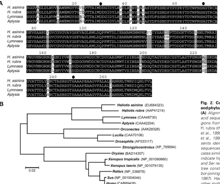
were compared with molluscan homologues and showed signifi-cant sequence identity with A. californica (Nagle et al., 1995) and L. stagnalis (Smit et al., 1992) PC2s (Fig. 2A). A phylogenetic
analysis clearly places the identified abalone genes within the PC2 class of enzyme, and most similar to Lymnaea and Aplysia
homologues (Fig. 2B). Predicted subtilisin-related serine pro-tease residues - D195, H236 and S410 - were present within the abalone PC2s, and there were three conserved consensus se-quences for putative N-linked glycosylation at NLT311, NCT403 and NLT431. As well, the abalone PC2s contain conserved mul-tiple dibasic residue sequences, KR. The end of the catalytic
region is likely to be directly after the L440.
Larval expression of H. asinina PC2 by RT-PCR
RT-PCR was performed to identify HasPC2 expression during
larval development (Fig. 3). The HasPC2 gene is expressed
continuously throughout development as HasPC2 was present
from 7 hours post-fertilization (hpf) to 24 hpf. The HasPC2
amplicon (242 bp) is also obtained from a cDNA preparation of mixed larvae at various stages in development and the adult pooled central nervous system (CNS). The control, in which no cDNA template was used (blank), was negative.
Fig. 2. Comparative alignment and phylogenetic analysis of PC2. (A) Alignment of predicted amino acid sequence of PC2 catalytic re-gions from H. asinina (this study), H. rubra (this study), Aplysia (Nagle et al., 1995), and Lymnaea (Kellett et al., 1994). Black shading repre-sents identical residues in all the sequences and grey shading indi-cates similar residues. Black circles indicate highly conserved Asp, His and Ser residues. (B) Phylogenetic tree constructed using the neigh-bor-joining method (Saitou and Nei, 1987). HasPC2 is most similar to other molluscan PC2’s.
Fig. 3. Temporal expression of HasPC2 during larval development.
In situ hybridisation analysis of HasPC2 expression during larval development
A 1021-bp digoxigenin-labeled antisense riboprobe was used to examine HasPC2 expression in whole mount larval
prepara-tions during development. HasPC2 transcripts were detected in
the vicinity of the developing central and peripheral nervous systems in all stages of larval development, from newly hatched trochophores to competent veligers (96 hpf). In the 9 hpf tro-chophore larva, cells in the region of the apical tuft expressed
HasPC2 (Fig. 4 A,B). In 12 hpf and 14 hpf pretorsional veliger
larvae and 17 hpf posttorsional veliger larvae, localised expres-sion of HasPC2 was detected in the head, foot, velum, and later,
B
C
D
A
Fig. 4 (Left).WMISH showing spatial expression of HasPC2 during larval development.(A,B) Lateral and anterior, respectively, 9 hpf trochophore larva showing HasPC2 expression in the apical tuft. (C-H) 12, 14 and 17 hpf larvae showing HasPC2 expression in areas of the cerebral (arrow), pedal, visceral, esophageal, branchial ganglia, and developing foot. Right schematics: Corresponding summary (lateral view) of spatial expression of HasPC2 from 9 hpf to 17 hpf. Areas represented by solid green circles were consistently stained. cg, cerebral ganglia; pg, pleural ganglia; pdg, pedal ganglia; ppg, fused pleuropedal ganglia; og, esophageal ganglia; brg, branchial ganglia; at, apical tuft; s, shell; v, velum; vm, visceral mass; f, foot; m, mantle; ct, cephalic tentacle. Scale bar, (A) 50 μm (same magnification in all panels).
Fig (Right). 5. WMISH showing spatial expression of HasPC2 in 96 hpf competent larvae.Representative micrographs showing HasPC2 expression in the regions of developing ganglia from the lateral (A), anterior (B), dorsal (C) and posterior (D) views. Expression is most prominent in or near cerebral ganglia (black arrows), pleuro-pedal ganglia (black arrow heads), putative brachial ganglia (white arrow head) and developing loop of visceral cells (white arrows). vm, visceral mass; r, radula; e, eye; m, mantle; o, operculum. Scale bar: (A)50 μm (same magnification in all panels). in the mantle (Fig. 4 C-H). Expression was restricted largely to the areas of the developing cerebral, pleural, pedal, esophageal and branchial ganglia, with the cerebral ganglia staining most in-tensely. Distinct expression was also observed in the region of the ganglia and commissures of 96 hpf competent larvae (Fig. 5).
Direct MALDI-MS-based peptide profiling of juvenile H. asinina ganglia
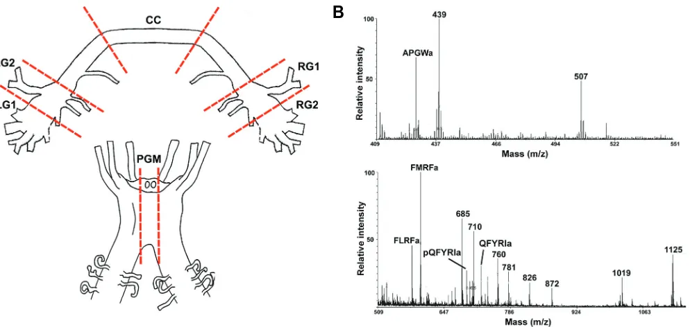
Direct MALDI-MS-based profiling of neurons and nerves al-lows the determination of many of the mature processed peptides present within a sample. As shown in Fig. 6A, neuronal samples were derived from areas of the juvenile abalone cerebral and pleuro-pedal ganglion. MALDI-MS of a sub-section of the right ganglion (RG1) reveals the presence of several previously char-acterized molluscan peptides including APGWamide (428 Da),
Peptide
Expected MW (Da)
Observed
MW (Da) Ganglion region identified
FMRFamide 598.7 598.8 CC, LG1, LG2, PGM, RG1, RG2 FLRFamide 580.7 580.9 CC, LG1, LG2, PGM, RG1, RG2
QFYRIamide 724.9 724.9 LG1, PGM, RG1, RG2
pQFYRIamide 707.8 707.8 LG1, RG1, RG2
APGWamide 428.2 428.9 CC, LG1, LG2, PGM, RG1, RG2 TABLE 1
PEPTIDE DISTRIBUTION IN CEREBRAL AND PLEURO-PEDAL GANGLIA
CC, cerebral commissure; LG, left cerebral ganglion; RG, right cerebral ganglion; PGM, pleuro-pedal ganglion middle.
G
B
C
D
E
F
FLRFamide (580 Da), FMRFamide (598 Da), pQFYRIamide (707 Da), and QFYRIamide (725 Da) (Table 1; Fig. 6B). APGWamide, FLRFamide, and FMRFamide, were detected in all regions exam-ined. pQFYRIamide and QFYRIamide were in the left and right cerebral and pleuro-pedal ganglion, but were not detected in cerebral commissure. Additional unassigned mass peaks likely represent other important neuropeptides processed from precur-sor proteins.
The Neuropred processing prediction web tool was applied to previously published EST sequences (Jackson and Degnan, 2006) to predict H. asinina larval peptides that may be released
from precursor proteins following convertase activity. Table 2 lists peptide sequences, including post-translational modifications (Amide, amidation; Ac-, acetylation; p-, pyroglutamation) and average masses obtained. Precursors are predicted to be pro-cessed at defined basic cleavage sites and subsequent
post-translational modifications added to generate peptides listed. Enzymes are expected to remove the N-terminal Arg, and those peptides ending in Gly are likely to be amidated, resulting in the final predicted average masses observed. The processing predic-tions imply that all precursors would be directed through the secretory pathway for extracellular release.
Discussion
This study shows that haliotids possess a PC2 gene.
Structur-ally, PCs contain defined regulatory regions including a pro-segment, a catalytic domain, a structural domain (P domain), and a highly variable C-terminal domain. The catalytic domain is characterised by the presence of a region similar to the subtilisin-related serine proteases, including active site residues aspartic acid, asparagine, histidine and serine. All conserved subtilisin
Clone* Sequence (amino acid) + post-translational modification MW (average) Probability extracellular**
EST 36 [Ac-]GADDCILLAGPCG[Amide] 1245.43 67%
EST 48 [p-]EQELVTLL[Amide] 925.09 67%
EST 54 [p-]QFPGGGYRNMRFRGQGINYGPGPGIGS[Amide] 2823.14 33%
EST 63 [p-]QIGLYNRYNRFGLLN 1824.07 56%
EST 153 [p-]ETDQVSMRNI[Amide] 1173.31 67%
EST 1372 [Ac-]ASFPINISDTTAGDIVFAQSGSDITVSLKYSVGATPFADNGNVVYAC 4826.32 56%
EST 1395 [Ac-]AVLHYDPLEITSRLESEMDDLMKLVMKPHTLS 3755.37 56%
EST 1482 [p-]QPAPNELPDKL 1204.3 56%
[Ac-]SDSVFYTSTDGQNGKGISFMATRHSFPSADLSLSFCQLMVL[Amide] 4488.04
EST 2823 [p-]QHIPLYPCETCVIEAFSD 2048.31 44%
EST 3345 DIGHSLTNVFQSLGHSLMHQLDTTGVDLLNAGINA[Amide] 3689.12 67%
TABLE 2
PEPTIDES PREDICTED TO BE GENERATED AND RELEASED FROM DEVELOPING ABALONE LARVAE
* (Jackson and Degnan, 2006). ** Based on precursor signal sequence (SignalP 3.0 and PSORTII)
Fig. 6. H. asinina ganglia preparation and MALDI-MS analysis. (A) Cerebral ganglia and pleuro-pedal ganglia were dissected and segments removed (shown by dashed lines). Sub-sections of LG (left cerebral ganglion), CC (cerebral commissue), RG (right cerebral ganglion) and pleuro-pedal ganglia middle (PGM) were spotted onto a MALDI sample plate. (B) Representative mass spectrums obtained from a sub-section of RG1. Mass peaks that match previously characterized molluscan peptides are labelled with peptide name.
sites are present in HasPC2. There is a high degree of sequence identity within this domain, thus reflecting the functional impor-tance of these residues in the catalytic activity of the enzyme.
Apart from the high degree of conservation for the convertase gene sequences, there are several additional common features, which are important for convertase maturation and activity, in-cluding the presence of putative post-translational modification sites. Post-translational modifications known to occur in PCs (Seidah et al., 1993) include tyrosine sulfation and
phosphoryla-tion, and N-linked glycosylaphosphoryla-tion, which prevents PC2 degradation in the endoplasmic reticulum (Benjannet et al., 1993). From the
sequences analysed, it appears that abalone PC2 is also a glycoprotein. In HasPC2, there are three conserved consensus sequences for N-linked glycosylation; although based on analysis of vertebrate PC2, it has been suggested that only one site, NCT403, is used (Smit et al., 1992). Multiple basic sites within PC
are also required for co- and post-translational processing. Analysis of a range of larval developmental stages and the juvenile brain ganglia of H. asinina reveals that the HasPC2 gene
is consistently expressed in the vicinity of ganglia and putative sensory cells. It has been shown that PC genes in other inverte-brates have a neuroendocrine-specific expression pattern (Smit
et al., 1992; Nagle et al., 1995). For instance, the Lymnaea PC2
mRNA is found exclusively in the central nervous system (Smit et al., 1992). HasPC2 transcripts are also present in the nervous
system (i.e. cerebral ganglia and pleuro-pedal ganglia). Within these ganglia, HasPC2 mRNA appears to be expressed in a
cell-specific manner from the earliest formation of the nervous system (Hinman et al., 2003). In fact, the cells labelled in the periphery
(foot and mantle) may also represent sensory cells (O’Brien and Degnan, 2002a; O’Brien and Degnan, 2002b). For example, peripheral sensory cells appear to be abundant in the foot of developing molluscs (Dickinson et al., 1999), and some of these
cells have been shown to contain FMRFamide-like peptides (Dickinson and Croll, 2003; Croll, 2006). These observations suggest that HasPC2 has a general role in processing neuropep-tides such as FMRFamide and related pepneuropep-tides throughout the life cycle of haliotids and possibly other gastropods. In all other animals studied, FMRFamide and related peptides are produced by proteolytic cleavage from a precursor protein with subsequent amidation of the individual peptides (Kellett et al., 1994; Favrel et al., 1998).
This is the first study to show a convertase is expressed early in molluscan development. While FMRFamide-like immunoreac-tivity has been reported widely in early larval stages of gastropod molluscs (Croll and Voronezhskaya, 1996; Dickinson et al., 2000;
Haszprunar et al., 2002; Dickinson and Croll, 2003; Croll, 2006),
neither the transcript for FMRFamide encoding genes nor the enzymes necessary for post-translational processing of the pre-cursor protein have previously been demonstrated in such early larval stages. The results from this study suggest that HasPC2 is contributing to the proteolytic activity necessary for the synthesis of FMRFamide and related peptides. Further supporting a role for HasPC2 in the generation of neuropeptides during larval develop-ment, is the bioinformatic detection of a range of peptides from a larvae EST set (Jackson and Degnan, 2006). This analysis combined with direct MALDI-MS-based profiling of a range of neuropeptides in the juvenile brain (e.g. FMRFamide, FLRFamide, QFYRIamide) is consistent with the requirement for convertase
activity early in the abalone’s life. Our direct demonstration of PC2 expression in the adult nervous system, when combined with immunocytochemical evidence (Chansela et al., 2008), and our
own findings in juvenile abalone of FMRFamide-related peptides and APGWamide, both of which are processed from larger precursor proteins, suggests that HasPC2 is functioning from the earliest stages of development onwards. Previous studies have additionally suggested the presence of other neuropeptides such as egg-laying hormone peptides (Saitongdee et al., 2005) in adult
abalone, expanding the potential role of HasPC2 to a range of physiological processes.
We conclude that HasPC2 is likely to be important throughout abalone ontogeny for successful neuropeptide activation neces-sary for regulation of ongoing physiological functions and possibly even the control of the large-scale remodelling that the nervous system undergoes during larval development. The expression pattern is consistent with the notion that genes encoding prohormone convertases are active where specific cleavages of precursor proteins are known to occur. This information will be of value in further investigations to determine the roles played by the various peptides released by prohormone convertases.
Materials and Methods
Animals
Gravid H. asinina were collected from Heron Island Reef, Great Barrier
Reef, Australia. Gametes were taken from a natural H. asinina spawning,
and the resultant eggs, embryos and larvae were reared at 24°C in“10“ μm-filtered sea water. Larvae of different developmental stages and one whole juvenile were prepared for either (1) total RNA extraction using the TriReagent (Molecular Research Centre), following the manufacturer’s instructions, or (2) fixed in 4% paraformaldehyde for 30 min, followed by serial dehydration in ethanol. The developmental stages of H. asinina
have been described previously (Hinman et al., 2003). Adult female H. rubra were collected from Port Phillip Bay (Victoria, Australia) under a
Fisheries research permit (97/R/049A). Total RNA was extracted from cerebral ganglia using Trizol (Invitrogen) following the manufacturer’s instructions.
Isolation and characterisation of abalone PC2 cDNAs
Conserved portions of the H. asinina and H. rubra PC2 were amplified
from cDNA reverse transcribed from RNAs isolated from larvae and cerebral ganglia, respectively. Degenerate oligonucleotide primers were AbPC2(1) 5′-AGCMATTATGGAYGAYGGWATYGA-3′;
AbPC2(2), 5′-CCANGTYTGNGTNGTCATYAANGG-3′, or as previously described (Selvamani et al., 1997). The full-length H. asinina PC2
(HasPC2) was isolated by RACE of same stage cDNA using a set of
gene-specific primers (available upon request). All RT-PCR products were size fractionated by gel electrophoresis, cloned into a pGEM-T vector (Promega), sequenced, and consensus sequences created in Sequenchert
3.1.1 (Gene Codes Corporation). Nucleotide sequences belonging to the targeted class were identified using the BLASTx algorithm to search against the National Center for Biotechnology Information (NCBI) data-base.
Phylogenetic analysis
Conceptual amino acid translations of the HasPC2 was aligned using
Clustal X 1.64b (Thompson et al., 1997) to a selection of protein
se-quences that closely matched during the BLAST analysis and repre-sented all the known families of PC2. Alignments were edited visually in the Sequence Alignment Program Se-Al v1.d1 (Rambaut et al., 1996) and
constructed using MEGA3 software (Kumar et al., 2004) with 1000
bootstrap trials using the neighbor-joining method (Saitou and Nei, 1987) and presented with a cutoff bootstrapping value of 50.
Temporal expression during H. asinina larval development H. asinina cDNAs were generated from collected larvae at stages 7 h,
8 h, 9 h, 10 h, 12 h, 15 h, 16 h, 17 h and 24 h post-fertilization, from a mixed larval preparation, and from adult pooled CNS total RNA. For PCR, 1 μl cDNA was used for amplification using the following sequence-specific primers derived from H. asinina PC2 to produce a 242-bp product –
PC2-sense 5′- ACCAGCCGTTCATGACAGACC -3′, PC2-antisense 5′- TCGTTGGTGGCTGAGTTGATG -3′.
PCR reactions had a final concentration of 1x PCR Buffer, 1.5 mM MgCl2, 200 μM dNTPs, 0.5 μM of sense and antisense primer, 0.25 units of Taq polymerase, and water to a final volume of 25 μl. As a negative control, the PCR reaction contained no cDNA. PCR consisted of 36 cycles, including 94°C/2 min denaturation, 62°C/1 min annealing and 72°C/1 min extension. Following PCR, 10 μl of amplification product was fractionated on a 2% agarose gel and visualized by ethidium bromide staining.
Probes and whole mount in situ hybridization (WMISH)
A 1021-bp amplification fragment was obtained spanning nucleotides 599-1619 of the HasPC2 cDNA following PCR with the sense primer
TTCAACTCACCGGATATTTACG-3’ and the antisense primer 5’-TTGGAGGTGAGAACAGTCAGG-3’. The amplicon was cloned into the transcription vector pGEM-T. After plasmid isolation and amplification of the template DNA using M13 forward and reverse primers, in vitro
transcription reactions were carried out in the presence of digoxigenin-UTP (DIG RNA Labeling Kit, Roche), with SP6 polymerase for antisense probes. The template was degraded with RNase-free DNase (Roche Molecular Biochemical). The DIG-labeled riboprobes were purified by ethanol and lithium chloride precipitation and stored at -80°C in RNase-free water until use for in situ hybridization.
WMISH was performed using DIG-labeled riboprobes with modifica-tions according to (Hinman et al., 2000). Fixed larvae (9 hours
post-fertilization, 12 hpf, 14 hpf, 17 hpf and 96 hpf) were rehydrated into phosphate-buffered solution (PBS) with 0.1% Tween20. After Proteinase K treatment (20 μg/ml in PBS plus 0.1% Tween20 at 37°C for 10–20 min) specimens were pre-hybridized 5 h in 50% formamide, 5x sodium saline citrate (SSC), 5 mM EDTA, 1% Denhardt’s solution, 100 μg/ml heparin, 100 μg/ml tRNA, 0.1% Tween20 at 55°C. Hybridization was performed using the same solution and by adding 200 ng/ml DIG-labeled riboprobe overnight at 55°C. Specimens were subsequently washed at 55°C twice in 50% formamide, 4xSSC, 0.1% Tween20, then twice in 50% formamide, 2xSSC 0.1% Tween20, then twice in 50% formamide, 1xSSC, 0.1% Tween20, for 15 min each, and then stepped into 0.1 M maleic acid, pH 7.5, 0.15 M NaCl, 0.1% Tween20. Antibody incubation for detection procedure was also performed overnight, followed by several washing steps (Shain and Zuber, 1996). Staining reactions were done by using Nitro Blue Tetrazolium/5-Bromo-4-Chloro-3- Indolyl Phosphate in glyc-erol. For documentation, specimens were dehydrated by stepwise etha-nol changes, cleared in benzyl benzoate: benzyl alcohol (2:1 v/v) and mounted in 70% glycerol.
Direct MALDI-MS-based peptide profiling of ganglia
Cerebral ganglia, including commissure, and pleuro-pedal ganglia were removed from a juvenile (14 mth) abalone after injection of 390 mM MgCl2. Segments were cut into left cerebral ganglion 1 and 2 (LG1, LG2), right cerebral ganglion 1 and 2 (RG1, RG2), cerebral commissure (CC) and pleuro-pedal ganglia middle (PGM). The method of direct MALDI-MS followed that described by Floyd et al. (Floyd et al., 1999), with
modifica-tions. Salts were eliminated by washing in an aqueous MALDI matrix solution, 20 mg/ml of 2,5-dihydroxybenzoic acid (Sigma) in 30% acetoni-trile/0.1% trifluoroacetic acid. Fine tweezers and needles were used to
desheath and isolate (<1 mm) sections from each segment. Each sub-section was then placed onto a MALDI-MS sample plate containing 0.5 μl of matrix solution. After drying at ambient temperature, samples were analyzed immediately. MALDI-TOF mass spectrometry was performed on a Voyager-DE STR Biospectrometry workstation (Applied Biosystems, Australia), equipped with a N2 laser and pulsed ion extraction accessory. The instrument was calibrated using a standard peptide mixture (Sigma). Final spectra resulted from 500 shots, recorded in the reflectron mode within a mass range from m/z 400 to m/z 1200.
Prediction of larval precursor processing
General rules for precursor protein cleavage recognition sites have been proposed (Southey et al., 2006b). The NeuroPred processing
prediction web tool (http://neuroproteomics.scs.uiuc.edu/neuropred.html) (Southey et al., 2006a) was used to predict cleavage sites at basic amino
acid locations, average masses and post-translational modifications to selected abalone larval EST precursor sequences (Jackson and Degnan, 2006). Signal sequence predictions were performed by SignalP 3.0 server (http://www.cbs.dtu.dk/services/SignalP) (Bendtsen et al., 2004)
and cellular pathway prediction determined by PSORTII (http://psort.ims.u-tokyo.ac.jp/form2.html) (Nakai and Horton, 1999).
Acknowledgements
We thank Robert J. Capon of the Institute for Molecular Bioscience, University of Queensland, for use to the facility for MALDI-MS and Alun Jones for valuable advice regarding its use. Much of this work was conducted while R.P.C. was on sabbatical leave at the University of Queensland. Research was supported by an Australian Research Coun-cil grant to B.M.D.
References
BENDTSEN, J., NIELSEN, H., VON HEIJNE, G. and BRUNAK, S. (2004). Improved prediction of signal peptides: SignalP 3.0. J Mol Biol 340: 783–795.
BENJANNET, S., RONDEAU, N., PAQUET, L., BOUDREAULT, A., LAZURE, C., CHRETIEN, M. and SEIDAH, N.G. (1993). Comparative biosynthesis, covalent post-translational modifications and efficiency of prosegment cleavage of the prohormone convertases PC1 and PC2: glycosylation, sulphation and identifi-cation of the intracellular site of prosegment cleavage of PC1 and PC2. Biochem J 294: 735-743.
BLOOMQUIST, B.T., EIPPER, B.A. and MAINS, R.E. (1991). Prohormone-convert-ing enzymes: regulation and evaluation of function usProhormone-convert-ing antisense RNA. Mol Endocrinol 5: 2014-2024.
BRAKS, J.A., GULDEMOND, K.C., VAN RIEL, M.C., COENEN, A.J. and MAR-TENS, G.J. (1992). Structure and expression of Xenopus prohormone convertase PC2. FEBS Lett 305: 45-50.
CHANSELA, P., SAITONGDEE, P., STEWART, P., SOONKLANG, N., STEWART, M., SUPHAMUNGMEE, W., POOMTONG, T. and SOBHON, P. (2008). Exist-ence of APGWamide in the testis and its induction of spermiation in Haliotis asinina Linnaeus. Aquaculture 279: 142-149.
CHASE, R.B. (2002). Behavior and its neural control in gastropod molluscs. Oxford University Press US.
CHUN, J.Y., KORNER, J., KREINER, T., SCHELLER, R.H. and AXEL, R. (1994). The function and differential sorting of a family of Aplysia prohormone process-ing enzymes. Neuron 12: 831-844.
CROLL, R.P. (2006). Development of embryonic and larval cells containing sero-tonin, catecholamines, and FMRFamide-related peptides in the gastropod mollusc Phestilla sibogae. Biol Bull 211: 232-247.
CROLL, R.P. and VORONEZHSKAYA, E.E. (1996). Early elements in gastropod neurogenesis. Dev Biol 173: 344-347.
DAVIDSON, H.W., RHODES, C.J. and HUTTON, J.C. (1988). Intraorganellar calcium and pH control proinsulin cleavage in the pancreatic beta cell via two distinct site-specific endopeptidases. Nature 333: 93-96.
FMRFamide-related peptides in Aplysia californica. Biol Bull 199: 305-315.
DICKINSON, A.J.G. and CROLL, R.P. (2003). Development of the larval nervous system of the gastropod Ilyanassa obsoleta. J Comp Neurol 466: 197-218.
DICKINSON, A.J.G., NASON, J. and CROLL, R.P. (1999). Histochemical localiza-tion of FMRFamide, serotonin and catecholamine in embryonic Crepidula fornicata (Prosobranchia: Gastropoda). Zoomorphology 119: 49-62.
FAVREL, P., LELONG, C. and MATHIEU, M. (1998). Structure of the cDNA encoding the precursor for the neuropeptide FMRFamide in the bivalve mollusc Mytilus edulis. Neuroreport 9: 2961-2965.
FISHER, J.M., SOSSIN, W., NEWCOMB, R. and SCHELLER, R.H. (1988). Multiple neuropeptides derived from a common precursor are differentially packaged and transported. Cell 54: 813-822.
FLOYD, P.D., LI, L., MOROZ, T.P. and SWEEDLER, J.V. (1999). Characterization of peptides from Aplysia using microbore liquid chromatography with matrix-assisted laser desorption/ionization time-of-flight mass spectrometry guided purification. J Chromatogr A 830: 105-113.
HASZPRUNAR, G., FRIEDRICH, S., WANNINGER, A. and RUTHENSTEINER, B. (2002). Fine structure and immunocytochemistry of a new chemosensory system in the Chiton larva (Mollusca: Polyplacophora). J Morphol 251: 210-218.
HINMAN, V.F., BECKER, E. and DEGNAN, B.M. (2000). Neuroectodermal and endodermal expression of the ascidian Cdx gene is separated by metamorpho-sis. Dev Genes Evol 210: 212-216.
HINMAN, V.F. and DEGNAN, B.M. (2002). Mox homeobox expression in muscle lineage of the gastropod Haliotis asinina: evidence for a conserved role in bilaterian myogenesis. Dev Genes Evol 212: 141-144.
HINMAN, V.F., O’BRIEN, E.K., RICHARDS, G.S. and DEGNAN, B.M. (2003). Expression of anterior Hox genes during larval development of the gastropod Haliotis asinina. Evol Dev 5: 508-521.
HWANG, J.R., SIEKHAUS, D.E., FULLER, R.S., TAGHERT, P.H. and LINDBERG, I. (2000a). Interaction of Drosophila melanogaster prohormone convertase 2 and 7B2. Insect cell-specific processing and secretion. J Biol Chem 275: 17886-17893.
HWANG, S.R., NG, S.M., STEINECKERT, B., SEIDAH, N.G. and HOOK, V.Y. (2000b). Molecular cloning demonstrates structural features of homologous bovine prohormone convertases 1 and 2. DNA Cell Biol 19: 409-419.
JACKSON, D.J. and DEGNAN, B.M. (2006). Expressed sequence tag analysis of genes expressed during development of the tropical abalone Haliotis asinina. J Shellfish Res 25: 225-231.
JULIUS, D., BRAKE, A., BLAIR, L., KUNISAWA, R. and THORNER, J. (1984). Isolation of the putative structural gene for the lysine-arginine-cleaving en-dopeptidase required for processing of yeast prepro-alpha-factor. Cell 37: 1075-1089.
KELLETT, E., SAUNDERS, S.E., LI, K.W., STADDON, J.W., BENJAMIN, P.R. and BURKE, J.F. (1994). Genomic organization of the FMRFamide gene in Lym-naea: multiple exons encoding novel neuropeptides. J Neurosci 14: 6564-6570.
KORNER, J., CHUN, J., HARTER, D. and AXEL, R. (1991). Isolation and functional expression of a mammalian prohormone processing enzyme, murine prohormone convertase 1. Proc Natl Acad Sci USA 88: 6834-6838.
KUMAR, S., TAMURA, K. and NEI, M. (2004). MEGA3: Integrated software for molecular evolutionary genetics analysis and sequence alignment. Brief Bioinform 5: 150-163.
MENTRUP, B., LONDERSHAUSEN, M., SPINDLER, K. and WEIDEMANN, W. (1999). Isolation and characterization of insect PC2-like prohormone convertase cDNA. Insect Mol Biol 8: 305-310.
NAGLE, G.T., GARCIA, A.T., KNOCK, S.L., GORHAM, E.L., VAN HEUMEN, W.R. and KUROSKY, A. (1995). Molecular cloning, cDNA sequence, and localization of a prohormone convertase (PC2) from the Aplysia atrial gland. DNA Cell Biol 14: 145-154.
NAKAI, K. and HORTON, P. (1999). PSORT: a program for detecting the sorting signals of proteins and predicting their subcellular localization. Trends Biochem Sci 24: 34-35.
O’BRIEN, E.K. and DEGNAN, B.M. (2000). Expression of POU, Sox, and Pax genes in the brain ganglia of the tropical abalone Haliotis asinina. Mar Biotech-nol (NY) 2: 545-557.
O’BRIEN, E.K. and DEGNAN, B.M. (2002a). Developmental expression of a class IV POU gene in the gastropod Haliotis asinina supports a conserved role in sensory cell development in bilaterians. Dev Genes Evol 212: 394-398.
O’BRIEN, E.K. and DEGNAN, B.M. (2002b). Pleiotropic developmental expression of HasPOU-III, a class III POU gene, in the gastropod Haliotis asinina. Mech Dev 114: 129-132.
PAGE, L.R. (2006). Early differentiating neuron in larval abalone (Haliotis kamtschatkana) reveals the relationship between ontogenetic torsion and crossing of the pleurovisceral nerve cords. Evol Dev 8: 458-467.
RAMBAUT, A., GRASSLY, N., NEE, S. and HARVEY, P. (1996). Bi-De: an application for simulating phylogenetic processes. Comput Appl Biosci 12: 469-471.
REUMER, A., VAN LOY, T., CLYNEN, E. and SCHOOFS, L. (2008). How functional genomics and genetics complements insect endocrinology. Gen Comp Endo-crinol 155: 22-30.
ROUILLE, Y., KANTENGWA, S., IRMINGER, J.C. and HALBAN, P.A. (1997). Role of the prohormone convertase PC3 in the processing of proglucagon to glucagon-like peptide 1. J Biol Chem 272: 32810-32816.
SAITONGDEE, P., APISAWETAKAN, S., ANUNRUANG, N., POOMTHONG, T., HANNA, P. and SOBHON, P. (2005). Egg-laying-hormone immunoreactivity in the neural ganglia and ovary of Haliotis asinina Linnaeus. Invert Neurosci. 5: 165-172.
SAITOU, N. and NEI, M. (1987). The neighbor-joining method: a new method for reconstructing phylogenetic trees. Mol Biol Evol 4: 406-425.
SEIDAH, N.G., DAY, R. and CHRETIEN, M. (1993). The family of pro-hormone and pro-protein convertases. Biochem Soc Trans 21 (Pt 3): 685-691.
SEIDAH, N.G., GASPAR, L., MION, P., MARCINKIEWICZ, M., MBIKAY, M. and CHRETIEN, M. (1990). cDNA sequence of two distinct pituitary proteins homologous to Kex2 and furin gene products: tissue-specific mRNAs encoding candidates for pro-hormone processing proteinases. DNA Cell Biol 9: 789.
SELVAMANI, M.J.P., COUNIHAN, R.T., VAN HEUMEN, W.R.A., JEBREEN, E.J., PRESTON, N.P. and DEGNAN, B.M. (1997). Regulation of reproduction in a tropical abalone Haliotis asinina: expression of a prohormone convertase gene in the central nervous system. In Proceedings of the Australian Coral Reef Society 75th Anniversary, (ed. GREENWOOD, J. G. and HALL, N. J.), pp. 119-124. Heron Island School of Marine Science, University of Queensland.
SHAIN, D.H. and ZUBER, M.X. (1996). Sodium dodecyl sulfate (SDS)-based whole-mount in situ hybridization of Xenopus laevis embryos. J Biochem Biophys Methods 31: 185-188.
SMEEKENS, S.P., AVRUCH, A.S., LAMENDOLA, J., CHAN, S.J. and STEINER, D.F. (1991). Identification of a cDNA encoding a second putative prohormone convertase related to PC2 in AtT20 cells and islets of Langerhans. Proc Natl Acad Sci USA 88: 340-344.
SMIT, A.B., SPIJKER, S. and GERAERTS, W.P. (1992). Molluscan putative prohormone convertases: structural diversity in the central nervous system of Lymnaea stagnalis. FEBS Lett 312: 213-218.
SOUTHEY, B., AMARE, A., ZIMMERMAN, T., RODRIGUEZ-ZAS, S. and SWEEDLER, J. (2006a). NeuroPred: a tool to predict cleavage sites in neu-ropeptide precursors and provide the masses of the resulting peptides. Nucl Acids Res 34: 267–272.
SOUTHEY, B., RODRIGUEZ-ZAS, S. and SWEEDLER, J. (2006b). Prediction of neuropeptide prohormone cleavages with application to RFamides. Peptides 27: 1087-1098.
Further Related Reading, published previously in the Int. J. Dev. Biol.
See our recent Special Issue Fertilization, in honor of David L. Garbers and edited by Paul M. Wassarman and Victor D. Vacquier at: http://www.ijdb.ehu.es/web/contents.php?vol=52&issue=5-6
Proprotein convertases modulate budding and branching morphogenesis of rat ventral prostate
Katsunori Uchida, Masahiro Kanai, Shigenori Yonemura, Kenichiro Ishii, Yoshifumi Hirokawa and Yoshiki Sugimura
Int. J. Dev. Biol. (2007) 51: 229-233
Development and function of bombesin-like peptides and their receptors Hiroko Ohki-Hamazaki, Maiko Iwabuchi1 and Fumihiko Maekawa
Int. J. Dev. Biol. (2005) 49: 293-300
The role of alpha-amidated neuropeptides in hydroid development—LWamides and metamorphosis in Hydractinia echinata
Günter Plickert, Eva Schetter, Nicole Verhey-Van-Wijk, Jörg Schlossherr, Marlis Steinbüchel and Martin Gajewski
Int. J. Dev. Biol. (2003) 47: 439-450
Ecological regulation of development: induction of marine invertebrate metamorphosis
Daniel Jackson, Sally P Leys, Veronica F Hinman, Rick Woods, Martin F Lavin and Bernard M Degnan
Int. J. Dev. Biol. (2002) 46: 679-686
Signals and signal-transduction systems in the control of development in Hydra and Hydractinia
M Hassel, T Leitz and W A Müller Int. J. Dev. Biol. (1996) 40: 323-330
Aminopeptidase activity levels during axonal and dendritic growth in the rat brain J M de Gandarias, J Irazusta, E Echevarría, E San Martín and L Casis
Int. J. Dev. Biol. (1992) 36: 583-585
Effects of activators and antagonists of the neuropeptides substance P and substance K on cell proliferation in planarians
J Baguñà, E Saló and R Romero Int. J. Dev. Biol. (1989) 33: 261-266