MP-09.01
Comparison of the new ShockPulse intracoroporeal lithotripter to three commercially available ultrasonic lithotripters
Matteliano, Andre A.1; Chew, Ben H.2; Paterson, Ryan F.2; De los Reyes, Thomas J.2; Lange, Dirk2
1Urology, University of Manitoba, Winnipeg, MB, Canada; 2Urologic Sciences, University of British Columbia, Vancouver, BC, Canada
Introduction and Objectives: Ultrasonic intracorporeal lithotripters are used
during percutaneous nephrolithotomy for stone fragmentation and removal. We performed standardized bench testing of the new ShockPulseTM stone eliminator against three commercially available systems to determine dif-ferences and nuances in performance against both hard and soft stones.
Methods: The new ShockPulse (Olympus) intracorporeal lithotripter was
tested against the LUS-2TM (Olympus), CyberwandTM (ACMI/Olympus) and EMS LithoClastTM in a standardized setting using hard (Utracal 30: U30) or soft (plaster of Paris: POP) stones. Using a rigid nephroscope, irrigation, camera, and video screen, the time to fragment equally sized U30 and POP stones in a rubber kidney model was assessed by three surgeons. The time needed to fragment each stone into pluckable fragments was first recorded, followed by the time to fully eliminate all fragments with the lithotripter. To determine the efficacy of each system at various pressures, a hands-free apparatus was used to transmit 1, 1.5, and 2 lbs of fixed force to both solid cylindrical stones and groups of six smaller stone fragments. The time required to fragment the stones was recorded at each fixed force.
Results: The time to create pluckable fragments in the kidney model was
similar among all four lithotripters for both POP stones (17-23 seconds) and U30 stones (25-33 seconds). The time to total fragmentation of free stones was similar for three of the lithotripters (45-66 seconds), which were all significantly faster than the Cyberwand system (112 seconds, p=0.046) for both U30 and POP stones (p=0.001). When fixed force testing was applied to solid cylindrical stones, the ShockPulse and Cyberwand were significantly faster at all fixed forces (p<0.0001). The LUS-2 was unable to fragment stones at 1 or 2 lbs of fixed force, and was only able to penetrate at 1.5 lbs of force. When fixed force testing was applied to the six smaller fragments, the ShockPulse was significantly faster than the other models at 1 lb (p<0.001) and 1.5 lbs (p<0.002). At 2 lbs, the Cyberwand was the slowest (p<0.0001), with no observed difference between the other three lithotripters (p=0.09).
Conclusions: The ShockPulse lithotripter is equally as effective as current
commercially available lithotripters. It was significantly faster at fragment-ing stones at lighter fixed forces, which are more in keepfragment-ing with those pressures applied clinically. The ShockPulse also performed equally well at greater fixed forces.
MP-09.02
Outcomes of establishing the acute stone clinic: A single centre review
Assmus, Mark1; De, Shubha K.2; Bochinski, Derek2; Schuler, Trevor D.2; Wollin, Timothy A.2
1Faculty of Medicine and Dentistry, University of Alberta, Edmonton, AB, Canada; 2Department of Surgery, Division of Urology, University of Alberta, Edmonton, AB, Canada
Introduction and Objectives: Patients with symptomatic urolithiasis often
present to emergency departments (ED) or family physicians preceding referral to urologists. Given significant pain, time of lost work, and poten-tial renal function decline, prompt transition to definitive management is
crucial. In 2014, the University of Alberta, Division of Urology established an acute stone clinic (ASC), with the goal of improving access to special-ist care for adults with symptomatic upper tract stones. Our hypothesis is that average time to urological consultation and definitive management is shorter following ASC implementation.
Methods: We retrospectively reviewed 337 adult patients referred to urology
for stone management at our institution. Three distinct cohorts were studied. Group 1 includes patients seen during two consecutive months (Feb-March 2009) prior to inception of a general urology emergency clinic (pre-EC). Group 2 were seen Feb-March 2012 after creating the EC and Group 3 was seen after establishing the ASC (Feb-March 2015). We examined time to consultation, management, and outcomes.
Results: 337 patients (75-pre-EC, 91-EC, 171-ASC) with mean age of 48
years (range 18-93) were reviewed. Referrals came from the ED in 67% (227/337), primary care physicians in 26% (88/337), urologists in 6% (20/337), and other in 1% (2/337) of cases. The median time to urology consultation for pre-EC, EC, and ASC cohorts was 20, seven and six days, respectively (p<0.05 between pre-EC and EC or ASC). On average, the number of patients seen per week in the pre-EC, EC, and ASC groups was nine, 11, and 20, respectively. The median time to resolution of the referred calculi from date of referral for the pre-EC, EC, and ASC cohorts was 42, 32, and 18 days, respectively (p<0.05 between all cohorts).
Conclusions: These outcomes reveal our improved institutional triaging
system, with more patients seen in conjunction with shorter time to con-sultation and treatment.
MP-09.03
Urinary sodium and hypercalciuria in metabolic stone clinic patients
Dion, Marie S.1; Olvera-Posada, Daniel1; Alenezi, Husain K.1; Denstedt, John D.1; Razvi, Hassan1
1Urology, Western University, London, ON, Canada
Introduction and Objectives: Moderate dietary sodium restriction has been
demonstrated in clinical trials to reduce the risk of nephrolithiasis. Urinary sodium as measured by 24-hour urine analysis correlates with daily sodium intake. High sodium intake is associated with hypercalciuria and hypoci-traturia. The purpose of our study was to determine the impact of elevated urinary sodium levels on hypercalciuria in patients at metabolic stone clinic.
Methods: A prospectively collected database of metabolic stone clinic
patients from September 2001 to October 2015 was reviewed. Patients were excluded if they had incomplete 24-hour urine collections based on urinary creatinine levels or missing data. The proportion of patients with elevated urinary sodium was determined. In patients with a second 24-hour urine collection we assessed the proportion of patients with resolution of hypercalciuria based on normalized urinary sodium levels. A multivari-ate logistic regression was completed to assess variables that significantly impacted hypercalciuria.
Results: In 914 patients with initial 24-hour urine analyses, 189 (20.7%) had
elevated urinary sodium levels. Of these patients, 81 (42.9%) also demon-strated hypercalciuria that was twice as high as the rate of hypercalciuria in the overall population (21.9%). On a second 24-hour urine analysis, 60 patients were able to achieve normal urinary sodium levels and in these patients, hypercalciuria resolved 85.0% of the time. On multivariate logistic regression of 1229 24-hour urine samples, elevated urinary sodium resulted in a statistically significant increase in hypercalciuria with an odds ratio of 2.54 (95% CI 1.74- 3.70; p<0.001).
Conclusions: Elevated urinary sodium is a common finding at metabolic
stone clinic and can have a significant impact on hypercalciuria and sub-sequent recurrence of stone disease. Patients should be counselled regard-ing the importance of dietary salt restriction and a high rate of resolution of hypercalciuria anticipated in those patients able to decrease salt intake.
MP-09.04
Natural history, complications, and re-intervention rates of asymptomatic residual stone fragments post-ureteroscopy: A report from the EDGE Research Consortium
Chew, Ben H.1; Brotherhood, Hilary1; Sur, Roger L.2; Knudsen, Bodo E.3; Miller, Nicole L.4; Yong, Courtney3; Marien, Tracy4; Wang, An-Qi2; Charchenko, Cameron5; Krambeck, Amy E.5; Humphreys, Mitchell R.6 1Urologic Sciences, University of British Columbia, Vancouver, BC, Canada; 2Urology, University of California San Diego, San Diego, CA, United States; 3Urology, Ohio State University, Columbus, OH, United States; 4Urology, Vanderbilt, Nashville, TN, United States; 5Urology, Mayo Clinic, Rochester, MN, United States; 6Urology, Mayo Clinic, Phoenix, AZ, United States
Introduction and Objectives: Non-obstructing fragments <4 mm are
termed “clinically insignificant residual fragments” (CIRFs) — a contro-versial term due to high rates of re-intervention. We examined the natural history, complication, and re-intervention rates of fragments following ureteroscopy.
Methods: Data was collected retrospectively from members of the
Endourology Disease Group for Excellence (EDGE) in 232 patients with residual fragments following ureteroscopy (URS) from 2006-2013. Patients with at least one KUB X-ray, ultrasound (US), or computed tomography (CT) within 12 months were studied for fragment location, size, growth, passage, complication, and re-intervention rates.
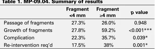
Results: Of the 232 subjects, 131 (56%) required no further intervention
and remained asymptomatic, 34 (15%) developed complications requir-ing no intervention, and 67 (29%) required intervention. Fragments >4 mm were more likely to grow over time (p<0.001) and have complications (p=0.039). Logistic regression shows the original stone size (p=0.0475) to be the only significant predictor of complication. Re-intervention was predictable based on the size (p=0.017) and location of fragments (p=0.02). There was a trend towards complication depending on the loca-tion of residual fragments (p=0.068) and re-intervenloca-tion with older age (p=0.075). Kaplan-Meier analysis found that dusting the stone and larger residual fragments (>4 mm) were more likely to require re-intervention (p=0.004). Re-interventions included URS (58), percutaneous nephroli-thotomy (PCNL) (4), and shock wave lithotripsy (SWL) (3).
Conclusions: This study suggests that fragment sizes >4 mm following
ureteroscopy is associated with significantly higher rates of stone growth, complications, and need for re-intervention. Even for fragments <4 mm, 28% underwent stone growth and 22% suffered a complication, chal-lenging the traditional description of CIRF. Ensuring complete stone-free status is the best way to reduce the rate of complications and interventions following ureteroscopy.
MP-09.05
Routine preoperative electrocardiograms in patients at low risk for cardiac complications during shockwave lithotripsy: Are they useful?
Sowerby, Robert J.1; Ghiculete, Daniela1; Hong, Aaron2; Farcas, Monica A.1; Barrett, Keith P.1; Lee, Jason Y.1; Ordon, Michael1; Pace, Kenneth T.1; Honey, R. John D.1
1Division of Urology, St. Michael’s Hospital, University of Toronto, Toronto, ON, Canada; 2Department of Anesthesia, St. Michael’s Hospital, University of Toronto, Toronto, ON, Canada
Introduction and Objectives: Routine preoperative electrocardiogram
(ECG) prior to shockwave lithotripsy (SWL) is frequently performed, despite recommendations against its use in asymptomatic patients under-going low-risk surgical procedures.1 This study aims to determine if routine preoperative ECG prior to SWL is useful in patients at low risk for cardiac complications.
Methods: A retrospective study of 30 892 patients referred for SWL
(2003-2013) reviewed all cardiac-related preoperative cancellations, intraopera-tive complications, postoperaintraopera-tive admissions, and emergency department (ED) presentations. Patients received SWL with sedation and continuous 5-lead ECG monitoring. Patients with <1% risk of major cardiac event (Revised Cardiac Risk Index) and no history of arrhythmia were classified as low risk for cardiac complications.2
Results: Preoperative ECG triggered 13 (0.04%) cancelations in low-risk
patients (one atrial fibrillation, 12 ischemia/previous infarction). Of these 12 patients, only one had subsequent abnormal cardiac workup and 10 patients underwent uncomplicated SWL without cardiac intervention (two history unknown). Of 27 722 SWL treatments, five (0.02%) were stopped prematurely in low-risk patients due to arrhythmia in which preopera-tive ECG was normal (three), abnormal (one), and not completed (one). Three patients developed arrhythmia with sedation and two patients were admitted postop due to cardiac complications (one atrial fibrillation, one hypertension), of whom all had normal preoperative ECG. No patients presented to our ED with cardiac complications after SWL.
Conclusions: In patients at low risk for cardiac complications,
preopera-tive ECG triggered few cancellations and did not predict arrhythmia during sedation, early termination of treatment, or cardiac complications after SWL. These findings suggest that in low-risk patients, routine preopera-tive ECG has little effect on treatment or complication rate and should be omitted, representing a significant cost savings.
1. Fleisher LA, Fleischmann KE, Auerbach AD, et al. 2014 ACC/ AHA guideline on perioperative cardiovascular evaluation and manage-ment of patients undergoing non-cardiac surgery: A report of the American College of Cardiology/American Heart Association Task Force on Practice Guidelines. Circulation 2014;130:e278 -333. 2. Lee TH, Marcantonio ER, Mangione CM, et al. Derivation and
pro-spective validation of a simple index for prediction of cardiac risk of major non-cardiac surgery. Circulation 1999;100:1043-9. http:// dx.doi.org/10.1161/01.CIR.100.10.1043
MP-09.06
Determining optimal laser settings for non-contact laser lithotripsy (“popcorning”)
Barrett, Keith P.1; Lee, Jason Y.1; Honey, R. John D.1
1Division of Urology, St. Michael’s Hospital, Toronto, ON, Canada
Introduction and Objectives: Intra-renal laser lithotripsy often
pro-duces fragments that may result in future episodes of colic or obstruc-tion. Basket extraction of these fragments may be time-consuming and inefficient. While non-contact laser lithotripsy (“popcorning”) is com-monly used to further reduce such fragments, optimal settings for this approach remain unclear. The objective of this study was to use and in-vitro model to determine the optimal laser settings for “popcorning.”
Methods: A 12 mm diameter test tube filled with 0.9% normal saline
was used to simulate a calyx, and a 365 µ holmium laser fiber was positioned 2 mm above 2 x 4 mm soda-lime phantoms. Two different Table 1. MP-09.04. Summary of results
Fragment <4 mm
Fragment
≥4 mm p value
was recorded prior to lithotripsy. Lithotripsy was carried out for durations of 30 and 90 seconds, with residual weight recorded after each run. In addition, the residual stone was run through a 2 mm sieve, and the weight of fragments >2 mm was recorded. Three runs were performed for each duration at each setting.
Results: Baseline wet weight of the samples were comparable between
the groups (0.176 g vs. 0.171 g; p=0.272). After 30 seconds of lasering, the reduction in residual weight was 0.021 g (-11.8%) in the H/L group and 0.035g (-20.6%) in the H/H group (p=0.154). The residual % of weight >2 mm was 88.2% in the H/L group and 66.7% in the H/H group (p=0.019). After 90 seconds of lasering, the reduction in residual weight was 0.052 g (-13.6%) in the H/L group and 0.051g (-29.3%) in the H/H group (p=0.009). The residual % of weight >2 mm was 68.2% in the H/L group and 43.2% in the H/H group (p=0.033).
Conclusions: The high-energy/high-rate setting resulted in significantly
greater vaporization at 90 seconds and significantly greater fragmentation at 30 and 90 seconds. Use of higher rates appears to be associated with greater efficiency of non-contact laser lithotripsy.
MP-09.07
A multicentred regional emergency department study of renal colic management using medical expulsion therapy
Bristow, Erin1; Kinnaird, Adam2; Schuler, Trevor D.2; Pang, Pamela1; Couperthwaite, Stephanie1; Villa-Roel, Cristina1; Rowe, Brian H.1 1Department of Emergency Medicine, University of Alberta, Edmonton, AB, Canada; 2Division of Urology, Department of Surgery, University of Alberta, Edmonton, AB, Canada
Introduction and Objectives: Patients with renal colic present frequently
to the emergency department (ED). Existing literature suggests manage-ment with medical expulsion therapy (MET) may improve outcomes, especially for those with stones >5 mm in size. This study evaluates the use of MET in the management of adult patients seen in regional EDs with a diagnosis of renal colic.
Methods: A multicentred medical chart review study was conducted in
seven Edmonton-Zone EDs. Approximately 100 cases from each site were randomly selected from administrative data from the 2014 calendar year; no repeat cases were permitted. Using a standardized data collection process and trained research assistance, data were abstracted from medi-cal charts. Medians and interquartile ranges (IQR), proportions, and odds ratios (OR) with 95% confidence intervals (CIs) are reported.
Results: Overall, 656 patient charts were included in the review; median
age was 46 years (IQR 35, 46) and 248 (38%) were female. Many (198 (30%)) received no initial ED imaging; computed tomography (CT) (223 (34%)) was favoured over ultrasound (34 (6%)) for initial imaging. Only 198 (31%) of charts contained documentation of the use of MET at dis-charge and the median duration of therapy was 10 days (IQR 7, 14). Intitiation of MET therapy did not vary based on older age (OR 1.29; 95% CI 0.91, 1.81); sex (OR 1.03; 95% CI 0.73, 1.45); resident involvement (OR 1.01; 95% CI 0.57, 1.82); or presentation to an academic centre (OR 0.80; 95% CI 0.56, 1.14); however, MET was used more for larger (>5 mm) stone size (OR 1.6; 95% CI 1.0, 2.64).
Conclusions: Management of renal colic with MET is uncommon in this
region and practice variation appears driven by physician preference rather than evidence. Practice guidelines with standardized order sets are urgently needed to improve care.
MP-09.08
The impact of body mass index on adverse outcomes in percutaneous nephrolithotomy
Azizi, Mounsif1; Paonessa, Jessica2; Lingeman, James E.2; Bhojani, Naeem1,2
1Department of Urology, Université de Montréal Health Centre, Montreal, QC, Canada; 2Department of Urology, Indiana University School of Medicine, Indianapolis, IN, United States
Introduction and Objectives: Obesity among patients in North America is
increasing and so is stone disease. Percutaneous nephrolithotomy (PCNL) remains the standard of care for renal calculi ≥2 cm. Whether body mass index (BMI) has an impact on adverse outcomes in renal percutaneous surgery is still not well-defined. This study sought to compare outcomes of PCNL in patients of various BMI.
Methods: A retrospective chart review was performed to include all
pro-cedures done at our institution between 2007 and 2011. Patients were categorized into four groups based on their BMI using the World Health Organization classification for obesity: normal weight (18.5-24.9 kg/ m2), obesity class I (30.0-34.9), obesity class II (35.0-39.9) and obesity class III (≥40). Preoperative, operative, and postoperative outcomes were compared. Complications were reported according to the Clavien-Dindo grading system. All procedures were performed in the prone position by a single surgeon.
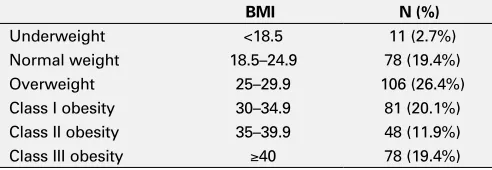
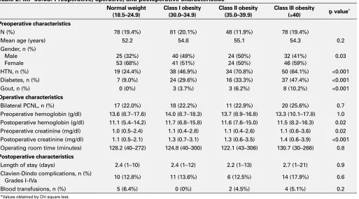
Results: 391 patients were included in this study: 78 (19.4%) with normal
weight and 207 (51.4%) with a BMI ≥30. Patients with obesity (class I-III) had a statistically significant higher baseline serum creatinine (p=0.02) and more hypertension, diabetes, and gout (all p<0.001) compared to patients with ideal body weight. Operating time and length of hospital stay were similar between groups. Postoperatively, patients with a BMI ≥30 had a greater serum creatinine (p<0.001) and hemoglobin (p=0.02) at 24 hours compared to patients with normal weight. There was no statistically significant difference in terms of blood transfusions or Clavien-Dindo complications between groups.
Conclusions: Over half of the patients in our cohort had a BMI ≥30. This study demonstrated that adverse outcomes of PCNL were statisti-cally independent of BMI. Therefore, obesity should not be considered a contraindication to PCNL.
Table 1. MP-09.08. Distribution of patients by body mass index
BMI N (%)
MP-09.09
Secondary diagnostic imaging findings significance in ureteric colic
Wong, Dean1; Innes, Grant2; Remondini, Taylor E.1; Dhaliwal, Navraj1; Dhaliwal, Ravneet1; Frusescu, Adrian1; Weber, Bryce A.1
1Urology, University of Calgary, Calgary, AB, Canada; 2Emergency Medicine, University of Calgary, Calgary, AB, Canada
Introduction and Objectives: Computed tomography (CT) is commonly
used to diagnose and characterize stones in the urinary tract. Stone size and location are strong predictors used to determine types of intervention used in the acute setting. However, the prognostic significance of hydrone-phrosis or peri-ureteral/peri-nephric stranding at the index visit is unclear. We aim to determine if the presence of hydronephrosis or stranding are associated with revisits to the emergency department (ED), admissions or surgical interventions in 60 days in both the medical therapy group and the surgical intervention group.
Methods: Imaging data was collected from ureteric colic patients from
multiple centres in Calgary, Alberta. CT scans were used to confirm the presence of ureteral stones, hydronephrosis, and stranding. Imaging data was linked with hospital data regarding ED revisits, admissions, and inter-ventions.
Results: 1850 patients from four hospitals had imaging confirmed ureteric
stones. A CT scan was used to confirm the presence of a stone for 1732 (93.6%) of these patients. Stranding was noted for 60.2% of CT patients in their imaging reports. Of the patients who had surgical intervention, 62.4% had stranding, while 58.1% of medically managed patients had stranding (p=0.078). There was no significant difference between patients who either had or did not have stranding in terms of revisiting the ED (p=0.21), admissions (p=0.80), and having to undergo a procedure within 60 days of the index visit (p=0.66). Hydronephrosis was seen on 85.3% of the CT imaging reports. 92.5% of surgical patients had hydronephro-sis, while 80.3% of medically managed patients had hydronephrosis
hydronephrosis cases was seen when examining surgically and medically managed patients separately.
Conclusions: For patients with acute renal colic, the secondary feature
of stranding on CT scan is not associated with increases in ED revisits, future admissions, or procedures following their index visit. The presence of hydronephrosis on CT scan, however, results in an increased likeli-hood of procedures being performed after the index visit. These findings are important to consider when counselling patients with symptomatic ureteric stones.
Acknowledgement: MSI Foundation, Edmonton, AB
MP-09.10
Understanding the predictors of negative ureteroscopy: Can we reduce the risk of unnecessary surgery?
Lavoie, Callum1; Levine, Max1; De, Shubha K.1; Schuler, Trevor D.1; Wollin, Timothy A.1; Bochinski, Derek1
1Department of Surgery, Division of Urology, University of Alberta, Edmonton, AB, Canada
Introduction and Objectives: Negative ureteroscopy (nURS) describes
when no stone is found in the kidney or ureter, despite imaging suggest-ing the presence of a stone. The objective of this study was to identify the prevalence of and factors predictive of nURS.
Methods: We performed a retrospective review of 245 URS procedures
for kidney stone treatment performed by three endourologists between January 1 and June 30, 2014. Variables assessed included: demographics, previous stone treatment, stented prior to URS, stone-specific characteris-tics, time from imaging to procedure, type of intervention, etc. Statistical analysis consisted of descriptive statistics, as well as univariate and mul-tivariate logistic regression analyses using SPSS software version 23.0.
Results: The overall rate of negative URS was 8.98%, and the average
patient was 53 years old, average body mass index (BMI) 30.3, and 52.7% were male. Smaller stones were more likely to result in nURS (OR 0.73; Table 2. MP-09.08. Preoperative, operative, and postoperative characteristics
Normal weight
(18.5–24.9) Class I obesity(30.0–34.9) Class II obesity(35.0–39.9) Class III obesity (≥40) p value* Preoperative characteristics
N (%) 78 (19.4%) 81 (20.1%) 48 (11.9%) 78 (19.4%)
Mean age (years) 52.2 54.8 55.1 54.3 0.2 Gender, n (%)
Male Female
25 (32%) 53 (68%)
40 (49%) 41 (51%)
24 (50%) 24 (50%)
32 (41%) 46 (59%)
0.03 HTN, n (%) 19 (24.4%) 38 (46.9%) 34 (70.8%) 50 (64.1%) <0.001 Diabetes, n (%) 7 (9.0%) 24 (29.6%) 16 (33.3%) 37 (47.4%) <0.001 Gout, n (%) 0 (0%) 3 (3.7%) 3 (6.2%) 8 (10.2%) <0.001
Operative characteristics
Bilateral PCNL, n (%) 17 (22.0%) 18 (22.2%) 11 (22.9%) 20 (25.6%) 0.7 Preoperative hemoglobin (g/dl) 13.6 (8.7–17.6) 14.0 (8.7–18.3) 13.7 (8.9–16.8) 13.3 (10.1–17.8) 1.0 Postoperative hemoglobin (g/dl) 11.1 (5.4–14.2) 11.7 (6.8–15.8) 11.6 (7.6–15.0) 11.5 (8.2–16.3) 0.02 Preoperative creatinine (mg/dl) 1.0 (0.5–2.4) 1.1 (0.4–2.8) 1.1 (0.4–2.6) 1.1 (0.6–3.6) 0.02 Postoperative creatinine (mg/dl) 1.1 (0.5–2.1) 1.3 (0.7–3.1) 1.3 (0.6–3.5) 1.4 (0.6–3.9) <0.001 Operating room time (minutes) 128.2 (40–272) 124.8 (40–300) 122.1 (43–306) 130.7 (30–266) 0.8
Postoperative characteristics
Length of stay (days) 2.4 (1–10) 2.4 (1–12) 2.2 (1–13) 2.7 (1–21) 0.9 Clavien-Dindo complications, n (%)
Grades I–IVa 10 (12.8%) 11 (13.6%) 6 (12.5%) 14 (17.9%) 0.6 Blood transfusions, n (%) 5 (6.4%) 0 (0%) 2 (4.5%) 4 (5.1%) 0.2
(57.1% of procedures) was associated with greater likelihood of nURS (OR 5.64; 1.62-19.65; p=0.007). Only stone size remained a significant predictor in multivariate analysis (OR 0.76; 0.62-0.94; p=0.011). More nURS stones were <4 mm (27.3% vs. 5.8%), while fewer were >10 mm (9.1% vs. 38.5%). Distal ureteral stones comprised 45.5% of nURS cases, with an average stone size of 6.5 mm, compared to 29.8% of +URS cases, with an average stone size of 7.7 mm.
Conclusions: The rate of nURS was relatively low in our patient
popu-lation, but not clinically insignificant. Intuitively, it appears as though smaller, distal stones are more likely to pass by the time patients make it to the operating room, suggesting updated preoperative imaging should help play a role in preventing unnecessary instrumentation of these patients. The use of semi-rigid URS being a predictor of passed stones is likely related to an ureterovesical junction (UVJ) position of the distal stone on prior imaging.
MP-09.11
Ambient temperature and the risk for renal colic: A population-based study of the impact of demographics and comorbidity Ordon, Michael1,2; Welk, Blayne K.2,3; Li, Qiongsi4; Wang, Jun4; Lavigne, Eric5,6; Yagouti, Abderrahmane7; Copes, Ray4,8; Burnett, Richard T.9; Cakmak, Sabit9; Chen, Hong3,4,8
1Urology, St. Michael’s Hospital, University of Toronto, Toronto, ON, Canada; 2Institute for Clinical Evaluative Sciences, Toronto, ON, Canada; 3Urology, St. Joseph’s Healthcare, Western University, London, ON, Canada; 4Public Health Ontario, Toronto, ON, Canada; 5Air Health Science Division, Health Canada, Ottawa, ON, Canada; 6Department of Epidemiology and Community Medicine, University of Ottawa, Ottawa, ON, Canada; 7Climate Change and Health Office, Health Canada, Ottawa, ON, Canada; 8Dalla Lana School of Public Health, University of Toronto, Toronto, ON, Canada; 9Population Studies Division, Health Canada, Ottawa, ON, Canada
Introduction and Objectives: To examine the impact of ambient
tem-perature on the incidence of emergency department (ED) admissions for acute renal colic (RC).
Methods: We conducted a population-based, time-series analysis using
linked healthcare databases in Ontario, Canada. The study population consisted of all residents of Ontario, age ≥19 years, who were admitted to the ED from April 2002 to December 2013. The primary outcome was daily number of RC admissions to the ED from each of Ontario’s 14 health regions. A distributed lag non-linear model with 21 days of lag was applied to estimate the cumulative effect of temperature on RC ED visits, controlling for humidity, holidays, long-term trends, and day of the week. We estimated risks for cold and heat, defined as temperatures below and above the optimal temperature, which corresponded to the point with minimum risk of RC admissions. We conducted stratified analyses by selected demographics (age, gender, socioeconomic status (SES)) and by comorbid conditions (hypertension (HTN), diabetes mellitus (DM)).
Results: Over the study period 423 396 patients presented to an ED with
RC. There was a significantly increased risk of RC requiring an ED visit, as ambient temperature increased (comparing daily temperature at 90th percentile vs. the optimal temperature at 10th percentile: RR 1.30; 95%CI 1.20-1.42). Subgroup analysis demonstrated an increased risk from heat for both genders, however, this risk was more pronounced in males (RR 1.36 vs. RR 1.20). In contrast to other age groups, there was an increased risk in those in their 40s (RR 1.42), 50s (RR 1.54) and 60s (RR 1.31). Only the middle SES revealed an increased risk. A history for HTN, but not DM, was associated with an increased risk of RC (RR 2.02).
Conclusions: Increasing ambient temperature was associated with
increased risk of ED visits for RC, particularly in certain demographic subgroups (males, age 40-69 and middle SES) and in those with HTN.
MP-09.12
Kidney stone analyses in Calgary
Burgess, Ellen1; Orton, Dennis1,2; Ward, David1; Sadrzadeh, Hossein1,2 1University of Calgary, Calgary, AB, Canada; 2Calgary Laboratory Services, Calgary, AB, Canada
Introduction and Objectives: As a preparation step to a quality
assur-ance project, an assessment of stone analysis use was conducted by the Clinical Biochemistry Laboratory of Calgary Laboratory Services (CLS).
Methods: In Calgary, CLS reports kidney stone analyses as percent
com-position, as determined by infrared spectroscopy. Categorization of the stones was based on composition and determined by which component made up >49% of the total stone content. An assessment of the use of stone analyses was conducted using de-identified data from specimens submitted from January 2010 to September 14, 2015. Descriptive statis-tics were used.
Results: During this time frame, there were 14 859 kidney stones
submit-ted for stone analysis from 12 433 patients; 1734 (13.95%) patients had more than one stone analysis performed, accounting for 2431 repeat stone analyses (16.36% of the stone analyses were repeat analyses). Of the patients having repeat stone analyses, 1175 (67.8%) patients had 1559 stones analyzed (64.1%) within one year of the first stone analysis. Within three years of the first stone analysis, 1617 patients (93.3%) had 1229 (92.9%) repeat stone analyses were performed. The majority, 76.2%, of the stones were calcium oxalate stones, 9.8% were calcium phosphate, 9.4% were urate, 0.8% were struvite, and 0.02% were cystine stones; the remainder had mixed composition with no single component making up >49% of the total content. 81.7% of the repeat stone analyses had the same dominant content of stone composition as the initial analysis.
Conclusions: Overall, the use of repeat stone analysis accounted for
one-sixth of the stone analyses and usually was not necessary since the dominant composition of the patient’s stone disease did not change.
MP-09.13
Efficacy and safety of percutaneous nephrolithotomy Khan, Faisal1; Motiwala, A.1; Keogan, R.1; Jones, R.1; MacDonagh, R.1 1Urology, Musgrove Park Hospital, Taunton, United Kingdom
Background: Percutaneous nephrolithotomy (PCNL) is the recommended
treatment option for large-volume renal and staghorn stone disease. To evaluate our success and overall complications rates of PCNL, we recorded complications as per European Association of Urology (EAU) guidelines by using Clavien-Dindo grading system. We also emphasized that even in low-volume centres with fewer number of PCNLs performed every year, we can still achieve the acceptable outcome with fewer com-plications.
Methods: A total of 34 patients underwent PCNL from January 2013
to January 2016 at our institute. Demographics, surgical details, and postoperative followup information were obtained to identify stone clear-ance rates and complications.
Results: The majority of PCNLs were performed on the left kidney (n=24),
with mean stone diameter of 2.2 cm. Overall, eight staghorn, 11 multiple, and 15 single stones were treated. The median body mass index (BMI)
Table 1. MP-09.13. Complications as per Clavien-Dindo grading system
Clavien-Dindo grades
No. of
patients % Complications
0 13 38%
I 13 38% Pain, temperature, nausea, bradycardia II 3 9% Sepsis, transfusion III 5 15% Stent, embolization IV 0 0
of patients was 30. Tract puncture were performed by radiologist with image intensifier. 27 cases were stone-free on fluoroscopy, while residual stones were noted in seven cases. Two were managed with extracorporeal shock wave lithotripsy (ESWL), giving an overall stone free rate of 85%. The remaining stones are managed conservatively. The median length of stay was three days. Complications were recorded as per Clavien-Dindo classification system (Table 1). Majority of patients have no or minor com-plications. Only five patients (15%) have Clavien Grade 3 comcom-plications.
Conclusion: The modified Clavien system provides a robust system to
record complications and their severity more accurately. Our results have shown comparable stone clearance and complication rateswith published evidence. We also have proved that even with smaller no of cases per-formed, it is safe to perform PCNL in a district general hospital. Therefore, a skilled surgical team and proper equipment are imperative.
MP-09.14
The effect of obesity on perioperative outcomes following percutaneous nephrolithotomy
Trudeau, Vincent1,2; Karakiewicz, Pierre1,2; Dell’Oglio, Paolo1; Tian, Zhe1; Valiquette, Luc2; Bhojani, Naeem2
1Cancer Prognostics and Health Outcomes Unit, Université de Montréal Health Centre, Montreal, QC, Canada; 2Department of Urology, Université de Montréal Health Centre, Montreal, QC, Canada
Introduction and Objectives: Obesity is on the rise and affects many
patients with kidney stones. We hypothesized that obesity predisposes to higher rates of complications and transfusions, longer length of stay (LOS), and higher total hospital charges (THC) after percutaneous neph-rolithotomy (PCNL).
Methods: Within the Nationwide Inpatient Sample (NIS), we identified
patients treated with PCNL for kidney stones between 1998 and 2010. Obesity was defined as a body mass index >30 using specific International Classification of Disease, 9th revision, clinical modification (ICD-9-CM) codes. We examined the temporal trends in PCNL use and charges among obese and non-obese patients. We then tested the effect of obesity on peri-operative complications, transfusions, LOS, and THC. LOS and THC were defined as a continuous variable and were also dichotomized according to the 75th percentile into prolonged LOS (pLOS) and increased THC (iTHC). Then, multivariable models adjusted for clustering were fitted.
Results: Within the NIS, a weighted sample of 90 529 individuals treated
with PCNL between 1998 and 2010 was examined. Of those patients, 9300 were obese (10.3%). The proportion of PCNLs performed in obese patients increased throughout the years from 7.4-16.7% (p<0.001). Overall complication rates were 21.6 vs. 22.0% (p=0.3) and transfu-sion rates were 4.3 vs. 4.0% (p=0.1) for obese and non-obese patients, respectively. Conversely, pLOS (20.9 vs. 18.8%; p<0.001) and iTHC (30.8 vs. 24.4%; p<0.001) were more frequently recorded in obese patients. In multivariable analyses, obesity was not associated with higher rates of overall complications (odds ratio (OR) 0.94; p= 0.3) nor with higher rates of transfusions (OR 0.89; p=0.3). However, obesity was associated with pLOS (OR 1.21; p=0.002), as well as iTHC (OR 1.17; p=0.002).
Conclusions: PCNL in obese patients did not result in higher rates of
indi-vidual complications or transfusions. However, from a global perspective, it resulted in higher rates of pLOS and iTHC.
MP-09.15
From lithotripters to lasers: History of endourology in Canada Beiko, Darren T.1; Razvi, Hassan2; Denstedt, John D.2; Pace, Kenneth T.3; Honey, R. John D.3; Wilson, James W.1
1Department of Urology, Queen’s University, Kingston, ON, Canada; 2Department of Surgery/Division of Urology, Western University, London, ON, Canada; 3Department of Surgery/Division of Urology, University of Toronto, Toronto, ON, Canada
Introduction and Objectives: The purpose of this study was to report
mile-stones in Canadian endourology, highlighting Canada’s historical contri-butions to advancements in the field of endourology.
recollections by various individuals, including some directly involved, were used in our quest for information on the history of endourology in Canada.
Results: Endourology was born in Canada when shock wave lithotripsy,
ureteroscopy, and percutaneous nephrolithotomy emerged as minimally invasive treatment options for stones in the early 1980s. Dr. Joachim Burhenne, a Harvard-trained radiologist from Germany, first used extra-corporeal shock wave lithotripsy in Canada at the University of British Columbia (UBC) for the treatment of biliary stones. It is believed that the first PCNL was performed at the University of Toronto in the early 1980s. The first use the Swiss Lithoclast for intracorporeal lithotripsy in North America and the first worldwide use of the holmium laser for lithotripsy of urinary tract calculi took place at Western University. Other endourology milestones in Canada include the formation of the Canadian Endourology Group and the emergence of Endourological Society-accredited fellowship programs at the University of Toronto and Western University in the 1990s. Canada hosted the 21st World Congress of Endourology and Shock Wave Lithotripsy annual meeting in Montreal.
Conclusions: Over the past three decades since endourology emerged
in Canada in the 1980s, there have been several milestones in Canadian endourology. Canadian urologists have contributed significantly to advances in the field of endourology and continue to do so. Through the training of the next generation of endourologists at Canadian institutions, the future of endourology in Canada is bright.
MP-09.16
Body composition and the relationship to patients with large stone burden
Zemp, Logan W.1; Baracos, Vickie2; Bochinski, Derek1; Schuler, Trevor D.1; Wollin, Timothy A.1; De, Shubha K.1
1Division of Urology, University of Alberta, Edmonton, AB, Canada; 2Department of Oncology, Cross Cancer Institute, Edmonton, AB, Canada
Introduction and Objectives: Computed tomography (CT) body
compo-sition (BC) analysis has not been previously described in patients with nephrolithiasis. Objective measurement of decreased muscle surface area (MSA) determines sarcopenia status. Sarcopenia is apredictor of morbid-ity in oncologic and emergency surgeries. BC has not been studied to determine the effects on 24-hour urine analysis and type stone produced. The objectives of this study were to determine the BC components may predict various stone types, trends in 24-hour urine metabolites, and complication rates.
Methods: Retrospective review of 152/178 percutaneous nephrolithotomy
cases from February 2011-2012. Body composition was analyzed from a single CT slice at the third lumbar level using Slice-O-Matic V4.3 software. Descriptive statistics were used to determine composition parameters and unpaired t-test was used to compare patient quartiles and differences between patient populations.
Results: Body mass index (BMI) of sarcopenic vs. non-sarcopenic was
28.1 vs. 31.7 (p=0.003). The most prominent type of stones were calcium oxalate (n=84) and uric acid stones (n=31), both of which had mean vis-ceral and subcutaneous adipose tissue cross-sectional areas were elevated compared to calcium phosphate stones. 24-hour urine studies revealed trends of increased Ca, Ox, Na, citrate, and volume in the most muscular quartiles. Increased total adipose tissue was more likely to decrease the pH of the urine. The overall complication rate was 38.2%. Complications occurred in patients with lower mean MSA (132cm2 vs. 149cm2; p=0.01) and lower mean visceral adipose tissue (151cm2 vs. 221cm2).
Conclusions: Advanced body composition analysis may be a more
MP-09.17
Communicating the scans: Are renal stone protocol CT reports giving us enough information?
Hogarth, David1; De, Shubha K.1; Schuler, Trevor D.1
1Division of Urology, University of Alberta, Edmonton, AB, Canada
Introduction and Objectives: Computed tomography (CT) imaging allows
for optimal stone management, as it has been shown to improve predic-tions for spontaneous stone passage and stone-free rates following surgical intervention.1 The objective of this study is to quantitatively assess CT reporting, in regards to the inclusion of clinically important variables.
Methods: A review of 100 consecutive renal stone protocol CT scans
performed on adult patients from the Alberta Urology Institute was con-ducted. Descriptive statistics were used to characterize the frequency of each variable included in the CT reports.
Results: Of 100 stone protocol CT scans, 66 were ordered by primary care
physicians (PCP) and 34 by urologists (Urol). Imaging for symptomatic patients were ordered more commonly by PCP (92% PCP, 21% Urol) where as followup scans were more commonly ordered by urologists (6% PCP, 79% Urol). 70 scans identified obstructing stones, 76% of which were ordered by PCP. All 70 reported stone location and size (47% one-dimension, 26% two-dimensions, 27% three-dimensions), two reported stone attenuation, and none reported skin to stone distance. 76 scans reported non-obstructing stone(s); exact number of stones was reported in 72%, estimated in 21%, and omitted in 7% of reports. All 76 reported stone location and size (68% one-dimension, 13% two-dimensions, 5% three-dimensions, 13% estimation only); none reported attenuation or skin to stone distances. Only three reported total radiation dose. Positive or negative findings of hydronephrosis (98%) and hydroureter (80%) were regularly reported, while other findings, such as perinephric standing (41%) and periureteric stranding (16%), were infrequently reported. Some non-urinary tract findings were often reported (bowel in 64%), while others were rarely reported (free air in 15%).
Conclusions: The indication for stone protocol CT scans change
dramati-cally depending on the ordering physician. Even though consistent report-ing of stone number, location, and hydronephrosis occur, other variables, such as stone density, skin to stone distance, and total radiation dose are infrequently reported.
1. White. American Urological Association, 2012. https://www.auanet. org/common/pdf/education/clinical-guidance/Imaging-Evidence-Report.pdf. Accessed April 27, 2016.
MP-09.18
Different incidence of uric acid stone formation in different nationalities
Naoum, Naimet1; Soryal, Magdy N.2
1Urology, SickKids, Toronto, ON, Canada; 2Urology, Oasis Hospital, Alain, United Arab Emirates
Introduction and Objectives: Different ethnic groups differ in their
abil-ity to form stones in different geographical locations. We aimed to study stone formers of different nationalities (Arabs and non-Arabs) living and working in the same city (Alain) in United Arab Emirates (UAE) for the types of stones they form.
Methods: A retrospective review of patient charts, who presented to a
single urology centre, from 2009-2014, with renal, ureteric,vesicle, or urethral stone(s), who were treated medically or surgically and ended with spontaneous passage or surgical extraction. Stones were analyzed using infrared analysis.The variables that were studied included: age, sex, nationality, stone location, and method of stone treatment. Descriptive and Chi-square categorical analysis were used.
Results: 811 stones studied in 712 patients. The average age was 37.5
year. Males constituted 94.2%. For all the studied groups, the most common stone location was the ureter in 58.1%, and the most com-mon treatment modality was extracorporeal shockwave lithotripsy in 46.2%. Mixed stones composed mainly of calcium oxalate monohydrate (CAOXMH) were the most common in 74.7%, followed by dihydrate in 16.3%, uric acid in 7.6%, and few other types. 77.4% of the patients were Asians of different non-Arabic nationalities, who formed mainly CAOX (monohydrate and dihydrate) stones in 91.6%, and uric acid stones
in 6.5%. Arabs formed CAOX (monohydrate and dihydrate) stones in 85.5%, and uric acid stones in 12.7%. Non-Arabs more significantly formed CAOXMH stones and less significantly uric acid stones than the Arabs (p=0.03, 0.006, respectively), who otherwise formed more signifi-cantly pure uric acid stones (p=0.02).
Conclusions: From a population who live and work in the same city in
UAE, Arabs were found to form more significantly mixed and pure uric acid stones compared with non-Arab stone formers, who more signifi-cantly formed CAOXMH stones. Food, type of work, or other personal predisposition factors may all play roles that needs to be further studied.
UP-09.01
Fluoroscopic radiation exposure in urology: The impact of surgeon-controlled vs. radiation technologist-controlled X-ray exposure
Setterfield, Jeremy G.1; Watterson, James D.1; Playfair, Matthew2; Roberts, Matthew T.1; Blew, Brian D.1; Oake, J. Stuart1
1Urology, The Ottawa Hospital, Ottawa, ON, Canada; 2Undergraduate Medical Education, College of Medical and Dental Sciences, University of Birmingham, Birmingham, United Kingdom
Introduction and Objectives: Our study explored the impact of a switch
from surgeon-controlled (SC) to radiation technologist (RT)-controlled fluoroscopy on fluoroscopy and operative times. We also aimed to identify other factors that impact fluoroscopy and operative times for ureteroscopy (URS) with laser lithotripsy.
Methods: Patients undergoing urological procedures requiring fluoroscopy
(excluding percutaneous nephrolithotomy (PCNL)) six months before and after the change were identified. Demographic data, disease character-istics, fluoroscopy, and operative times were collected via chart review. Median fluoroscopy and operative times were compared between cohorts. A multivariate analysis was performed to identify factors correlating with fluoroscopy and operative times for URS and laser lithotripsy.
Results: No significant difference was found between SC (n=206) and RT
(n=230) cohorts in regards to median fluoroscopy (58 vs. 56.7 seconds; p=0.3428) or operative (38.5 vs. 36 minutes; p=0.1434) times. For URS with laser lithotripsy, median fluoroscopy and operative times were sig-nificantly reduced in the RT cohort (54 vs. 76 seconds; p=0.0028 and 40 vs. 48 minutes, respectively; p<0.0001). Patients undergoing URS alone had a significantly decreased median fluoroscopy time in the SC cohort (47 vs. 73 seconds; p=0.0097). In patients undergoing URS with laser lithotripsy, factors associated with significantly increased fluoroscopy time included male sex, ureteric stent insertion, and flexible ureteroscope or hydrophilic glidewire use. Factors associated with significantly increased operative time included flexible ureteroscope use, large stone size, and difficult ureteric stent insertion.
Conclusions: Fluoroscopy and operative times are not significantly
influ-enced by who controls fluoroscopy during urological procedures. Patients undergoing URS with laser lithotripsy have decreased fluoroscopy and operative times with RT-controlled fluoroscopy exposure. Patients under-going URS alone have decreased fluoroscopy times with SC-fluoroscopy exposure.
UP-09.02
Understanding our failures in getting it up: The prevalence and predictors of failed ureteral access in ureteroscopy
Levine, Max1; Lavoie, Callum1; Wollin, Timothy A.1; Schuler, Trevor D.1; Bochinski, Derek1; De, Shubha K.1
1Division of Urology, University of Alberta, Edmonton, AB, Canada
Introduction and Objectives: Failed access (FA) in ureteroscopy (URS)
occurs when one is unable to reach the anatomic area of interest for treat-ing symptomatic calculi. This results in aborttreat-ing a procedure and a return to the operating room, increasing cost and prolonging discomfort. This study’s purpose was to assess rates of FA among our institution’s stone specialists, while attempting to identify preoperative factors associated with FA.
Methods: Retrospective analysis of 245 consecutive URS performed by
exclu-sion of diagnostic URS or treatment of non-stone pathology. FA was defined as an inability to advance an ureteroscope (semi-rigid or flexible) to an adequate position for treatment of the stone of interest, requiring the procedure to be aborted. Demographic, anatomic, stone-specific, and intraoperative variables were collected. Descriptive statistics, χ2 analysis, and Student’s t-test were used to assess for differences in preoperative fac-tors between successful and FA. Logistic regression was used to identify predictive factors.
Results: From 245 patients reviewed, 16 (6.5%) FA occurrences were
documented. Mean age, gender distribution, and stone size did not differ between the two groups (56 vs. 53 yrs; 56% vs. 52% male; 9.5 vs. 9.1 mm, respectively, N.S.). On χ2 or t-test, no variables differed between the groups; rates of stents in situ at the time of URS (6% vs. 25%, p=0.08) approached significance. 19% of FA stones were located in the distal ureter, 37% proximal ureter, 12.5% ureteropelvic junction (UPJ)/renal pelvis, and 30% were intra-renal (6% upper-, 6% mid-, 18% lower-pole). Between surgeons, the rate of FA ranged from 2-9% (p>0.05).
Conclusions: FA rates in URS are low, but not insignificant (6.5%) among
the three stone specialists in our institution. Stones associated with FA were mostly proximal. Our study failed to identify any preoperative parameters predictive of FA. Limitations include the retrospective nature of our data collection, small sample size from a single centre.
1. Cetti RJ, Biers S, Keoghane SR. The difficult ureter: What is the inci-dence of pre-stenting? Ann R Coll Surg Engl 2011;93:31-3. http:// dx.doi.org/10.1308/003588411X12851639106990
2. Ji C, Gan W, Guo H, et al. A prospective trial on ureteral stenting combined with secondary ureteroscopy after an initial failed pro-cedure. Urol Res 2012;40:593-8. http://dx.doi.org/10.1007/s00240-012-0476-0
3. Viers BR, Viers LD, Hull NC, et al. The difficult ureter: Clinical and radiographic characteristics associated with upper urinary tract access at the time of ureteroscopic stone treatment. Urology 2015;86:878-84. http://dx.doi.org/10.1016/j.urology.2015.08.007
UP-09.03
DaVinci surgical system’s error during robotic surgeries: Result from largest Canadian multispecialty experience at a single academic institute
Rajih, Emad S.1,2; Cormier, Beatrice1; Samoulien, Vanessa1; Lierman, Moishe1; Widmer, Hugues1; Lattouf, Jean-Baptiste1; Alenizi, Abdullah M.1; Valdivieso, Roger1; Hueber, Pierre-Alain1; El-Hakim, Assaad1; Zorn, Kevin C.1
1Department of Surgery, Université de Montréal Hospital Centre (CHUM), Montreal, QC, Canada; 2Urology Department, Taibah University, Madinah, Saudi Arabia
Introduction and Objectives: Several studies have reported on the robotic
error and faults in the initial era of widespread usage in different surgical disciplines. During that time of first-generation robotic system uses and
learning curves, physicians were faced with concerns about surgical robot reliability and errors leading to adverse outcomes.1 Herein, we report on the latest fourth-generation da Vinci Surgical (Si) System malfunctions and errors.
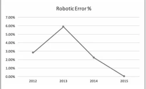
Methods: A total of 844 robotic surgeries were performed between
October 2012 and October 2015 at our academic centre. All cases were also performed with the same dedicated robotic nursing staff. The three subspecialties using the robot included urology, gynecology, and thoracic surgery. Studied outcomes included the robotic surgical error types, occur-rence year, and clinical consequences.
Results: Overall robotic malfunctions were documented on the DaVinci
Si computer console in 2.72% (23/844). The most common error was arm-collision-related in 18 cases (2.13%). Other errors include battery-related in two cases (0.23%), unrecoverable electronic-battery-related in two (0.23%), and illuminator-related in one case (0.11%). Surgical delay was reported only in one patient. No conversion to either open or laparoscopic occurred due to robotic malfunctions. Furthermore, there was no reported patient injury or death directly related to robotic malfunctions.
Conclusions: For all the advanced features the da Vinci system offers,
it continues to be surprisingly reliable. Throughout our multi-users and multi-specialties experience, device failures did not result in any case conversion or procedure abortion. As such, a low device failure rate can be reported when counselling patients undergoing current robotic surgery. Most importantly, while mechanical and electronic errors can happen, they do not appear to impact surgical outcomes or patient safety. 1. Zorn KC, Gofrit ON, Orvieto MA, et al. Da Vinci robot error and
failure rates: Single institution experience on a single three-arm robot unit of more than 700 consecutive robot-assisted laparoscopic radical prostatectomies. J Endourol 2007;21:1341-4. http://dx.doi. org/10.1089/end.2006.0455
UP-09.04
External lower abdominal pressure to aid semi-rigid ureteroscopy in the proximal ureter: Opinion of modern era endourologists, is it safe and effective?
Farcas, Monica A.1,2; Ghiculete, Daniela1; Barrett, Keith P.1,2; Sowerby, Robert J.1,2; Pace, Kenneth T.1,2; Honey, R. John D.1,2
1Department of Urology, St. Michael’s Hospital, Toronto, ON, Canada; 2Department of Urology, University of Toronto, Toronto, ON, Canada
Introduction and Objectives: In 2005, we described the technique of
applying external abdominal pressure (EAP) to allow semirigid ureteros-copy (SURS) above the iliac vessels.1 This led to attempted treatment of all ureteric calculi at our centre with SURS. In this study, we survey modern-era endourologists about their use of this technique. Furthermore, we present our experience with the technique’s safety and efficacy.
Methods: In 2009, a survey was circulated to the Endourological Society
inquiring about use of EAP in SURS. In 2013, the survey was re-circulated to those unfamiliar with the technique in 2009, following provision of its reference. Retrospective chart review included all upper- and mid-ureteric calculi treated with SURS at our centre from 2012-2014 with radiologic followup of at least three months. Access difficulties, intraoperative
com-Table 1. UP-09.03. Type of instruments failure (Was the arm a failure and could not be used or just a recoverable fault/error?)
Type of failure Cases (%)
who had not used this technique yielded a 43% response rate, with 23 having attempted it and 16 planning to continue to using it. Of 519 URSs performed between 2012 and 2014 at our centre, 75 met study criteria. EAP was used in all cases as deemed necessary. In 91% of cases, the ureter was accessed without difficulty. Five (6.7%) conversions to flexible URS were required due to a tortuous or narrow ureter. Two patients (2.7%) were stented due to narrow ureter, and planned for SURS at a later date. There were no ureteric injuries due to SURS over the iliac vessels or psoas muscle. No patients developed ureteric strictures requiring intervention.
Conclusions: Eight years after publication of a technique for using EAP
to aid SURS in the upper ureter, only a minority of endourologists have adopted it. At our centre, this technique continues to be the standard of practice, with excellent success rates and minimal complications. 1. Dagnone AJ, Blew BD, Pace KT, et al. Semirigid ureteroscopy of
the proximal ureter can be aided by external lower-abdominal pressure. J Endourol 2005;19:342-7. http://dx.doi.org/10.1089/ end.2005.19.342
UP-09.05
Compliance of the recurrent renal stone former with current best practice guidelines
Bos, Derek1; Hoogenes, Jennifer1; Kim, Kevin1; Lambe, Shahid A.1; Shayegan, Bobby1; Matsumoto, Edward D.1
1Division of Urology, McMaster Univeristy, Hamilton, ON, Canada
Introduction and Objectives: Patient compliance to best practice
guide-lines is a significant factor in renal stone prevention. Patient compliance has historically been poor. As a result, it has seen increasing attention in the medical literature. However, there remains a paucity of data in the renal stone setting. We evaluated the recurrent renal stone formers’ compliance with current Canadian Urological Association (CUA) best practice guidelines.
Methods: Recurrent renal stone former patients were consecutively
recruited during urology clinic at our institution. Participants completed a one-time questionnaire that was developed in accordance with CUA renal stone best practice guidelines. Questionnaire sections included: 1) demographics; 2) interaction(s) with and satisfaction of encounters with primary care physicians and urologists; and 3) knowledge and compli-ance with best practices.
Results: A total of 191 patients were enrolled in the study; 56% were men,
67% had a positive history of stone surgery, and 22% had a family history of stone disease. Participants perceived satisfactory knowledge translation with their urologist and primary care physician 83.5% and 62.1% of the time, respectively (p<0.05). Patients perceived their disease as severe 22% of the time, while their belief in the utility of preventative stone measures was 65%. Overall, patient compliance with CUA best practice guidelines was 46%; the majority of patients (70%) complied with high fluid intake, the most critical stone-inhibitory practice.
Conclusions: Practice compliance of the recurrent stone former was
rela-tively low in this patient sample, which is consistent with previous studies. The low compliance rate may be attributed to insufficient knowledge translation, lack of perceived disease severity, and/or patient uncertainty regarding the value of preventative stone practices.
UP-09.06
60-day outcomes after early surgical intervention vs. medical treatment of ureteric stones causing acute renal colic
Remondini, Taylor E.1; Innes, Grant2; Cook, Anthony J.1; Amin, Parthiv1; Sami, Samir1; Simister, Riley1; Wong, Dean1; Weber, Bryce A.1
1Urology, University of Calgary, Calgary, AB, Canada; 2Emergency Medicine, University of Calgary, Calgary, AB, Canada
Introduction and Objectives: Renal colic is a common condition,
affect-ing up to 10% of the population. Patients frequently present to the emer-gency department (ED) requiring rapid care and aggressive symptom management. Therefore, we wish to evaluate a large cohort of renal colic patients in a high-intervention setting to determine their characteristics.
Methods: This multicentre administrative database study retrospectively
reviewed the all Calgary patients with an ED diagnosis of renal colic
between January 1, 2014 and December 31, 2014. Tests and treatments were captured from the order entry database, and ED revisits, admissions, and interventions from the discharge abstract database.
Results: Of 3104 renal colic visits, 921 (29.7%) had an index surgical
intervention and 2183 (70.3%) were managed medically. 1850 (59.6%) had imaging-confirmed ureteric stones, with 752 (40.6%) of these patients receiving surgery at an index visit. Patients were more likely to have sur-gery if they had a proximal ureteric stone (OR 2.18; p<0.001), stone larger than 5 mm (OR 4.38; p<0.001), or hydronephrosis (OR 2.81; p<0.001). Of these proximal stone patients, the ones that received surgical intervention at the index visit were less likely to require surgical procedures within 60 days of the index visit (OR 0.52; p=0.006). Patients with stones that are larger than 5 mm in diameter are more likely to revisit the ED (p=0.003), be admitted to the hospital (p=0.06), and undergo a procedure within 60 days (p=0.008). Out of patients with these larger ureteric stones, 412 (63.7%) went on to receive surgery at the index visit. If a patient with a larger stone received a surgery at the index visit, they were less likely to have surgery (OR 0.48; p=0.002), yet more likely to be admitted (OR 1.91; p=0.007) within 60 days of the index visit.
Conclusions: Patients with proximally located ureteric stones or stones
larger than 5 mm in diameter were more likely to have surgeries to treat their colic. Proximal stone patients treated with surgery were less likely to have to return to the hospital to have a procedure performed within 60 days of the index visit. Patients with stones larger than 5 mm who received surgical intervention at the index visit were less likely to have to undergo a future surgery.
Acknowledgement: MSI Foundation, Edmonton, AB
UP-09.07
Intraoperative radiographic determination of ureteric length as a method of determining ideal stent length
Barrett, Keith P.1; Ghiculete, Daniela1; Farcas, Monica A.1; Sowerby, Robert J.1; Pace, Kenneth T.1; Honey, R. John D.1
1Division of Urology, St. Michael’s Hospital, Toronto, ON, Canada
Introduction and Objectives: Accurate determination of ureteric length
and appropriate stent length remains a challenge. The objective of this study was to describe an intraoperative technique to measure ureteric length and determine appropriate stent length, and to compare this tech-nique to other methods of determining ureteric length.
Methods: Patients undergoing ureteroscopy requiring postoperative
stent-ing were prospectively identified. Gender, age, height, body mass index (BMI), and lumbar height on computed tomography (CT) were recorded. Ureteric length was measured using four methods: direct measurement with a ureteric catheter (ULdir), ureteropelvic junction (UPJ) to uretero-vesical junction (UVJ) distance on axial and coronal CT, and using an intraoperative radiographic technique. To perform the radiographic mea-surement, the cystoscope was positioned at the UVJ. A metal bead was affixed to the skin over the UVJ. An angiographic catheter with radiopaque markings at 1 cm intervals was positioned at the UPJ. Ureteric length was the distance from the UPJ to the bead measured using the catheter mark-ers. Correlation between ULdir and the recorded variables and methods of ureteric length measurement were calculated. Stent length was chosen based on radiographic measurement, and was appropriate if it demon-strated a proximal coil in the renal pelvis and a distal coil in the bladder not crossing midline.
Results: 18 ureters were included. Radiographically measured ureteric
length was most strongly correlated with ULdir (r=0.833; p< 0.01). Coronal CT ureteric length was also significantly associated with ULdir (r=0.569; p=0.01). Height, CT axial ureteric length, and lumbar height were not significantly associated with ULdir. Stents were of appropriate length in 17/18 cases.
Conclusions: Radiographic ureteric length measurement was strongly