R E V I E W
Current and emerging therapy for the management
of vitiligo
Alicia Cecile Borderé Jo Lambert
Nanny van Geel
University Hospital of Ghent, Department of Dermatology, Ghent, Belgium
Correspondence: Nanny van Geel Department of Dermatology, De Pintelaan 185, 9000 Ghent, Belgium Tel +32 9 332 22 87
Fax +32 9 332 49 96 Email nanny.vangeel@ugent.be
Abstract: Vitiligo is an acquired cutaneous disorder of pigmentation, with an incidence of 0.5% to 2% worldwide. There are three major hypotheses for the pathogenesis of vitiligo that are not exclusive of each other: biochemical/cytotoxic, neural and autoimmune. Recent data provide strong evidence supporting an autoimmune pathogenesis of vitiligo. As vitiligo can have a major effect on quality of life, treatment can be considered and should preferably begin early when the disease is active. Current treatment modalities are directed towards stopping progression of the disease and achieving repigmentation. Therapies include corticosteroids, topical immunomodu-lators, photo(chemo)therapy, surgery, combination therapies and depigmentation of normally pigmented skin. Topical class 3 corticosteroids can be used for localized vitiligo. The use of topical immunomodulators (TIMs) in vitiligo seems to be equally effective as topical steroids, especially when used in the face and neck region. In photo(chemo)therapy, narrowband ultra-violet-B therapy (NB-UVB) seems to be superior to psoralen ultraviolet-A therapy (PUVA) and broadband UVB. In surgical techniques, split-thickness grafting and epidermal blister grafting were shown to be effective methods, although the non-cultured epidermal suspension technique has many advantages and seems to be a promising development. Depigmentation therapy can be considered if vitiligo affects more than 60% to 80% of the body. Complementary therapies such as Polypodium leucotomos show promising results in combination with UVB therapy. No causative treatment for vitiligo is currently available. More randomized controlled trials on the treatment of vitiligo are necessary.
Keywords: vitiligo, non surgical treatment, surgical treatment
Introduction
Vitiligo is an acquired cutaneous disorder of pigmentation, with a 0.5% to 2% incidence worldwide, without predilection for sex or race. Besides skin, oral mucosa and hair may also be depigmented. Classically, vitiligo is divided into segmental and general-ized forms. The age of onset of vitiligo is variable, but peaks in the second and third decades. The depigmentation has a predilection for acral areas and around body orifi ces (mouth, eyes, nose, anogenital region).1
The Vitiligo European Task Force (VETF) was founded in 2003 during the ESPCR meeting in Ghent and formulated the following defi nition:
“Generalized vitiligo or non-segmental vitiligo (NSV) is an acquired chronic pig-mentation disorder characterized by white patches, often symmetrical, which usually increase in size with time, corresponding to a substantial loss of functioning epidermal and sometimes hair follicle melanocytes”.2
“Segmental vitiligo is an acquired chronic pigmentation disorder characterized by white patches with a unilateral distribution that may totally or partially match a dermatome, but not necessarily. Other distribution patterns can be encountered that cross several dermatomes, or correspond to large areas delineated by Blaschko’s lines”.2
Clinical, Cosmetic and Investigational Dermatology downloaded from https://www.dovepress.com/ by 118.70.13.36 on 21-Aug-2020
Borderé et al
Pathogenesis
The precise cause of vitiligo is unknown. There are 3 major hypotheses for the pathogenesis of vitiligo that are not exclusive of each other: biochemical/cytotoxic, neural and autoimmune. The autoimmune hypothesis is based on genetic data and the fact that vitiligo is associated with other auto-immune diseases. The cytotoxic theory postulates that cell death of melanocytes is caused by cytotoxic precursors to melanin synthesis. The neural hypothesis is based on case reports were patients with nerve injury develop vitiligo at the affected sites and links segmental vitiligo with neurons that interact with melanocytes and release melanocytotoxic substrates.3
As none of the three hypotheses is suffi cient to explain fully the mechanisms of vitiligo, the convergence theory
is proposed. This theory states that stress, accumulation of toxic compounds, infection, autoimmunity, mutations, altered cellular environment and impaired melanocyte migration and proliferation can all contribute in varying proportions to the etiopathogenesis of vitiligo.4
There is increasing evidence for an autoimmune patho-genesis of vitiligo. In 2000, van den Wijngaard et al analyzed lesional, perilesional, and non-lesional skin biopsies from patients with vitiligo and compared these with immune infi l-trates found in the skin of normal healthy donors and relevant disease controls and found a major role for skin-homing T cells in the death of melanocytes seen in vitiligo.5
An autoimmune process is also suggested by the con-comitant occurrence of other autoimmune diseases like autoimmune thyroiditis, diabetes mellitus, pernicious ane-mia, Addison’s disease, primary adrenal insuffi ciency and hypopituitarism in patients with vitiligo.6
Genetic factors also appear to play a role in the etio-pathogenesis of vitiligo as 20% to 30% of patients have a family history of the disorder.7 Vitiligo does not follow
a normal Mendelian pattern of inheritance, and it has been suggested that the disease most likely has a multifactorial, polygenic basis.8
However, many patients with vitiligo have neither a family history of vitiligo nor a history of other autoimmune diseases. A number of other hypotheses have been proposed to explain the pathogenesis of this disorder, like an intrinsic abnormality of melanocytes, increased local catecholamine release, hypoxia, oxidative stress and melanocytorrhagia.9
Treatment
Since a causative treatment is not available, current treatment modalities are directed towards stopping the progression of
vitiligo and achieving repigmentation in order to repair the morphology and functional defi ciencies of the depigmented skin areas.10 Current treatment modalities aim to stimulate
melanocyte proliferation or interfere with infl ammatory factors affecting melanocyte structure or function. No single treatment method has yet been found that is consistently effective with relatively few side-effects.11
Current repigmentation therapies include corticosteroids, topical immunomodulators, photo(chemo)therapy, surgery and depigmentation of normally pigmented skin.
Non-surgical repigmentation treatment
CorticosteroidsDifferent classes of topical corticosteroids were studied. Topical steroids are most effective for lesions on the face, elbows and knees, the distal extremities respond poorly. Factors contrib-uting to the variation in repigmentation rate by anatomic site include skin permeability, migration of residual melanocytes from uninvolved skin, reversibility of melanocyte damage and especially preservation and density of follicular reservoirs.13
In 1977, Bleehen performed a placebo controlled study and treated 20 patients with vitiligo either with 0.1% beta-methasone valerate or with 0.05% clobetasol propionate creams and similar control areas with placebo preparations and concluded that both topical corticosteroids could be used for the treatment of selected patients with vitiligo and can induce repigmentation of the skin.14 Clayton conducted
a double blind study and compared 0,05% clobetasol propri-onate in a cream base with the cream base alone and came to the conclusion that the active product was signifi cantly supe-rior to the cream base alone in the treatment of vitiligo.15
In a controlled randomized trial by Kandil, 0.1% beta-methasone valerate in 50% isopropyl alcohol was compared to the alcohol base alone in 19 patients with vitiligo. A higher percentage of lesions had complete repigmentation when treated with the active product as compared to placebo.16
A study by Lepe et al compared (left-right) 0.05% clo-betasol proprionate with 0.1% tacrolimus for the treatment of vitiligo in children and showed a similar repigmentation rate of 49.3% for clobetasol and 41.3% for tacrolimus.17
Sanclemente et al compared the effect of topical 0.05% betamethasone valerate versus catalase/dismutase superoxide (C/DSO) in a randomized, matched-paired, double-blind trial with 25 patients and concluded that vitiligo repigmentation with topical C/DSO at 10 months is similar to repigmenta-tion with topical 0.05% betamethasone valerate.18
In 1999, Njoo et al performed a meta-analysis of 20 ran-domized controlled trials and 75 patient series on the treatment
Clinical, Cosmetic and Investigational Dermatology downloaded from https://www.dovepress.com/ by 118.70.13.36 on 21-Aug-2020
Vitiligo therapies
of vitiligo and concluded that topical class 3 corticosteroids and ultraviolet-B therapy (UVB therapy) are the most effec-tive and safest therapies for localized and for generalized vitiligo, respectively.19
The biggest side-effect of topical corticosteroids is skin atrophy, more seen in class 4 than in class 3 corticosteroids. Other side-effects were striae, hypertrichosis, acneiform eruption and telangiectasia.
High and low doses of oral corticosteroids in pulse therapy have been shown to be successful in patients with progressive disease.20,21 Recently, Rath et al compared
dif-ferent phototherapy methods (psoralen ultraviolet-A PUVA [therapy], narrowband UVB and broadband UVB) with an oral minipulse of steroids (OMP) as an adjunct to determine the method with the best tolerability and effi cacy. They ran-domly assigned 68 patients with progressive vitiligo to differ-ent study groups (OMP, OMP + PUVA, OMP + narrowband UVB, OMP + broadband UVB). Each patient was followed up for 6 months and then the treatment was discontinued. Clinical evaluation was made at the end of 3 and 6 months. They concluded that the oral steroids only had an adjunct value and were not very effective by themselves. Narrowband UVB is preferred over broadband UVB. Narrowband UVB and PUVA showed comparable effi cacy.22
No placebo controlled studies have been performed for the use of oral corticosteroids in vitiligo yet. So their use is still considered controversial since systemic side-effects (eg, moon face, weight gain) are associated with this therapy.
Photo(chemo)therapy
The ultraviolet (UV)-spectrum is divided into 3 major groups: UVA, UVB en UVC. UVC-radiation (200–280 nm) is fi ltered by the atmosphere and has no place in phototherapy. UVB-radiation (280–320 nm) is biologically the most active. UVA-radiation (320–400 nm) has the longest wavelength of the UV-spectrum and is biologically less active than UVB-radiation. However, it is partly responsible for erythema and pigmentation induced by sunlight.23
(P)UVA
Only a few studies (no randomized, controlled trials) have been performed, studying UVA-therapy in vitiligo. El Mofty et al studied the effect of broadband UVA on vitiligo lesions in 20 patients, 15 J/cm2 in group I and 5 J/cm2 in group II,
a total of 48 sessions over 16 weeks. All patients received 3-weekly sessions of UVA. Overall pigmentation of 60% and above was recorded in 50% and 10% of patients in groups I and II, respectively. They concluded that broadband UVA
alone, without psoralens, may have an important therapeutic value in the treatment of vitiligo.24
Westerhof et al performed a left-right comparison study of the combination of fl uticasone propionate and UVA versus either fl uticasone propionate or UVA for the long-term treat-ment of vitiligo and concluded that the combination treattreat-ment with fl uticasone proprionate and UVA is much more effective in reaching complete repigmentation than are fl uticasone proprionate and UVA used alone, but large inter-individual differences occurred in the study.25
PUVA therapy
This is the combination of body irradiation with UVA-light and a psoralenderivate. The psoralens most commonly used are 8-methoxypsoralen (8-MOP), 5-methoxypsoralen (5-MOP) and 4,5’, 8-trimethylpsoralen (TMP). Already in ancient Egyptian times, plant extracts containing psoralens were used to treat patients with vitiligo.26 In the 1940’s,
PUVA was introduced to treat vitiligo.27 In PUVA therapy,
the psoralens can be applied topically or can be taken orally. In vitro studies demonstrated that oral PUVA stimulates melanocytes to proliferate, differentiate and migrate. It also has a local immunomodulatory effect.28 Ermis et al
performed a placebo-controlled double-blind study to inves-tigate whether the effectiveness of PUVA therapy could be enhanced by combination with topical calcipotriol in the treatment of vitiligo and concluded that concurrent topical calcipotriol potentiates the effi cacy of PUVA therapy in the treatment of vitiligo, and that this combination achieves earlier pigmentation with a lower total UVA dosage.29
Oral PUVA therapy has a lot of side-effects like nausea, pruritus, phototoxic reactions and a large contrast in pig-mentation between normal and repigmented skin. Therefore, PUVA therapy is not the fi rst choice in the treatment of vitiligo.
UVB
The exact mechanism of vitiligo repigmentation due to UVB is not known. It is probably related to the positive effect of UVB on immunomodulation, growth stimulation and pro-liferation of melanocytes.
The traditional broadband UVB sources were the fi rst to be used. Broadband UVB therapy, which implies signifi cant exposure to wavelength spectra of less than 300 nm and even less than 280 nm (UVC), carries in theory a greater risk of long-term development of nonmelanoma skin cancer com-pared with narrowband UVB (UVB 311 nm), although there is no clinical study that proves this in practice.30
Clinical, Cosmetic and Investigational Dermatology downloaded from https://www.dovepress.com/ by 118.70.13.36 on 21-Aug-2020
Borderé et al
In 1990, Köster et al reported repigmentation of more than 75% in 8 of 14 patients suffering from extensive vitiligo, who underwent phototherapy with broadband UVB. Espe-cially the patients with facial lesions of the skin types V and VI achieved a nearly complete and cosmetically satisfying repigmentation after 12 months of treatment.31
Recently, Asawanonda et al performed a small ran-domized, controlled trial (20 patients) comparing targeted narrowband UVB with targeted broadband UVB and concluded that the clinical responses were similar in both groups. However, the device used in this trial was a targeted UVB device, this implicates that the results cannot be applied to other light sources (eg, excimer laser or total-body irradia-tion cabinets).32
Narrowband UVB (NB-UVB) is a more recent form of phototherapy that uses wavelengths between 305 and 311 nm. Short-time side-effects include pruritus and xerosis, long-term side-effects (eg, carcinogenesis) are unknown. NB-UVB can be used in children, pregnant or lactat-ing women and in individuals with hepatic or kidney dysfunction.23
In 1997, Westerhof fi rst reported the use of NB-UVB phototherapy for the treatment of vitiligo and concluded the treatment of patients with vitiligo with 311-nm UV-B radiation is as effi cient as with topical PUVA and has fewer adverse effects.33 In a recent study by Rath et al narrowband
UVB was preferred over broadband UVB. Narrowband UVB and PUVA showed comparable effi cacy.22
Esfandiarpour et al studied 68 patients with vitiligo. The patients were divided into two groups and treated with NB-UVB in combination with pimecrolimus or with NB-UVB in combination with a placebo cream for 3 months. The authors concluded that, on the face, NB-UVB works better if combined with 1% pimecrolimus cream rather than used alone.34
El Mofty et al performed an evaluation of NB-UVB 311 nm in the treatment of vitiligo by two independent studies. The fi rst study compared NB-UVB with PUVA therapy, and the second study compared NB-UVB with NB-UVB + psoralens. They showed no statistical differ-ence in repigmentation or side-effects between NB-UVB and PUVA therapy. In the second study, there was no statistical difference in repigmentation between NB-UVB and NB-UVB + psoralens, but the group with psoralens had more side- effects.35
In 2007, Yones et al performed a randomized double blind trial in 56 patients with nonsegmental vitiligo, comparing PUVA therapy with NB-UVB therapy and concluded that,
in the treatment of nonsegmental vitiligo, NB-UVB therapy is superior to oral PUVA therapy.36
Arca et al compared NB-UVB as monotherapy to the combination of NB-UVB + calcipotriol in a randomized study and concluded that NB-UVB is an effective therapy for vitiligo but they could not show an additional effect of topical calcipotriol.37
We can conclude that narrowband UVB is as effective (or more effective) than PUVA therapy in the treatment of vitiligo but has fewer side-effects. It is preferred over broad-band UVB and is a promising treatment for vitiligo.
Excimer laser
The xenon chloride excimer laser emits a wavelength of 308 nm. The excimer laser has some useful technical char-acteristics like an articulated arm and variable spot size. It is possible to selectively turn the beam of light and to treat only the involved area, sparing healthy skin. In vitiligo, this selectivity limits the unwanted hyperpigmentation of non involved skin, which is commonly observed with the other phototherapies. The articulated arm makes it possible to treat body regions that are otherwise diffi cult to reach (eg, folds and mucosa). Disadvantages include the fact that the limited spot size means that large surfaces (⬎20% of total surface body area) cannot be treated and that purchase and mainte-nance costs of these devices remain quite expensive.38
Hadi et al performed a retrospective chart review of 97 patients (a total of 221 vitiligo patches) with chronic stable vitiligo treated with the excimer laser and concluded that it is an effective and safe modality for the treatment of vitiligo, with good results achieved in a relatively short duration of time. Lesions on the face responded better than lesions elsewhere.39
In 2006, Passeron and Ortonne performed a review of 10 studies on the use of the 308-nm excimer laser for pso-riasis and vitiligo. For vitiligo, low fl uencies (from 50 to 200 mJ/cm2) were used in 1 to 3 sessions a week for 1 to
6 months, depending on the study. The number of plaques with repigmentation at the end of the treatment was high (57%–100%). On average, 20% to 30% of treated plaques reach more than 75% repigmentation, but some series report confl icting results (from 0% to 75%). The clinical response to the treatment is especially dependent of the localization of the lesions. UV-resistant areas (eg, extremities and bony prominences) were more diffi cult to treat.38
Passeron et al examined the effi cacy of combined treat-ment with 0.1% tacrolimus ointtreat-ment plus 308 nm excimer laser versus excimer laser monotherapy. They concluded that the combined therapy was only superior to the excimer laser
Clinical, Cosmetic and Investigational Dermatology downloaded from https://www.dovepress.com/ by 118.70.13.36 on 21-Aug-2020
Vitiligo therapies
monotherapy for the treatment of UV-resistant vitiliginous lesions.40
Goldinger et al did a prospective (left-right) comparative, single blinded study to compare the effectiveness of the excimer laser and the combination of the excimer laser with topical calcipotriol in the treatment of vitiligo. They showed no signifi cant difference in overall repigmentation between the two groups.41
Just recently, Sassi et al did a randomized, controlled trial to compare the effectiveness of the 308 nm excimer laser alone or in combination with topical hydrocortisone 17-butyrate cream in patients with vitiligo, unresponsive to previous treatment with topical steroids or narrowband ultra-violet B phototherapy. They found that recalcitrant vitiligo of the face and neck may benefi t from the combination of excimer laser phototherapy with topical hydrocortisone 17-butyrate cream.42
The excimer laser is a promising therapy for vitiligo, with good clinical results, especially for the treatment of vitiligo lesions on the face.
Calcipotriol
Calcipotriol is a vitamin D3 analogue that might be effective on immunomodulatory systems, infl ammatory mediators and melanocytes.43 Tomita et al showed that vitamin D3 induced
features similar to those noted in UV-radiated skin (such as swelling of melanocytes in the epidermis and increased tyrosinase activity resulting in a deposition of melanin in the epidermis).44
Kumaran et al conducted a randomized trial that studied the effect of 0,005% topical calcipotriol, 0.05% betametha-sone dipropionate and their combination in the treatment of localized vitiligo. When used individually, the betamethasone dipropionate and the calcipotriol were found to be equally effective but the combination of the two appeared to give a signifi cantly faster onset of repigmentation along with better stability of the achieved pigmentation and with lesser number of side-effects.45
Ameen et al conducted an open study to investigate the effi cacy and tolerability of calcipotriol cream as mono-therapy and in combination with PUVA mono-therapy. They found that topical calcipotriol appears to be an effective and well-tolerated treatment for vitiligo and can be safely used in combination with PUVA.46 Chiavérini et al performed a
prospective, right-left comparative, open study and exam-ined the effi ciency of topical calcipotriol as a monotherapy for the treatment of vitiligo. They concluded that it was not effective.47
However, no randomized controlled trials have been performed yet with calcipotriol as a monotherapy for vitiligo. The combination of calcipotriol and PUVA or UVB has also been studied in other trials, but the results are contradicting as some authors describe a benefi t as others do not.41,43
Topical immunomodulators (tacrolimus/ pimecrolimus)
Tacrolimus and pimecrolimus (macrolide immunomodulators) can be used topically and are called topical immunomodu-lators (TIMs). TIMs inhibit the action of calcineurin, and consequently prevent the transcription of infl ammatory cytokines dependent on the transcription factor nuclear fac-tor of activated T-cells (NF-AT). This reversible inhibition of cytokine synthesis affects both T helper type 1 (Th-1) cytokines (interleukin-2, interferon-γ) and Th-2 cytokines (interleukin-4, interleukin-10). This is considered the work-ing mechanism of action of TIMs in vitiligo.48,49
The use of TIMs in vitiligo has been documented in randomized controlled trials and in several case reports and open studies. One randomized controlled study compared 0.1% tacrolimus and 0.05% clobetasol cream in 20 children with vitiligo. The level of repigmentation was 49.3% with clobetasol and 41.3% with tacrolimus.17 Dawid et al
per-formed a double-blind, intra-patient comparison of 1% pimecrolimus cream with placebo cream in 20 patients with vitiligo predominantly situated on the extremities and not on the face and found no signifi cant change in mean target lesion.49
Since 2002, clinical studies have favored the use of calci-neurin inhibitors in vitiligo, especially in children, in whom they were shown to be equally effective as corticosteroids. However, in adult patients the effect was mainly restricted to the face and neck, whereas lesions in other regions of the body showed variable responses.50 Just recently, Choi et al
performed a retrospective review of 79 patients and showed that topical immunomodulators are as effective as topical steroids in repigmentation of vitiligo. They also showed that the duration between start of treatment and onset of repigmentation was signifi cantly shorter in the topical immu-nomodulator group.51
In a prospective placebo-controlled right-left comparison study, Hartmann et al investigated the effi cacy and safety of 0.1% tacrolimus ointment for up to 12 months in 30 adult patients with vitiligo, and tested the infl uence of additional occlusion. They concluded that 0.1% tacrolimus ointment proved an effective and safe treatment option for adult patients with vitiligo. Beyond the face and neck areas, repigmentation
Clinical, Cosmetic and Investigational Dermatology downloaded from https://www.dovepress.com/ by 118.70.13.36 on 21-Aug-2020
Borderé et al
could be achieved only by additional occlusion (eg, household foil, hydrocolloid dressing or polyurethane foil).50
A recent randomized, double-blind, placebo-controlled study compared combination of 1% pimecrolimus with NB-UVB versus placebo with NB-UVB and concluded that, on the face, NB-UVB works better if combined with 1% pimecrolimus cream than used alone.52
Topical calcineurin inhibitors are effective in the treat-ment of vitiligo, especially when the lesions are situated on the face.
Unlike topical corticosteroids, topical calcineurin inhibi-tors do not cause skin atrophy. A noted side-effect was a burning sensation at the site of application. In 2005, the Pediatric Advisory Committee of the US FDA implemented a black box warning for tacrolimus and pimecrolimus due to the lack of long-term safety data and the potential risk of the development of malignancies. However, evidence of a causal relation to the development of skin cancer or lymphoma is still missing. More studies are needed to understand this risk.53,54
Pseudocatalase cream
One study showed that patients with vitiligo have low catalase levels in their epidermis in association with accumulation of hydrogen peroxide (H2O2). A topical pseudocatalase cream and calcium used twice daily with UVB therapy, twice weekly, resulted in repigmentation in the majority of cases in a case study with 33 patients after 2–4 months. However, it was not known whether the repigmentation was due to the UVB or the pseudocatalase.55 Sanclemente et al compared the effect
of topical 0.05% betamethasone valerate versus catalase/ dismutase superoxide (C/DSO) and concluded that vitiligo repigmentation with topical C/DSO at 10 months is similar to repigmentation with topical 0.05% betamethasone valerate.18
More studies on the use of pseudocatalase cream in the treatment of vitiligo are necessary to prove its possible therapeutic effect.
Comparison
Recently, Lotti et al performed an open-label study compar-ing the current non-surgical treatments for vitiligo (311-nm narrowband microphototherapy, 0.1% tacrolimus ointment twice a day, 1% pimecrolimus cream twice a day, 0.05% betamethasone dipropionate cream twice a day, 0.005% calci-potriol ointment twice a day and 10% l-phenylalanine cream twice a day as a monotherapy or the NB-UVB therapy in com-bination with the above mentioned local treatments). They concluded that when the single treatments are considered
alone, 311-nm narrowband UVB microphototherapy and 0.05% betamethasone dipropionate cream are the most effective treatments. In combination therapies, 0.05% betamethasone dipropionate cream plus 311-nm narrowband UVB microphototherapy gave the best repigmentation rate. In this study, the only side-effects registered were cutane-ous atrophy with the corticosteroid cream, and stinging and burning with the topical immunomodulators.56
Surgical repigmentation therapy
Surgical repigmentation can be considered in stabilized viti-ligo, if other repigmentation therapies fail. All surgical tech-niques have the same basic principle: to transplant autologous melanocytes from pigmented donor skin to regions without melanocytes. However, patients need to be carefully selected as not every vitiligo patch is suitable for transplantation. Sur-gical techniques may be considered in stable, non-progressive disease only (there is no consensus on the exact period of stability, various recommendations suggest stable vitiligo for 6 months to 2 years).57 Contraindications are a positive
Koebner phenomenon and hypertrophic scars or keloids in the past. The site of transplantation is also important, as some areas such as joints, lips, eyelids, genitalia, cutaneous folds, the dorsum of hands and feet, fi ngers and toes, are diffi cult to repigment.58
There are several methods for surgical repigmentation.
Autologous minipunchgrafting
The fi rst method is autologous minipunchgrafting were multiple punch biopsies are obtained from a normally pig-mented donor site and implanted within achromic areas, separated from each other by 5 to 8 mm. To determine if this technique works for a specifi c patient, a test-graft is performed (3–5 minigrafts). If pigment has spread from the donor tissue after a period of time and no Koebner phenomenon exists at the donor site, the entire region is grafted. This procedure is useful for limited areas of vitiligo, but a cobblestone appearance, sinking pits, infection or scarring are potential side-effects.3,59 In 1988, Falabella treated 22 patients with
localized vitiligo with autologous minigrafting and a 90% to 100% repigmentation was achieved in 13 patients.60 Another
study by Sarkar et al showed a repigmentation rate of 40% to 60% in the majority of the treated patients who has segmental vitiligo.61 Boersma et al performed minigrafts in patients
with stable vitiligo vulgaris (stable for 6 months). 14 out of 36 lesions showed 80% to 99% repigmentation after 6 months. They state that repigmentation rates of more than 80% are generally not reached before 6 months after grafting.62
Clinical, Cosmetic and Investigational Dermatology downloaded from https://www.dovepress.com/ by 118.70.13.36 on 21-Aug-2020
Vitiligo therapies
Epidermal suction blister grafting
Another technique is epidermal suction blister grafting. Blisters are formed at donor and recipient sites with a suction machine or by freezing with liquid nitrogen for 20 to 25 seconds. The roof of the blister is removed from both sites and the donor graft is placed on top of the denuded recipient site. The grafts are held in place with bandages for 7 days. Repigmentation usually occurs in 1 to 2 weeks, and total repigmentation can occur within 1 to 3 months.3 Gupta et al compared epidermal
suction blister grafting to punch skin grafting in 49 patients and reported a repigmentation rate of more than 75% in 67% of the lesions in the punch graft group and in 82% of the lesions in the epidermal suction blister group. This difference was not signifi -cant. However, the latter technique gives cosmetically better results. 63 The overall success rate of epidermal suction blister
grafting varies between 73% and 88% in different studies and is increased if the donor site is treated with PUVA therapy before the procedure. Epidermal grafting shows excellent cosmetic results and is an easy, inexpensive, safe and effi cient treatment, however it is a time consuming technique.59
Split-thickness grafting
In split-thickness skin grafting, the whole epidermis and a part of the upper papillary dermis is grafted at the donor site using a hand dermatome or shaving blade. Grafts are posi-tioned on abraded recipient areas that have been prepared previously. The graft is secured with pressure and immobi-lization. This technique has a success rate of 78% to 90%.59
Ozdemir et al were the fi rst to compare 2 surgical techniques in a blinded matter (epidermal suction blister grafting versus split-thickness skin grafting). In their study, 25% of patients showed repigmentation with suction blister erosion with-out grafting, 45% of patients showed repigmentation with suction blister grafting. Thin split-thickness grafting led to repigmentation in 90% of patients.64 Success rates depend on
using a very thin graft to prevent additional cosmetic damage to both recipient and donor areas. Side-effects of this tech-nique are stuck-on effects, persistent hyperpigmentation, milia, perigraft halo of depigmentation and scars on donor areas, and it is not suitable for vitiliginous lesions on the palms, soles, and skin folds. The benefi ts are the immediate results, the highest success mean rate and the ability to treat diffi cult areas like eyelids, penis, vagina and lips.65,66
Transplantation of non-cultured epidermal cellsuspension
In 1992, Gauthier and Surleve-Bazeille described a technique in which non-cultured melanocytes were isolated from samples
of skin of the hair scalp, then trypsinized and transplanted. The grafting method is carried out in two steps: fi rst, blisters are formed on the depigmented lesions by freezing with liquid nitrogen, then, each blister is injected with a suspension of epidermal cells (mainly keratinocytes and melanocytes). Repigmentation occurred within 25 to 30 days and they concluded that this technique is an effective and simple transplantation method.67 With this technique, large areas can
be treated (5- to 10-fold the donor area). Disadvantages are a long-lasting erythema (up to 6 months) at the recipient site due to dermabrasion and the need for specialized laboratories with trained personnel.59
Van Geel et al performed the fi rst double-blind placebo-controlled study on autologous transplanted epidermal cell suspensions for repigmenting vitiligo. To improve the viscosity and fi xation of the cellsuspension, they investi-gated a modifi ed approach adding hyaluronic acid to the cellular grafting procedure. In this study, transplantation resulted in repigmentation of at least 70% of the treated area in the majority of patients. They could demonstrate that repigmentation was primarily caused by the transplanted melanocytes, as repigmentation at the placebo treated site was negligible.68
Mulekar performed a long-term follow-up study in 142 patients treated with a non-cultured melanocyte–keratinocyte cell transplantation. More than half of the treated patients had a complete repigmentation that was fully retained for a 6-year follow-up period.69
Transplantation of in vitro-cultured epidermis
A shave biopsy of normally pigmented skin is taken. After separating the epidermis from the dermis, the cells are seeded in a medium that allows co-cultivation of melanocytes and keratinocytes. After 3 weeks a cultured sheet is obtained, released by treatment with dispase and attached to a gauze as support. The gauze (to which the epithelium adheres) is placed onto the recipient site after it is treated with derm-abrasia.55 This technique has the advantage that a large area
can be treated (10 fold the donor site). Studies have shown a good repigmentation in 33% to 54% of treated patients.59,70,71
A side-effect of this technique can be hyperpigmentation, but this tends to disappear after a few months. Another disadvantage is failure of culture and the fact that it is an expensive procedure.
Transplantation of in vitro-cultured melanocytes
Here, melanocytes are obtained from shave biopsies and cultured in vitro with addition of growth factors and chemical
Clinical, Cosmetic and Investigational Dermatology downloaded from https://www.dovepress.com/ by 118.70.13.36 on 21-Aug-2020
Borderé et al
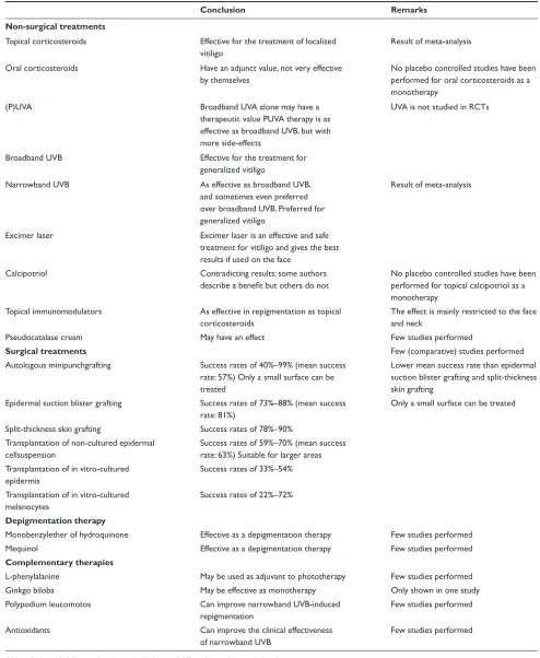
Table 1 Conclusions on treatment for vitiligo
Conclusion Remarks
Non-surgical treatments
Topical corticosteroids Effective for the treatment of localized vitiligo
Result of meta-analysis
Oral corticosteroids Have an adjunct value, not very effective by themselves
No placebo controlled studies have been performed for oral corticosteroids as a monotherapy
(P)UVA Broadband UVA alone may have a
therapeutic value PUVA therapy is as effective as broadband UVB, but with more side-effects
UVA is not studied in RCTs
Broadband UVB Effective for the treatment for generalized vitiligo
Narrowband UVB As effective as broadband UVB, and sometimes even preferred over broadband UVB. Preferred for generalized vitiligo
Result of meta-analysis
Excimer laser Excimer laser is an effective and safe treatment for vitiligo and gives the best results if used on the face
Calcipotriol Contradicting results: some authors describe a benefi t but others do not
No placebo controlled studies have been performed for topical calcipotriol as a monotherapy
Topical immunomodulators As effective in repigmentation as topical corticosteroids
The effect is mainly restricted to the face and neck
Pseudocatalase cream May have an effect Few studies performed
Surgical treatments Few (comparative) studies performed
Autologous minipunchgrafting Success rates of 40%–99% (mean success rate: 57%) Only a small surface can be treated
Lower mean success rate than epidermal suction blister grafting and split-thickness skin grafting
Epidermal suction blister grafting Success rates of 73%–88% (mean success rate: 81%)
Only a small surface can be treated
Split-thickness skin grafting Success rates of 78%–90% Transplantation of non-cultured epidermal
cellsuspension
Success rates of 59%–70% (mean success rate: 63%) Suitable for larger areas Transplantation of in vitro-cultured
epidermis
Success rates of 33%–54%
Transplantation of in vitro-cultured melanocytes
Success rates of 22%–72%
Depigmentation therapy
Monobenzylether of hydroquinone Effective as a depigmentation therapy Few studies performed
Mequinol Effective as a depigmentation therapy Few studies performed
Complementary therapies
L-phenylalanine May be used as adjuvant to phototherapy Few studies performed
Ginkgo biloba May be effective as monotherapy Only shown in one study
Polypodium leucomotos Can improve narrowband UVB-induced repigmentation
Few studies performed
Antioxidants Can improve the clinical effectiveness of narrowband UVB
Few studies performed
Abbreviations: PUVA, psoralen ultraviolet-A therapy; RCT, randomized controlled trial.
Clinical, Cosmetic and Investigational Dermatology downloaded from https://www.dovepress.com/ by 118.70.13.36 on 21-Aug-2020
Vitiligo therapies
media. The suspension is then transplanted on abrased recipient areas. Success rates vary between 22% and 72%. Disadvantages are that it is a time consuming technique and that there are only a few trials published that study this technique.57 In 2004, Chen et al performed an analysis of
120 patients with vitiligo who were treated with autologous cultured pure melanocyte suspension combined with carbon dioxide laser abrasion. They concluded that this technique is an effective treatment for patients with stable vitiligo who fail to respond to medical treatments, especially for those with stable localized vitiligo.72
As there are no uniform and acceptable criteria for the evaluation of the outcome after surgical repigmentation, it is diffi cult to compare the results of different studies on dif-ferent transplantation methods.
In 1998, Njoo et al performed a systematic review of the effectiveness, safety, and applicability of autologous transplanta-tion methods in vitiligo. Recently developed techniques using noncultured epidermal cellsuspension were not included in the review. Split-thickness grafting and epidermal blister grafting were the most effective and safest methods in this review. Mini-grafting showed a relatively lower success rate, which could be explained by variations in the size of pigment spread of the punch grafts. They state that postoperative PUVA therapy or exposition to sunlight may improve the repigmentation grade in minigraft-ing.73 However, as no randomized controlled trials were included
in the review, results should be interpreted with caution.
Depigmentation therapy
Depigmentation with monobenzylether of hydroquinone can be considered if vitiligo affects more than 60% to 80% of the face or body. The major side-effect is contact or irritant con-tact dermatitis. Only a few studies (no randomized controlled trials) with hydroquinone for vitiligo have been performed. In a retrospective study of 18 vitiligo patients who used 20% monobenzylether of hydroquinone as a depigmenting agent, 8 achieved complete depigmentation after 10 months of use and 3 achieved dramatic but no complete depigmentation.74
In 1996, Oakley described a rapid repigmentation after depigmentation with monobenzylether of hydroquinone, the mechanism was unknown.75
4-methoxyphenol (4-MP) or mequinol is another phe-nol derivate and has similar melanocytotoxic properties as monobenzylether of hydroquinone. Njoo et al performed a retrospective study on depigmentation therapy in 16 patients and concluded that depigmentation therapy using a 4-MP cream and/or Q-switched ruby laser therapy can be an effective and safe method to remove residual pigment in
patients with vitiligo universalis.76 However, because there
are no randomized controlled trials performed, results should be considered with caution.
Complementary therapies
Just recently, Szczurko and Boon performed a comprehen-sive review of literature on different natural health products that may have an effect on vitiligo. The most commonly studied product was L-phenylalanine, not in monotherapy but in combination with phototherapy or other products. Although it was not possible to pool the collected data, the overall outcome was that there is moderate evidence for effi cacy of L-phenylalanine as adjuvant to phototherapy. There was no convincing evidence that vitamins have a place in vitiligo treatment.77 Ginkgo biloba also seems to be
promising as monotherapy for vitiligo in a double-blind, pla-cebo-controlled trial, performed by Parsad et al but more and controlled studies are necessary to confi rm this effect.78
In 2007, Middelkamp-Hup et al performed a randomized double-blind placebo-controlled study to determine if polypo-dium leucotomos, an antioxidative and immunomodulatory plant extract, improves narrowband UVB-induced repigmen-tation. There was a trend towards an increased repigmentation of head and neck (not other body areas) in the group that was treated with the combination of polypodium leucomotos and UVB. This effect was more pronounced in patients with light skin types (type II and III).79 Another study by Dell’Anna et al
evaluated the combination treatment of antioxidants (contain-ing alpha-lipoid acid, vitamins C and E and polyunsaturated fatty acids) and narrowband UVB in a double-blind placebo controlled trial and concluded that the antioxidants sig-nifi cantly improved the clinical effectiveness of narrowband UVB, reducing vitiligo-associated oxidative stress.80
Conclusion
Many studies have been performed to determine which treat-ment is the best for vitiligo (table 1). Since there is no consen-sus on the pathogenesis of vitiligo, a treatment to completely cure vitiligo does not exist. More randomized controlled trials on the treatment of vitiligo are necessary.
Disclosures
The authors have no confl icts of interest to disclose.
References
1. Bolognia JL, Jorizzo JL, Rapini RP. Dermatology. 2nd Revised edition: Elsevier Health Sciences; 2007. Chapter 65.
2. Taïeb A, Picardo M; VETF Members. The defi nition and assessment of vitiligo: a consensus report of the Vitiligo European Task Force.
Pigment Cell Res. 2007;20(1):27–35.
Clinical, Cosmetic and Investigational Dermatology downloaded from https://www.dovepress.com/ by 118.70.13.36 on 21-Aug-2020
Borderé et al
3. Kovacs SO. Vitiligo. J Am Acad Dermatol. 1998;38(1):647–666. 4. Le Poole IC, Das PK, van den Wijngaard RM, Bos JD, Westerhof W.
Review of the etiopathomechanism of vitiligo: a convergence theory.
Exp Dermatol. 1993;2(4):145–153.
5. Oakley AM. Rapid repigmentation after depigmentation therapy: vitiligo treated with monobenzyl ether of hydroquinone. Australas J Dermatol. 1996;37(2):96–98.
6. Spritz RA. The genetics of generalized vitiligo and associated autoim-mune diseases. Pigment Cell Res. 2007;20(4):271–278.
7. Passeron T, Ortonne JP. Physiopathology and genetics of vitiligo.
J Autoimmun. 2005;25 Suppl:S63–S68.
8. Alkhateeb A, Fain PR, Thody A, Bennett DC, et al. Epidemiology of vitiligo and associated autoimmune diseases in Caucasian probands and their families. Pigment cell res. 2003;16(3):208–214.
9. Gauthier Y, Cario Andre M, Taïeb A. A critical appraisal of vitiligo etiologic theories. Is melanocyte loss a melanocytorrhagy? Pigment Cell Res. 2003;16(4):322–332.
10. Njoo MD, Spuls PI, Bos JD, Westerhof W, et al. Nonsurgical repig-mentation therapies in vitiligo. Meta-analysis of the literature. Arch Dermatol. 1998;134(12):1532–1540.
11. Kwinter J, Pelletier J, Khambalia A, Pope E. High-potency steroid use in children with vitiligo: A retrospective study. J Am Acad Dermatol. 2007;56(2):236–241.
12. Schaffer J, Bolognia J. The treatment of hypopigmentation in children.
Clinics in Dermatology. 2003;21(4):296–310.
13. Whitton ME, Ashcroft DM, Barrett C, González U. Interventions for vitiligo. Cochrane Database Syst Rev. 2006 Jan 25; (1):CD003263. 14. Bleehen SS. The treatment of vitiligo with topical corticosteroids. Light
and electronmicroscopic studies. Br J Dermatol. 1976;94(Suppl 12): S43–S50.
15. Clayton R. A double-blind trial of 0%–05% clobetasol proprionate in the treatment of vitiligo. Br J Dermatol. 1977;96(1):71–73.
16. Kandil E. Treatment of vitiligo with 0–1 per cent betamethasone 17-valerate in isopropyl alcohol-a double-blind trial. Br J Dermatol. 1974;91(4):457–460.
17. Lepe V, Moncada B, Castanedo-Cazares JP, et al. A double-blind randomized trial of 0.1% tacrolimus vs 0.05% clobetasol for the treat-ment of childhood vitiligo. Arch Dermatol. 2003;139(5):581–585. 18. Sanclemente G, Garcia J, Zuleta J, et al. A double-blind, randomized
trial of 0.05% betamethasone vs. topical catalase/dismutase superoxide in vitiligo. J Eur Acad Dermatol Venereol. In press 2008.
19. Njoo MD, Spuls PI, Bos JD, Westerhof W, et al. Nonsurgical repig-mentation therapies in vitiligo. Meta-analysis of the literature. Arch Dermatol. 1998;134(12):1532–1540.
20. Seiter S, Ugurel S, Tilgen W, Reinhold U. Use of high-dose meth-ylprednisolone pulse therapy in patients with progressive and stable vitiligo. Int J Dermatol. 2000;39(8):624–627.
21. Kim SM, Lee HS, Hann SK. The effi cacy of low-dose oral corticoste-roids in the treatment of vitiligo patients. Int J Dermatol. 1999;38(7): 546–550.
22. Rath N, Kar HK, Sabhnani S. An open labeled, comparative clini-cal study on effi cacy and tolerability of oral minipulse of steroid (OMP) alone, OMP with PUVA and broad/narrow band UVB pho-totherapy in progressive vitiligo. Indian J Dermatol Venereol Leprol.
2008;74(4):357–360.
23. Bolognia JL, Jorizzo JL, Rapini RP. Dermatology. 2nd Revised edition: Elsevier Health Sciences; 2007. Chapter 134.
24. El-Mofty M, Mostafa W, Youssef R, et al. Ultraviolet A in vitiligo.
Photodermatol Photoimmunol Photomed. 2006;22(4):214–216. 25. Westerhof W, Nieuweboer-Krobotova L, Mulder PG, Glazenburg EJ.
Left-right comparison study of the combination of fl uticasone propionate and UV-A vs either fl uticasone propionate or UV-A alone for the long-term treatment of vitiligo. Arch Dermatol. 1999;135(9):1061–1066. 26. Schneider LA, Hinrichs R, Scharffetter-Kochanek K. Phototherapy and
photochemotherapy. Clin Dermatol. 2008;26(5):464–476.
27. El Mofty A. A preliminary clinical report on the treatment of leukoderma with Ammi Majus Linn. J Royal Egypt Med Assoc. 1948;31:651–655.
28. Fitzpatrick TB. Mechanisms of phototherapy of vitiligo. Arch Dermatol.
1997;133(12):1591–1592.
29. Ermis O, Alpsoy E, Cetin L, Yilmaz E. Is the effi cacy of psoralen plus ultraviolet A therapy for vitiligo enhanced by concurrent topical calcipotriol? A placebo-controlled double-blind study. Br J Dermatol. 2001;145(3):472–475.
30. Schneider LA, Hinrichs R, Scharffetter-Kochanek K. Phototherapy and photochemotherapy. Clinics in Dermatology. 2008;26(5):464–476. 31. Köster W, Wiskemann A. Phototherapy with UV-B in vitiligo. Zeitschr
Hautkr. 1990;65(11):1022–1024.
32. Asawanonda P, Kijluakiat J, Korkij W, Sindhupak W. Targeted broad-band ultraviolet b phototherapy produces similar responses to targeted narrowband ultraviolet B phototherapy for vitiligo: a randomized, double-blind study. Acta Derm Venereol. 2008;88(4):376–381. 33. Westerhof W, Nieuweboer-Krobotova L. Treatment of vitiligo with
UV-B radiation vs topical psoralen plus UV-A. Arch Dermatol. 1997;133(12):1525–1528.
34. Esfandiarpour I, Ekhlasi A, Farajzadeh S, Shamsadini S. The effi cacy of pimecrolimus 1% cream plus narrow-band ultraviolet B in the treatment of vitiligo: A double-blind, placebo-controlled clinical trial.
J Dermatolog Treat. In press 2008.
35. El Mofty M, Mostafa W, Esmat S. Narrow band Ultraviolet B 311 nm in the treatment of vitiligo: two right-left comparison studies.
Photodermatol Photoimmunol Photomed. 2006;22(1):6–11.
36. Yones SS, Palmer RA, Garibaldinos TM, Hawk JL. Randomized double-blind trial of treatment of vitiligo: effi cacy of psoralen-UV-A therapy vs Narrowband-UV-B therapy. Arch Dermatol. 2007;143(5): 578–584.
37. Arca E, Tastan HB, Erbil AH, Sezer E, Koc E, Kurumlu Z. Narrow-band ultraviolet B as monotherapy and in combination with topical calcipot-riol in the treatment of vitiligo. J Dermatol. 2006;33(5):338–343. 38. Passeron T, Ortonne JP. Use of the 308-nm excimer laser for psoriasis
and vitiligo. Clin Dermatol. 2006;24(1):33–42.
39. Hadi S, Tinio P, Al-Ghaithi K, et al. Treatment of vitiligo using the 308-nm excimer laser. Photomed Laser Surg. 2006;24(3):354–357. 40. Passeron T, Ostovari N, Zakaria W. Topical tacrolimus and the 308-nm
excimer laser: a synergistic combination for the treatment of vitiligo.
Arch Dermatol. 2004;140(9):1065–1069.
41. Goldinger SM, Dummer R, Schmid P, et al. Combination of 308-nm xenon chloride excimer laser and topical calcipotriol in vitiligo. J Eur Acad Dermatol Venereol. 2007;21(4):504–508.
42. Sassi F, Cazzaniga S, Tessari G, et al. Randomized controlled trial comparing the effectiveness of 308-nm excimer laser alone or in combination with topical hydrocortisone 17-butyrate cream in the treatment of vitiligo of the face and neck. Br J Dermatol. 2008;159(5): 1186–1191.
43. Goktas EO, Aydin F, Senturk N, Canturk MT, Turanli AY. Combina-tion of narrow band UVB and topical calcipotriol for the treatment of vitiligo. J Eur Acad Dermatol Venereol. 2006;20(5):553–557. 44. Tomita Y, Torinuki W, Tagami H. Stimulation of human melanocytes
by vitamin D3 possibly mediates skin pigmentation after sun exposure.
J Invest Dermatol. 1988;90(6):882–884.
45. Kumaran MS, Kaur I, Kumar B. Effect of topical calcipotriol, betamethasone dipropionate and their combination in the treatment of localized vitiligo. J Eur Acad Dermatol Venereol. 2006;20(3): 269–273.
46. Ameen M, Exarchou V, Chu AC. Topical calcipotriol as monotherapy and in combination with psoralen plus ultraviolet A in the treatment of vitiligo. Br J Dermatol. 2001;145:476–479.
47. Chiavérini C, Passeron T, Ortonne JP. Treatment of vitiligo by topical calcipotriol. J Eur Acad Dermatol Venereol. 2002;16(2):137–138. 48. Luger T, Paul T. Potential new indications of topical calcineurin
inhibi-tors. Dermatology. 2007;215 Suppl 1:S45–S54.
49. Dawid M, Veensalu M, Grassberger M, Wolff K. Effi cacy and safety of pimecrolimus cream 1% in adult patients with vitiligo: Results of a randomized, double-blind, vehicle-controlled study. J Dtsch Dermatol Ges. 2006 Nov; 4(11):942–946.
Clinical, Cosmetic and Investigational Dermatology downloaded from https://www.dovepress.com/ by 118.70.13.36 on 21-Aug-2020
Vitiligo therapies
50. Hartmannv A, Bröcker EB, Hamm H. Occlusive Treatment Enhances Effi cacy of Tacrolimus 0.1% Ointment in Adult Patients with Vitiligo: Results of a Placebocontrolled 12-month Prospective Study. Acta Derm Venereol. 2008;88(5):474–479.
51. Choi CW, Chang SE, Bak H, et al. Topical immunomodulators are effective for treatment of vitiligo. J Dermatol. 2008;35(8):503–507. 52. Esfandiarpour I, Ekhlasi A, Farajzadeh S, Shamsadini S. The effi cacy
of pimecrolimus 1% cream plus narrow-band ultraviolet B in the treatment of vitiligo: A double-blind, placebo-controlled clinical trial.
J Dermatolog Treat. 2008;16:1–5.
53. Bieber T, Cork M, Ellis C. Consensus statement on the safety profi le of topical calcineurin inhibitors. Dermatology. 2005;211(2):77–78. 54. US Food and Drug Administration. FDA Public Health Advisory: Elidel
(pimecrolimus) cream and Protopic (tacrolimus) ointment. 2005 Mar 10. Available from: www.fda.gov/medwatch/SAFETY/2005/safety05. htm#Elidel.
55. Schallreuter KU, Wood JM, Lemke KR, Levenig C. Treatment of vitiligo with a topical application of pseudocatalase and calcium in combination with short-term UVB exposure: a case study on 33 patients.
Dermatology. 1995;190(3):223–229.
56. Lotti T, Buggiani G, Troiano M, et al. Targeted and combination treat-ments for vitiligo. Comparative evaluation of different current modali-ties in 458 subjects. Dermatol Ther. 2008;21(Suppl 1):S20–26. 57. Parsad D, Gupta S. Standard guidelines of care for vitiligo surgery.
Indian J Dermatol Venereol Leprol. 2008;74 Suppl:S37–S45. 58. Falabella R. Surgical Approaches for Stable Vitiligo. Dermatol Surg.
2006;31(10):1277–1284.
59. van Geel N, Ongenae K, Naeyaert JM. Surgical techniques for vitiligo: A review. Dermatology. 2001;202(2):162–166.
60. Falabella R. Treatment of localized vitiligo by autologous minigrafting.
Arch Dermatol. 1988;124(11):1649–1655.
61. Sarkar R, Mehta SD, Kanwar AJ. Repigmentation after autologous miniature punch grafting in segmental vitiligo in North Indian patients.
J Dermatol. 2001;28(10):540–546.
62. Boersma BR, Westerhof W, Bos JD. Repigmentation in vitiligo vulgaris by autologous minigrafting: results in nineteen patients. J Am Acad Dermatol. 1995;33(6):990–995.
63. Gupta S, Jain VK, Saraswat PK. Suction blister epidermal grafting versus punch skin grafting in recalcitrant and stable vitiligo. Dermatol Surg. 1999;25(12):955–958.
64. Ozdemir M, Cetinkale O, Wolf R et al. Comparison of two surgical approaches for treating vitiligo: a preliminary study. Int J Dermatol. 2002;41(3):135–138.
65. Parsad D, Gupta S. IADVL Dermatosurgery Task Force. Standard guidelines of care for vitiligo surgery. Indian J Dermatol Venereol Leprol. 2008;74 Suppl:S37–S45.
66. Rusfi anti M, Wirohadidjodjo YW. Dermatosurgical techniques for repigmentation of vitiligo. Int J Dermatol. 2006;45(4):411–417. 67. Gauthier Y, Surleve-Bazeille JE. Autologous grafting with noncultured
melanocytes: A simplifi ed method for treatment of depigmented lesions.
J Am Acad Dermatol. 1992;26(2 Patiënt 1):191–194.
68. van Geel N, Ongenae K, De Mil M, Haeghen YV, Vervaet C, Naeyaert JM. Double-blind placebo-controlled study of autologous transplanted epidermal cell suspensions for repigmenting vitiligo. Arch Dermatol.
2004;140(10):1203–1208.
69. Mulekar SV. Long-term follow-up study of 142 patients with vitiligo vulgaris treated by autologous, non-cultured melanocyte-keratinocyte cell transplantation. Int J Dermatol. 2005;44(10):841–845.
70. Andreassi L, Pianigiani E, Andreassi A, Taddeucci P, Biagioli M. A new model of epidermal culture for the surgical treatment of vitiligo. Int J Dermatol. 1998;37(8):595–598.
71. Falabella R, Escobar C, Borrero I. Treatment of refractory and stable vitiligo by transplantation of in vitro cultured epidermal autograft bear-ing melanocytes. J Am Acad Dermatol. 1992;26(2 Pt 1): 230–236. 72. Chen YF, Yang PY, Hu DN, Kuo FS, Hung CS, Hung CM. Treatment
of vitiligo by transplantation of cultured pure melanocyte suspension: analysis of 120 cases. J Am Acad Dermatol. 2004;51(1):68–74. 73. Njoo MD, Westerhof W, Bos JD, Bossuyt PM. A systematic review
of autologous transplantation methods in vitiligo. Arch Dermatol.
1998;134(12):1543–1549.
74. Mosher DB, Parrish JA, Fitzpatrick TB. Monobenzylether of hydro-quinone. A retrospective study of treatment of 18 vitiligo patients and a review of the literature. Br J Dermatol. 1977;97(6):669–679. 75. Oakley AM. Rapid repigmentation after depigmentation therapy:
vitiligo treated with monobenzyl ether of hydroquinone. Australas J Dermatol. 1996 May;37(2):96–98.
76. Njoo MD, Vodegel RM, Westerhof W. Depigmentation therapy in vitiligo universalis with topical 4-methoxyphenol and the Q-switched ruby laser. J Am Acad Dermatol. 2000;42(5 Pt 1):760–769.
77. Szczurko O, Boon HS. A systematic review of natural health product treatment for vitiligo. BMC Dermatology. 2008;22(8):2.
78. Parsad D, Pandhi R, Juneja A. Effectiveness of oral Ginkgo biloba in treating limited, slowly spreading vitiligo. Clin Exp Dermatol. 2003;28(3):285–287.
79. Middelkamp-Hup MA, Bos JD, Rius-Diaz F, Gonzalez S, Westerhof W. Treatment of vitiligo vulgaris with narrow-band UVB and oral Polypo-dium leucotomos extract: a randomized double-blind placebo-controlled study. J Eur Acad Dermatol Venereol. 2007;21(7):942–950. 80. Dell’Anna ML, Mastrofrancesco A, Sala R, et al. Antioxidants and
narrow band-UVB in the treatment of vitiligo: a double-blind placebo controlled trial. Clin Exp Dermatol. 2007;32(6):631–636.
Clinical, Cosmetic and Investigational Dermatology downloaded from https://www.dovepress.com/ by 118.70.13.36 on 21-Aug-2020
Clinical, Cosmetic and Investigational Dermatology downloaded from https://www.dovepress.com/ by 118.70.13.36 on 21-Aug-2020