Drug Design, Development and Therapy
Dove
press
R e v i e w
open access to scientific and medical research
Open Access Full Text Article
Clinical development of galunisertib (LY2157299
monohydrate), a small molecule inhibitor of
transforming growth factor-beta signaling pathway
Stephan Herbertz,1 J Scott
Sawyer,2 Anja J Stauber,2
ivelina Gueorguieva,3
Kyla e Driscoll,4 Shawn T
estrem,2 Ann L Cleverly3
Durisala Desaiah,2 Susan C
Guba,2 Karim A Benhadji,2
Christopher A Slapak,2
Michael M Lahn2
1Lilly Deutschland GmbH, Bad Homburg, Germany; 2Lilly Research Laboratories, eli Lilly and Company, indianapolis, iN, USA; 3Lilly Research Laboratories, eli Lilly and Company, windlesham, Surrey, UK; 4Lilly Research Laboratories, eli Lilly and Company, New York, NY, USA
Abstract: Transforming growth factor-beta (TGF-β) signaling regulates a wide range of biological processes. TGF-β plays an important role in tumorigenesis and contributes to the hallmarks of cancer, including tumor proliferation, invasion and metastasis, inflammation, angiogenesis, and escape of immune surveillance. There are several pharmacological approaches to block TGF-β signaling, such as monoclonal antibodies, vaccines, antisense oligonucleotides, and small molecule inhibitors. Galunisertib (LY2157299 monohydrate) is an oral small mol-ecule inhibitor of the TGF-β receptor I kinase that specifically downregulates the phosphory-lation of SMAD2, abrogating activation of the canonical pathway. Furthermore, galunisertib has antitumor activity in tumor-bearing animal models such as breast, colon, lung cancers, and hepatocellular carcinoma. Continuous long-term exposure to galunisertib caused cardiac toxicities in animals requiring adoption of a pharmacokinetic/pharmacodynamic-based dosing strategy to allow further development. The use of such a pharmacokinetic/pharmacodynamic model defined a therapeutic window with an appropriate safety profile that enabled the clinical investigation of galunisertib. These efforts resulted in an intermittent dosing regimen (14 days on/14 days off, on a 28-day cycle) of galunisertib for all ongoing trials. Galunisertib is being investigated either as monotherapy or in combination with standard antitumor regimens (includ-ing nivolumab) in patients with cancer with high unmet medical needs such as glioblastoma, pancreatic cancer, and hepatocellular carcinoma. The present review summarizes the past and current experiences with different pharmacological treatments that enabled galunisertib to be investigated in patients.
Keywords: TGF-β, TGF-βRI kinase inhibitor, ALK5, galunisertib, LY2157299, cancer, clinical trials
Introduction
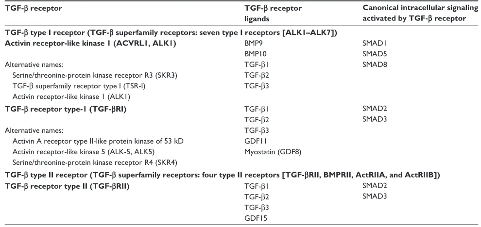
Since the discovery of the cytokine transforming growth factor-beta 1 (TGF-β1),1 a diverse family of ligands, corresponding receptors, and signal transduction proteins known as the TGF-β superfamily were identified (Table 1).2 Although the members of the TGF-β superfamily share genetic and protein structures, some of these members have different physiological functions and play distinct roles in diseases, including cancer (Figure 1). For example, the activin receptor-like kinase 1 (ALK1) receptor and the ALK5 receptor, also known as TGF-β receptor type I (TGF-βRI), can both promote angiogenesis but have different intracellular activation pathways with different effects on angiogenesis (for details, see the section TGF-β/ALK5 signaling pathway and its role in cancer). Here, we will focus on the role of the TGF-β/ALK5-mediated pathway and describe the experiences of developing galunisertib.
Correspondence: Michael M Lahn 6820 wisconsin Avenue, Condo 8008, Bethesda, MD 20815, USA
Tel +1 240 899 9415 email michalahn@aol.com
Journal name: Drug Design, Development and Therapy Article Designation: Review
Year: 2015 Volume: 9
Running head verso: Herbertz et al
Running head recto: Clinical development of galunisertib DOI: http://dx.doi.org/10.2147/DDDT.S86621
Point your SmartPhone at the code above. If you have a QR code reader the video abstract will appear. Or use:
http://youtu.be/cN-vG9YO22I video abstract
Drug Design, Development and Therapy downloaded from https://www.dovepress.com/ by 118.70.13.36 on 22-Aug-2020
For personal use only.
Number of times this article has been viewed
This article was published in the following Dove Press journal: Drug Design, Development and Therapy
Dovepress Herbertz et al
TGF-
β
/ALK5 signaling pathway
and its role in cancer
In the early 1980s, TGF-β was biochemically isolated from tumor cells and was named after its ability to transform normal rat kidney fibroblasts.1 This approximately 13 kDa polypeptide was later designated as TGF-β1.3 TGF-β ligands include TGF-β1, TGF-β2, and TGF-β3 (Table 1), all of which regulate diverse biological functions.4,5 All three ligands can
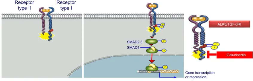
independently engage the specific receptor TGF-βRI/ALK5, which then undergoes dimerization with TGF-β receptor type II (TGF-βRII).6 Recent studies have shown that two TGF-β ligand molecules bind as homodimers to two TGF-βRI and two TGF-βRII molecules (ie, a homodimer bound to a het-erotetramer), creating a heterodimer complex consisting of six distinct molecules.7 For simplicity reasons, figures often do not show the duplicity of the heterodimer (Figure 2). This heterodimer complex phosphorylates the intracellular proteins SMAD2 and SMAD3, activating a signaling cascade to induce several nuclear transduction proteins. The induction of these proteins leads to cellular proliferation, differentiation, motil-ity, survival, and apoptosis in tumor cells.8 This TGF-β/ALK5 signaling pathway is commonly referred to as the canonical signaling pathway (Figure 1). In addition to this canonical pathway, a number of accessory receptors have been identi-fied, which can separately interact with the RI and TGF-RII. One example is the TGF-βRIII, to which the glycoprotein endoglin (CD105) can bind with high affinity.9
Given the complexity of the TGF-β signaling pathway, its physiological role is diverse and appears to be depen-dent on the disease setting and cellular context. In cancer, TGF-β can affect tumor growth directly (referred to as intrinsic effect of TGF-β signaling) or indirectly (referred to as extrinsic effect) by promoting tumor growth, inducing epithelial-mesenchymal transition (EMT), blocking antitumor immune responses, increasing tumor-associated fibrosis and Table 1 TGF-β receptors, their respective ligands, and canonical intracellular signaling proteins currently targeted by clinical investigation
TGF-β receptor TGF-β receptor
ligands
Canonical intracellular signaling activated by TGF-β receptor TGF-β type I receptor (TGF-β superfamily receptors: seven type I receptors [ALK1–ALK7])
Activin receptor-like kinase 1 (ACVRL1, ALK1)
Alternative names:
Serine/threonine-protein kinase receptor R3 (SKR3) TGF-β superfamily receptor type i (TSR-i) Activin receptor-like kinase 1 (ALK1)
BMP9 BMP10 TGF-β1 TGF-β2 TGF-β3
SMAD1 SMAD5 SMAD8
TGF-β receptor type-1 (TGF-βRI)
Alternative names:
Activin A receptor type ii-like protein kinase of 53 kD Activin receptor-like kinase 5 (ALK-5, ALK5) Serine/threonine-protein kinase receptor R4 (SKR4)
TGF-β1 TGF-β2 TGF-β3 GDF11
Myostatin (GDF8)
SMAD2 SMAD3
TGF-β type II receptor (TGF-β superfamily receptors: four type II receptors [TGF-βRII, BMPRII, ActRIIA, and ActRIIB])
TGF-β receptor type II (TGF-βRII) TGF-β1
TGF-β2 TGF-β3 GDF15
SMAD2 SMAD3
Notes: The TGF-β superfamily includes the following ligands: TGF-β1, TGF-β2, and TGF-β3; BMPs (BMP1–10); GDFs (GDF1–10); activins/inhibins (activin 1–5); nodal; leftys
(lefty1 and lefty2); and anti-muellerian hormone.
Abbreviations: TGF-β, transforming growth factor-beta; BMP, bone morphogenetic protein; GDF, growth and differentiation factor.
7*)β
,QYDVLRQDQG PHWDVWDVLV
,QIODPPDWLRQ
,PPXQHHVFDSH $QJLRJHQHVLV
Figure 1 TGF-β signaling and hallmarks of cancer.
Note: TGF-β signaling plays an important role in inducing angiogenesis, inflammation,
invasion/metastasis, and immune escape.
Drug Design, Development and Therapy downloaded from https://www.dovepress.com/ by 118.70.13.36 on 22-Aug-2020
Dovepress Clinical development of galunisertib
enhancing angiogenesis (Figure 1).10 Although TGF-β1 was originally found to transform normal cells,1 subsequent studies revealed that it also behaved as a tumor suppressor in epithelial cells. The factors determining whether TGF-β
signaling has a tumor promoter or suppressor function are a matter of intense research.8 Currently, it is postulated that the tumor suppressor function of TGF-β signaling is lost in early stages of cancer similar to recessive loss-of-function mutations in other tumor suppressors.5 Such a loss of func-tion has been associated with drug resistance in tumors and reported in colon and lung cancer cell lines.11–14 The switch to becoming a tumor promoter with resulting drug resistance often occurs in the presence of EMT.15 TGF-β signaling plays a prominent role in EMT by influencing key transcription factors, including Snail, zinc-finger E-box-binding, and basic helix–loop–helix transcription factors. Besides these genetic and physiological changes, TGF-β signaling plays a critical role in the regulation of immune cell function in normal and tumor-associated lymphocytes, in particular, the activation of T-regulatory cells.16,17
In addition to its role in regulating immune cells, TGF-β
signaling is a strong inducer of fibrosis.18 It is an activator of cancer-associated fibroblasts (CAFs)19 that can be a major source of collagen type I and other fibrogenic factors. CAFs and their associated production of fibrogenic factors may further foster a microenvironment that decreases immune responses and increases drug resistance and tumor angiogenesis.20
The role of TGF-β signaling in angiogenesis is well rec-ognized during ontogeny and in tumor growth.21 For example, murine embryos deficient in TGF-βRI exhibit severe defects in vascular development; TGF-β signaling acts as a key regulator of development of both vascular endothelial and smooth muscle cells.21
In summary, TGF-β signaling plays a key role in pathways that are considered hallmarks of cancer by aiding cancer
cells in their evasion of immunosurveillance, increasing inflammation (in part by modulating cytokine responses, tumor migration, and angiogenesis) (Figure 1).5,23,24 Thus, targeting this TGF-β-mediated signal transduction in tumors may provide a novel approach to controlling tumor growth by blocking a central activation node with its connected downstream signaling pathways.25
Pharmacological approaches:
blocking the TGF-
β
signaling
pathway
Specific inhibitors are key for the pharmacological development of ALK5 inhibitors:
Given the structural and genetic similarities of the different TGF-β signaling pathways, inhibitors must be highly spe-cific to block the intended activation pathway. For example, ALK5 and ALK1 pathways both increase tumor angiogen-esis (Table 1).26 ALK1 inhibitors block the interaction of bone morphogenetic protein (BMP)-9 and -10 with ALK1, which interrupts the subsequent phosphorylation of SMAD1 (pSMAD1)/pSMAD5/pSMAD8 (Table 1). PF-03446962 is an ALK1 neutralizing antibody that does not bind other ALKs. Currently, PF-03446962 is being evaluated in Phase II trials in patients with solid tumors to determine its ability to block angiogenesis (Table 2). Another molecule directed against ALK1 is dalantercept/ACE-041, a chimeric protein consisting of the ALK1 ligand binding extracellular domain and an Fc portion (ALK1-Fc). This ligand trap is a soluble receptor that can bind circulating ligands and prevents their engagement with cell surface receptors. Dalantercept efficiently blocks BMP-9 and BMP-10-induced SMAD1 phosphorylation and SMAD1-dependent transcription. Dalantercept is currently in Phase II trials as monotherapy and in combination with vascular endothelial growth factor
P P
ALK5/TGF-βRI
Galunisertib
Receptor type I
PP Receptor
type II
Gene transcription or repression P
SMAD2,3 P
SMAD4
P
Figure 2 TGF-βRi (ALK5) and TGF-βRii canonical activation.
Note: Galunisertib blocks the kinase of the ALK5 pathway reducing the pSMAD2 activation in cells.
Abbreviations: ALK5, activin receptor-like kinase 5; pSMAD2, phosphorylation of SMAD2; TGF-β, transforming growth factor-beta.
Drug Design, Development and Therapy downloaded from https://www.dovepress.com/ by 118.70.13.36 on 22-Aug-2020
Dovepress Herbertz et al
Table 2
TGF-β
inhibitors in clinical development
Target/mode of action
Generic name
Clinical development phase
Indications in clinical trials
Company
References
TGF-β
receptor serine/threonine kinase inhibitors
TGF-β
R
i
ALK5
Galunisertib LY2157299
ii/
iii
Pancreatic carcinoma, glioblastoma, hepatocellular carcinoma, myelodysplastic syndromes (MDS) eli Lilly and Company
NCT01746004 NCT01965808 NCT01582269 NCT01722825 NCT02008318 NCT01220271 NCT01246986 NCT01373164 NCT01682187
TGF-β
R
i
ALK5
T
ew
-7197
i
Melanoma, breast cancer, hepatocellular carcinoma, prostate cancer
MedPacto
inc.
(South Korea)
NCT02160106
Antibodies and protein therapeutics against
TGF-β
and
TGF-β
receptors ALK1, ALK5, ALK6
TGF-β
R
ii
LY3022859 iMC-TR1
i
Advanced solid tumors
eli Lilly and Company
NCT01646203
TGF-β
-1
LY2382770
ii
Diabetic kidney disease, diabetic nephropathy, diabetic glomerulosclerosis eli Lilly and Company
NCT01113801
Pan
TGF-β
Fresolimumab (GC-1008)
ii
Systemic sclerosis, metastatic breast cancer, focal segmental glomerulosclerosis, glioma, renal cell carcinoma, melanoma, malignant pleural mesothelioma, myelofibrosis Genzyme, a Sanofi Company
NCT01284322 NCT01401062 NCT01665391 NCT01472731 NCT00356460 NCT00923169 NCT01112293
ALK-1
PF-03446962
ii
Hepatocellular carcinoma, urothelial cancer, malignant pleural mesothelioma
Pfizer
NCT01911273 NCT01620970 NCT01486368
BMP9/BMP10 ALK-1 interaction (soluble form of ALK-1) Dalantercept AC
e-041
ii
Squamous cell carcinoma of the head and neck, renal cell carcinoma, ovarian epithelial, fallopian tube, or primary peritoneal cavity cancer
Acceleron Pharma
inc.
NCT01458392 NCT01727336 NCT01720173 http://www.acceleronpharma.com/ products/dalantercept
/
Soluble fusion protein of receptor type
iiA
(ActR
iiA) linked to the
Fc protein of human
igG1
Sotatercept AC
e-011
ii
Anemia in rare blood diseases, including β-thalassemia, MDS, and diamond blackfan; chronic kidney disease; multiple myeloma
Acceleron Pharma
inc.
and Celgene Corporation
NCT01736683 NCT01571635 NCT01146574 NCT01999582 NCT01464164 NCT01712308 NCT01562405 http://www.acceleronpharma.com/ products/sotatercept
/
Drug Design, Development and Therapy downloaded from https://www.dovepress.com/ by 118.70.13.36 on 22-Aug-2020
Dovepress Clinical development of galunisertib
Modified type II activin receptor fusion protein that acts as a ligand trap for members in the
TGF-β
superfamily
Luspartecept AC
e-536
ii
β
-thalassemia; anemia in patients with MDS
Acceleron Pharma
inc.
and Celgene Corporation
NCT01749540 NCT01749514 http://www.acceleronpharma.com/ products/luspatercept
/
TGF-β
-1 blocking peptide
P144 Peptide 144 Disirtertide
ii
Skin fibrosis in systemic sclerosis
Digna Biotech S.L.
NCT00781053 NCT00574613 NCT00656825
Direct and indirect inhibitors of
TGF-β
synthesis and secretion
TGF-β
mRNA antisense
Trabedersen (AP-12009), intravenous (convection enhanced delivery formulation terminated)
ii
Pancreatic neoplasms, melanoma, colorectal neoplasms
isarna Therapeutics GmbH
NCT00844064 NCT00431561 NCT00761280 http://www.isar
na-the
rapeutics.
com/fileadmin/user_upload/ documents/pre
ss-releases/
2013/201302
26_EN_Press
Release_CorporateStrategy_2013. pdf
TGF-β
synthesis
Belagenpumatucel-l
Cancer vaccine
(TGF-β
antisense)
Lucanix
iii
Nonsmall-cell lung cancer
NovaRx Corporation
NCT00676507
TGF-β
synthesis
Pirfenidone etuary F-647 esbriet
®
Pirespa
®
iii Approved for ideopathic pulmonary fibrosis in europe, Canada, and Japan Idiopathic pulmonary fibrosis, pulmonary fibrosis, focal glomerulosclerosis, diabetic nephropathy, neurofibromatosis type I and progressive plexiform neurofibromas, systemic sclerosis-related interstitial lung disease, hypertrophic cardiomyopathy, radiation-induced fibrosis, uterine leiomyoma (fibroids)
interMune
inc.
NCT00063583 NCT00076102 NCT00287716 NCT01933334 NCT01366209 NCT00011076 NCT00754780 NCT00020631 NCT00053937 NCT02009293 NCT00332033
“bi-shRNAi(furin)/GMCSF DNA/autologous tumor cell” vaccine (FANG)
FANG™ vaccine
ii
Melanoma,
ewing’s sarcoma, ovarian cancer,
colorectal carcinoma
Gradalis
inc.
NCT01453361 NCT01061840 NCT01505166 NCT01309230 NCT01867086 NCT01551745 NCT01505153
Abbreviations:
ALK, activin receptor-like kinase; BMP, bone morphogenetic protein;
TGF-β
, transforming growth factor-beta; NCT, national clinical trial.
Drug Design, Development and Therapy downloaded from https://www.dovepress.com/ by 118.70.13.36 on 22-Aug-2020
Dovepress Herbertz et al
inhibitors.27 In contrast to the ALK1 inhibitors, the inhibi-tion of the ALK5 pathway blocks activainhibi-tion of different intracellular proteins (eg, SMAD2/3) and alters the vascular and smooth muscle cell compartment.21 This may improve delivery of chemotherapy28 although it may have a reduced inhibitory effect on pro-angiogenic factors, such as vascular endothelial growth factor and basic fibroblast growth factor. In contrast to ALK1 inhibitors, ALK5 inhibitors increase angiogenesis in cell cultures of normal endothelial cells.29
Monoclonal antibodies: Arguably, monoclonal anti-bodies are considered highly specific and can provide the best approach to develop selective inhibitors to the TGF-β
signaling pathway. Fresolimumab (formerly GC1008) is one of the first pan-TGF-β ligand monoclonal antibodies to be investigated in cancer patients.30 Fresolimumab is being developed for patients with renal fibrosis and until recently, for patients with metastatic cancer.30–33 In a Phase I trial, fresolimumab was given to 29 patients with malignant melanoma and renal cell carcinoma administered in doses up to 15 mg/kg given first every 28 days then subsequently by biweekly dosing to patients with stable disease. Revers-ible cutaneous keratoacanthomas/squamous cell carcinoma (four patients) and hyperkeratosis were reported as major drug-related adverse events. One patient with malignant melanoma achieved a partial response lasting 44.4 weeks, and six melanoma patients had stable disease. Of the seven patients with partial response or stable disease, six patients had received 3 mg/kg of fresolimumab. Given that patients with higher doses (up to 15 mg/kg) had no responses or stable disease, the authors postulated that clinical activity was associated with lower doses.30 In a second Phase II study, fresolimumab was given to 13 patients with malignant pleural mesothelioma at 3 mg/kg given every 21 days, the anticipated most active dose. None of the patients had radiographic responses, and three patients had stable disease at 3 months. However, five patients developed new antibodies against tumor lysates isolated from malignant pleural mesothelioma, suggesting a possible immune response. After the 13 patients were treated, the manufacturer terminated further develop-ment of fresolimumab for oncology indications.33 Eli Lilly and Company also developed a pan-TGF-β ligand inhibitor but did not pursue its development due to severe toxicity in animals (data on file, Eli Lilly and Company, Indianapolis, IN, USA). In addition, a TGF-β1-specific monoclonal antibody (TβM1 or LY2382770) was evaluated in a Phase I study in patients with cancer. LY2382770 showed no radiographic responses once escalated to the predefined dose of 240 mg (flat dose) given every 28 days, which was thought to be efficacious
based on a pharmacokinetic/pharmacodynamic (PK/PD) model for LY2382770.34 There are also monoclonal antibod-ies that were designed to block the TGF-β receptors, such as the monoclonal antibody TR1.35 TR1 blocks the TGF-βRII; this monoclonal antibody is currently being evaluated in a first-in-human dose (FHD) study. Given the toxicity observed with small molecule inhibitors (SMIs) blocking ALK5 (see section SMIs of the TGF-bRI/ALK5 and the early develop-ment of galunisertib), specific inhibitors of TGF-βRII using monoclonal antibodies were thought to have more manage-able toxicity or perhaps even a reduced toxicity profile.
Antisense oligonucleotides (ASOs): Another approach to selectively blocking the TGF-β signaling is the use of ASOs. For example, the first-generation ASO trabedersen (formerly AP12009) was developed to block the TGF-β2 production in glioma cells and was advanced to clinical investigation.36 Although the safety profile was manageable, the antitumor activ-ity of trabedersen seemed to be limited to a subset of patients, mainly those with World Health Organization grade III glioma. Similar to fresolimumab, lower doses appeared to be associated with better radiographic responses. However, the clinical devel-opment of trabedersen with its convection-enhanced delivery of compound in patients with glioblastoma was terminated. The convection-enhanced delivery was necessary for trabedersen because first-generation ASOs generally achieve their optimal pharmacological activity if they are continuously applied and administered directly to the tumor. Because of this added com-plexity for the administration of an ASO, newer ASOs with improved chemistry are being generated for future investigation in a range of indications, including cancer.37
vaccines strengthening the patient’s microenvironment:
Another approach to blocking the TGF-β signaling pathway is based on vaccines. For example, belagenpumatucel-l is a cancer vaccine (NovaRX Corporation, San Diego, CA, USA) that inhibits the synthesis of TGF-β2. Genetically modified whole tumor cells stimulate the patient’s own immune sys-tem to attack the tumor. The vaccine contains four allogenic and irradiated nonsmall-cell lung cancer (NSCLC) cell lines expressing a TGF-β2 synthetic antisense gene. The recently reported Phase III “STOP” trial in patients with stage III/IV NSCLC did not meet its predefined primary endpoint of overall survival (OS). However, an increase in OS was observed in a subgroup of patients who had started treatment with belagenpumatucel-l within 12 weeks of completing frontline chemotherapy. Also, patients with nonadenocar-cinoma NSCLC (= squamous cell carcinoma and large cell
Drug Design, Development and Therapy downloaded from https://www.dovepress.com/ by 118.70.13.36 on 22-Aug-2020
Dovepress Clinical development of galunisertib
lung cancer) had an OS of 19.9 months when treated with belagenpumatucel-l compared with an OS of 12.3 months for patients treated with placebo (hazard ratio 0.55, P=0.036). The authors of the study concluded that belagenpumatucel-l should be further evaluated in lung cancer.38
SMIs of the TGF-
β
RI/ALK5 and the
early development of galunisertib
(LY2157299 monohydrate)
Among the TGF-β inhibitors, SMIs represent a large and diverse group of chemical entities that are designed to block the activation of the signaling cascade downstream of the TGF-β receptor type I kinase (TGF-βRI or ALK5) or type II (TGF-βRII) by inhibiting the serine/threonine kinase. There is a growing list of SMIs blocking the TGF-βRI,39–42 including recent SMIs such as Ki2689443 and TEW-7197.44,45
At Eli Lilly and Company, several SMIs were identi-fied in the past years (Table 3). A large library of SMIs was screened in vitro using a TGF-β-dependent cell-based assay. Selected compounds were further evaluated for their ability to inhibit autophosphorylation of the isolated human TGF-βR-I kinase domain. The TGF-βR-I kinase domain was expressed as a constitutively active construct (T204D muta-tion) by Sf9 insect cells.46,47 For example, the diheteroaryl-substituted pyrazole 1 (LY364947) was identified as a potent inhibitor (IC50=51 nM) (Table 3). Compounds were further evaluated by measuring their inhibitory effect in a TGF-β- dependent luciferase assay produced in mink lung cells (p3TP Lux) and their growth inhibition in mouse fibroblasts (NIH3T3).48 Compounds such as LY580276,49 LY364947,50 and LY210976151 share with LY2157299 monohydrate the selectivity profile and inhibition of the ALK5.52–54 Overall, the kinase selectivity profile of galunisertib met the desired characteristics (Table 4).
Compared with other SMIs, galunisertib (LY2157299) monohydrate had reduced cardiovascular toxicity in ani-mals and appeared to be less potent in inhibiting pSMAD2 levels in vitro (Table 3).49,55–57 Galunisertib (pronounced: gal-ue’ ni-ser-tib) is now the United States Adopted Name and International Nonproprietary Name designation for LY2157299 monohydrate. The first part of the name refers to the Greek-Roman physician Aelius Galenus (born: 129 AD; died: c.200/c.219 AD), also known as Galen of Pergamon.58 Apart from galunisertib that started its clinical development in 2006, only TEW-7197 is known to be in clinical investiga-tion. In mid-2014, a Phase I study of TEW-7197 was initiated in patients with breast cancer, melanoma, hepatocellular
carcinoma (HCC), and glioblastoma.59 Table 3
Clinical and surrogate compounds developed by Eli Lilly and Company (data on file)
Ki (
µ
M)
TGF-β
RI kinase IC
50 , µ M Mean values ± SEM for
a minimum of three determinations (n)
p3TP lux, IC
50 , µ M Mean values ± SEM for
a minimum of three determinations (n)
NIH3T3, IC 50 , µ M Mean values ± SEM for
a minimum of three determinations (n)
p38MAPK, IC
50
,
µ
M
Single-point determination
Half-life mean (hours)
Toxicity other than known toxicities for ALK5 SMIs in animals
Rat Mice Dog Galunisertib (LY2157299) 0.086 0.051 ± 0.005 (258) 0.047 ± 0.016 (55) 0.089 ± 0.010 (68) 0.53 0.3 2.26 – LY2109761 0.038 0.069 ± 0.031 (2) 0.18 ± 0.093 (5) 0.21 ± 0.170 (4) Not done 1.8 Not done 1–2 Not done LY364947 0.028 0.051 ± 0.005 (258) 0.047 ± 0.016 (55) 0.089 ± 0.010 (68) 0.74 Not done Not done LY580276 0.037 0.175 ± 0.088 (4) 0.096 ± 0.016 (3) 0.339 ± 0.349 (4) 20 Not done Not done Abbreviations: ALK, activin receptor-like kinase; iC50 , half maximal inhibitory concentra tion; MAPK, mitogen-activated protein kinases; SM is, small molecule inhibitors; Se M, standard error of the mean; TGF-β , transforming growth factor-beta.
Drug Design, Development and Therapy downloaded from https://www.dovepress.com/ by 118.70.13.36 on 22-Aug-2020
Dovepress Herbertz et al
Table 4 Kinase selectivity profile of galunisertib (LY2157299) (data on file, Eli Lilly and Company)
Kinase IC50 (µM)
TGF-βRi 0.17
TGF-βRii 0.21
ALK4 0.08
MiNK 0.19
RiPK2 0.22
CSNK1A1 0.26
MAP4K4 0.28
GAK 0.31
CSNK1e1 0.4
BMPR1B 0.47
BRAF 0.50
TNiK 0.51
RSK4 0.72
Abl1 0.86
ZAK 0.86
NLK 0.91
Abbreviations: iC50, half maximal inhibitory concentration; TGF-β, transforming
growth factor-beta; TGF-βRi, TGF-β receptor type i; TGF-βRii, TGF-β receptor type ii; ALK, activin receptor-like kinase; MiNK, misshapen/NiK-related kinase; RiPK2, receptor-interacting serine-threonine kinase; CSNKiA1, casein kinase alpha 1; MAP4K4, mitogen-activated protein kinase kinase kinase kinase 4; CSNKie1, casein kinase epsilon 1; BMPR, bone morphogenetic protein receptor type 1B; TNiK, TRAF2 And NCK interacting Kinase; RSK4, ribosomal S6 kinase 4; ABi1, Abelson interactor 1; ZAK, Sterile alpha motif and leucine zipper containing kinase AZK; NLK, Nemo-Like Kinase.
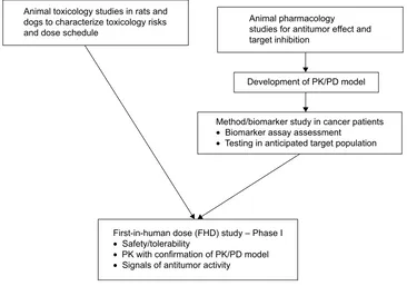
the following: (a) running preclinical toxicology studies with administration schedules that identified a sufficient margin of safety, (b) developing a predictive PK/PD model using animal pharmacology information, and (c) developing assays of PD markers in patients with cancer to confirm the therapeutic window as predicted by the PK/PD model (Figure 3).
Toxicology
In animal studies, several ALK5 inhibitors showed an increased incidence of hemorrhagic, degenerative, and inflam-matory lesions in heart valves. Galunisertib was selected for clinical investigation based on its profile in animal toxicol-ogy studies in rats and dogs.49,50,57,61,62 The lesions appeared either at very high doses (1 month of continuous dosing) or during continuous dosing for 6 months. Intermittent dosing provided a sufficient margin of safety to advance galunisertib into clinical development.57 In general, rats appeared to be more sensitive to galunisertib toxicity than dogs. Because of the difference in heart rate and also intravascular blood pressure, there is a possibility that the cardiac lesions were related to shear stress-associated intravascular remodeling. Under such conditions, the TGF-β signaling pathway is activated in endothelial and smooth muscle cells.63,64 Based upon these findings, it is possible that the lesions observed in the valves resulted from an inhibition of a physiologic
TGF-β-dependent mechanism, which is normally needed to repair
Animal toxicology studies in rats and dogs to characterize toxicology risks and dose schedule
Animal pharmacology studies for antitumor effect and target inhibition
Development of PK/PD model
Method/biomarker study in cancer patients
• Biomarker assay assessment
• Testing in anticipated target population
First-in-human dose (FHD) study – Phase I
• Safety/tolerability
• PK with confirmation of PK/PD model
• Signals of antitumor activity
Figure 3 Preparing for the first-in-human dose (FHD) study.
Note: Standard Good-Laboratory Practice (GLP) animal toxicology studies were performed as well as a pharmacokinetic/pharmacodynamic (PK/PD) model established
prior to the FHD study.
Despite the long list of ALK5 SMI inhibitors, few ALK5 inhibitors have been moved to clinical investigation, perhaps because of the observed severe cardiac toxicities in animals.41,60 Galunisertib overcame this barrier by addressing
Drug Design, Development and Therapy downloaded from https://www.dovepress.com/ by 118.70.13.36 on 22-Aug-2020
Dovepress Clinical development of galunisertib
the injury caused by shear/stress at the outflow of the heart. When valves and ascending aorta were examined, they showed activation of the TGF-β pathway after administration of a TGF-β inhibitor as measured by pSMAD2 staining. Patients with Loeys–Dietz syndrome have a similar paradox where a loss of functional mutation in the TGF-β signaling gene is present; however, in the tissue, an activation of the TGF-β
pathway is observed.65,66 One hypothesis for this paradox is that alternative pathways are activated.67 A second hypothesis proposes a changed receptor/ligand processing as observed in studies with cells.68,69 If the second hypothesis based on a possible pathway adaptation is correct, then the intermittent dosing of TGF-β inhibitors provides a safe dosing regimen.
Galunisertib affects bone development and alters inflam-matory responses in the skin or gut of rats and dogs. It is well known that blocking TGF-β signaling can cause chronic inflammation in skin and gut, which in turn can lead to pre-cancerous conditions.70,71 Intermittent or continuous regimens with low doses may reduce such a risk of developing chronic inflammation and thus allow long-term administration of a TGF-β inhibitor.
Pharmacology in cellular assays
and animals
In addition to the animal toxicology studies, evidence of target inhibition and preclinical antitumor efficacy was needed for developing a predictive PK/PD model.72,73 The initial antitumor activity of galunisertib was characterized in three different cancer models: two breast cancer models, MX1 and 4T1, and a NSCLC model using Calu6. Dosing (75 mg/kg twice daily by oral gavage) was initiated 4–6 days postinoculation and continued for 20 days in the xenograft models (MX1 and Calu6) for the entire length of the sur-vival study with the 4T1 model.74 A statistically significant tumor growth delay of 10.1 days was observed in the MX1 model, and the 4T1 tumor model exhibited a 4.5-day survival advantage. In the Calu6 xenograft, galunisertib significantly delayed tumor growth.
Despite these encouraging observations with some cell lines in standard xenografts (eg, Calu6 and U87MG), it appears that only select tumor cell lines respond to galuni-sertib treatment in standard, immune-deficient xenografts. In patient-derived xenografts (PDXs), the antitumor response to galunisertib treatment is also limited.75 Furthermore, in some immune-deficient PDX models, tumor growth appeared to be enhanced, which was previously raised as a potential risk for administering TGF-β inhibitors to patients.76,77 However, it is possible that such growth is only observed
in immune-compromised animals; in immune-competent murine models, no such tumor growth augmentation has been observed. Tumor-bearing, immune-competent mice treated with the monoclonal antibody against TGF-βRII, TR1, had antitumor effects that were at least partly dependent on the presence of T-cell subsets.35 Finally, mice receiving long-term exposure (over 1 year of administration) of a soluble TGF-β antagonist had no adverse events.78 All these observa-tions imply that immune-competent animal models may be more predictive to evaluate the activity of TGF-β inhibitors. It appears that an active immune response is essential to assess the effect of TGF-β signaling inhibition in animal models; thus, models using immune-compromised animals may have limited use in screening for TGF-β inhibitors.
Because of the difficulties of using animal models as a screening method for identifying novel agents, alternative in vitro models were considered. Tumor clonogenic assays with PDX were evaluated, but it became evident that many PDX had a disrupted canonical pathway.75 Hence, this assay was found not to be adequate to screen large libraries of novel SMI TGF-β inhibitors. Recently, experiments with a 3D in vitro model were used to assess the effect of galunisertib.79 In such 3D culture systems, galunisertib had activity whereas it had none in standard monolayer assays. An advantage of the 3D culture model was the possibility to evaluate some aspects of the immune response. Whether such 3D in vitro assays may also offer an alternative to in vivo models is not yet determined.
Drug resistance mechanism and
implications on drug combination
Based on the biology of TGF-β signaling, the associated mechanisms that affect drug resistance are likely to be depen-dent on the tumor microenvironment. One such example are the CAFs: these produce large amounts of the TGF-βligands and are associated with fibrosis and with resistance to 5-fluorouracil (5-FU).20 Recently, drug resistance related to TGF-β signaling was reported to be associated with loss of the MED12 gene in tumors.80 MED12 loss induces not only an EMT-like phenotype that results in chemotherapy resistance to 5-FU but also resistance to the epidermal growth factor receptor tyrosine kinase inhibitor (EGFR TKI) gefi-tinib. Treatment with galunisertib in MED12-deficient cells restored the sensitivity to both chemotherapy and EGFR TKI. In addition to drug resistance to 5-FU and EGFR TKIs, there were reports connecting TGF-β signaling to paclitaxel resistance in triple-negative breast cancer.81 In all these obser-vations, it appears that EMT or EMT-like phenotype of the
Drug Design, Development and Therapy downloaded from https://www.dovepress.com/ by 118.70.13.36 on 22-Aug-2020
Dovepress Herbertz et al
tumor cells plays a critical role to drug resistance associated with TGF-β signaling.
PK/PD model – predicting a
therapeutic window in patients with
an acceptable safety profile
The development of preclinical PK/PD models have been invaluable in guiding early clinical trial design.82,83 A similar model was built using preclinical data on pSMAD2 inhibi-tion, antitumor activity of galunisertib in Calu6 xenografts, and the observed PK in mice, rats, and dogs.72,73 The half-life of galunisertib in animals was less than 3 hours (Table 3). An observed moderate variation in PK was, in part, attrib-utable to the formulation of galunisertib.84 Allometric PK scaling of galunisertib allowed a reliable prediction of both the exposure in humans within the expected range to produce antitumor activity. The drug effect continued even after the systemic disappearance of the drug: the PD effect of reducing pSMAD2 was still detectable in tumor tissue and peripheral blood mononuclear cells (PBMCs) up to 7 days after stopping galunisertib and when galunisertib was no longer detected in the plasma. This delayed PD effect was also seen when treated with the monoclonal antibody against TGF-βRII, TR1, suggesting that this phenomenon is not limited to SMIs (data on file, Eli Lilly and Company).
The simultaneous inhibition of pSMAD2 inhibition in tumor and surrogate tissue (ie, PBMCs) led to the develop-ment of a PD detection assay using peripheral blood. This assay was developed to monitor and confirm the PK/PD relationship during the FHD study. To avoid toxicity and maintain antitumor activity, the galunisertib exposure had to be limited to a pSMAD2 inhibition of approximately 30% over 24 hours, combined with a maximum inhibition of 50%. This was achieved by a twice-daily (BID) dose schedule that produced a modulatory exposure.85
Dosing considerations for
galunisertib
Based on the PK/PD modeling and the toxicity observa-tion, we decided to use a BID dosing schedule and a 14-day on/14-day off schedule. In preclinical models and later in the Phase I study, we had observed that pSMAD2 inhibition was extended up to 7 days after galunisertib was stopped. Given that continuous dosing may increase the risk for chronic toxicity, the 14-day treatment with an anticipated prolonged pSMAD2 inhibition of 7 days was the most acceptable regimen for long-term treatment. To avoid high single-day exposures, a morning and evening dosing schedule was
instituted. All these interventions were designed to avoid a steady-state or continuous on-target inhibition.
Early biomarker development
The biomarker work early in development focused on two main objectives: a) biomarkers for patient selection and b) pharmacodynamic response markers. For patient selection, three groups were considered: those whose tumors produced high amounts of TGF-β1, (eg, in renal cell carcinoma,86 prostate cancer,87 and breast cancer25); those in whom TGF-β inhibition had shown clinical responses with other TGF-β
inhibitors (such as glioma36), and those with skeletal metas-tasis. In such conditions, TGF-β is being mobilized from the bone matrix, and increased TGF-β1 can serve as a marker of tumor progression.88
A pSMAD2 assay to measure the reduction of pSMAD2 in PBMCs during the FHD trial was established.89 This pSMAD2 enzyme-linked immunosorbent assay (ELISA) used a poly-clonal antisera and was tested on serum from patients with skeletal metastasis (a nondrug interventional trial). The intra-patient variability was determined to be less than 30%.90 This variability was considered acceptable for the FHD trial.
A plasma TGF-β1 ELISA was developed and sub-sequently used for PD assessments. This assay provided reliable information on plasma TGF-β1 levels when citrate-theophylline-adenosine-dipyridamole tubes were used for collection.91 The assay detects the activated TGF-β1 form in a standard ELISA format (R&D Systems, DB100B). The original hypothesis assumed that TGF-β1 levels will be reduced if galunisertib interrupts a possible autocrine or paracrine growth signal.
Other plasma proteins such as parathyroid hormone-related protein, von Willebrand factor, and interleukin-10 correlated with TGF-β1 levels.90 Given this connection, the measure-ments of such indirect plasma markers were thought to help with understanding of the downstream effect of the TGF-β
signaling inhibition with galunisertib. Thus, they were consid-ered as alternative PD markers for future clinical trials.
Gene expression profiling of PBMCs was initially pur-sued as an additional option to assess PD changes,92,93 but previously observed changes in PBMCs in ex vivo experi-ments were not able to be reproduced in subsequent clinical trials. In earlier ex vivo studies, T-cells were activated with CD3/CD28 costimulation to obtain reliable gene expression profiles.92 By contrast, PBMCs obtained from patients with skeletal metastasis were only stimulated with exogenous TGF-β1.93 The stimulation with exogenous TGF-β1 led to a rapid and subtle change in gene expression, which may
Drug Design, Development and Therapy downloaded from https://www.dovepress.com/ by 118.70.13.36 on 22-Aug-2020
Dovepress Clinical development of galunisertib
have been too transient to be detected in patients treated with galunisertib.
Several attempts were also made to develop patient selection strategies based on gene expression profiles from tumor-bearing animal or PDX models.75,94,95 These profiles were compared with publicly available gene expression data sets, but it was not possible to define a clear profile. In part, this was due to the minimal antitumor activity with galuni-sertib in immune-deficient animal models or monolayer cell line experiments.
FHD study and early clinical
development of galunisertib in
cancer patients, including patients
with glioblastoma
Given the potential for toxicity and general safety concerns, throughout the course of the FHD study the predictive PK/ PD model was continuously updated, and results were shared with the scientific community (a summary of all clinical stud-ies with galunisertib is presented in Table 5). The decision to select galunisertib as a clinical candidate was presented in 2005,72 and the FHD study was disclosed at a conference in 2006.56 Over the subsequent years, the progress of the FHD study and the patient safety information were routinely presented.96–99 Additionally, preclinical studies of galunisertib were regularly updated.72,74,94
One of the most important objectives of the FHD study was to assess the safety of galunisertib when administered to reach the predicted therapeutic window. Based on the PK/PD model, the therapeutic window was anticipated to be achieved when compound was dosed between 160 and 360 mg.73 Within the initial cohorts of the FHD study, galuni-sertib was given daily continuously based on the standard 1-month animal toxicology studies. Although not generally required for oncology Phase I trials, 6-month animal toxicol-ogy studies were begun to assess the risk of more prolonged exposures. This study started concurrently with enrollment of the patients into the FHD study. Unexpectedly, new toxicities emerged after the evaluation of 6-month animal toxicology studies. As a result of these new findings, the FHD study was placed on clinical hold and additional animal toxicology studies began using an intermittent dosing schedule. The intermittent dosing schedule in the most sensitive species (rats) provided an acceptable margin of safety. These data supported lifting the clinical hold; the FHD study resumed but was restricted to patients with glioblastoma, in part because of the previously reported activity of trabedersen.36
During the FHD study, all patients underwent com-prehensive cardiac monitoring. These studies consisted of baseline and every-other-month echocardiography/Doppler imaging, baseline and monthly serial plasma assessment for Troponin I, brain natriuretic peptide, and additional cardiac measurements.100 No drug-related cardiovascular toxicity was observed during the entire course of the FHD study. The use of a central echocardiography/Doppler assessment along with cardiac serial serum marker evaluations proved valuable and was supplemented by the help of dedicated cardiologists on call at each site. In this study, one postmortem examination was performed on a patient who died from progressive malig-nant glioma: although the aorta was found to be abnormal, the ante-mortem echocardiography/Doppler was normal. There was an absence of pathological cardiac serum markers, and there were no histopathologic changes in the heart valves. The case was also peer reviewed by an external vascular patholo-gist who found the changes to be consistent with routine degenerative findings in age-matched patients.101
During the course of the FHD study, radiographic responses were observed; some subjects obtained complete responses that proved durable with patients on study drug more than 2 years. Some of these individuals with glioblas-toma included patients with relapsed disease (World Health Organization grade II and III).102,103 Responses tended to occur after a minimum of two cycles of galunisertib. Also, in two patients with radiographic responses, a site-specific imaging study found that tumor blood flow was changed, similar to that seen with antiangiogenic agents.104 However, compared with other antiangiogenic drugs, the blood flow changes occurred at a later time.
Tissue was available from 8 of 20 patients with secondary/low-grade glioma. Of these eight patients, five had an IDH1/2 mutated tumor; within this cohort, four patients (80%) had either radiographic response or stable disease
six cycles. This suggests that the TGF-β pathway plays an important role in this subgroup of glioma patients.103 This observation is intriguing because of the similarities described with trabedersen.36 Furthermore, in vitro, the IDH1 R131H variant has been associated with induction of mesenchymal gene expression phenotype.105 In addition to the patients with an IDH1 mutation and response, the other patients who appeared to benefit had no clear genetic pattern of a muta-tion. The recently defined mesenchymal subtype of glioma may be driven by TGF-β signaling given the well-recognized association between TGF-β and the EMT.106 As such, the mesenchymal phenotype or IDH1 variant may enrich for patients likely to respond to galunisertib.
Drug Design, Development and Therapy downloaded from https://www.dovepress.com/ by 118.70.13.36 on 22-Aug-2020
Dovepress Herbertz et al
Table 5
Clinical trials with galunisertib (LY2157299)
Trial
Phase
Disease
Number of patients
Treatment
Primary endpoint
NCT#
Phase
ii/
iii
study of monotherapy
LY2157299 monohydrate in very low-, low-, and intermediate-risk patients with myelodysplastic syndromes
ii/
iii
v
ery low-, low-, and intermediate- risk myelodysplastic syndromes
140
LY2157299 plus BSC vs placebo plus BSC
•
Percentage of participants with hematological improvement (H
i) based on
international
w
orking Group (
iw
G) 2006
criteria
•
Percentage of participants who are transfusion free or have hemoglobin (Hb) increase
1.5 g/dL maintained for 8 weeks
NCT02008318
Phase
ii study of LY2157299
in patients with hepatocellular carcinoma
ii
Hepatocellular carcinoma
190
LY2157299 vs LY2157299 plus sorafenib
•
Relationship of change in response biomarker to clinical benefit Time to progression
NCT01246986
A Phase
ii study of LY2157299
monohydrate monotherapy or LY2157299 monohydrate plus lomustine therapy compared with lomustine monotherapy in patients with recurrent glioblastoma
ii
Recurrent glioblastoma
180
LY2157299 plus lomustine vs LY2157299 vs lomustine plus placebo
Overall survival
NCT01582269
Phase
ib/
iia study combining
LY2157299 with standard temozolomide-based radiochemotherapy in patients with newly diagnosed malignant glioma
ib/
iia
Newly diagnosed glioblastoma
62
LY2157299 plus radiation plus temozolomide
•
Phase
i: recommended dose for Phase
ii
portion
•
Phase
ii: relationship of change in response
biomarker to clinical benefit
NCT01220271
A Phase
ib/
ii study with gemcitabine
and LY2157299 for patients with metastatic cancer (Phase
ib) and
advanced or metastatic unresectable pancreatic cancer (Phase
ii)
ib/
iia
Metastatic cancer and advanced or metastatic unresectable pancreatic cancer
168
LY2157299
+
gemcitabine
•
Phase
ib: recommended Phase
ii dose
•
Phase 2: overall survival
NCT01373164
Disposition of [14c]-LY2157299 monohydrate following oral administration in healthy subjects
1
Healthy volunteers
8
[14C]-LY2157299
Urinary and fecal excretion of LY2157299 radioactivity
NCT01746004
effect of food on the pharmacokinetics of LY2157299 monohydrate in healthy subjects
1
Healthy volunteers
16
LY2157299
Pharmacokinetics
NCT01965808
Phase
i dose-escalation study of
LY2157299 monotherapy in Japanese patients with solid tumors
1
Advanced metastatic cancer
12
LY2157299 monohydrate
Safety and side effects of LY2157299 in Japanese patients
NCT01722825
Abbreviations:
BSC, best supportive care; NCT, national clinical trial; vs, versus.
Drug Design, Development and Therapy downloaded from https://www.dovepress.com/ by 118.70.13.36 on 22-Aug-2020
Dovepress Clinical development of galunisertib
In PBMCs, pSMAD2 was reduced in most patients treated with galunisertib.97 A post-treatment biopsy in one patient showed a reduction in Id1 and CD44 gene expres-sion, both of which are upregulated by TGF-β signaling.107 These observations support that galunisertib achieved its pharmacologic effect by its presumed central mode of action (by blocking the TGF-β signaling). The data from PBMCs further indicated that peripheral blood cells could be used to determine the PD effect of TGF-β inhibitors; however, we did not identify whether this effect is mainly related to the effect on neutrophils, macrophages or lymphocyte subsets.
Using continuous assessment of PK and PD, the preclini-cal prediction of PK and PD was confirmed in humans, and the therapeutic window was deemed to be safe for further development.85 For the Phase II studies, a 150-mg BID dose (300 mg/day; given intermittently 14 days on/14 days off on a 28-day schedule) was selected, with few Grade 3 or 4 toxicities observed to date.103
Glioblastoma (glioma)
Nonclinical observations
The TGF-β signaling pathway and, especially, the pSMAD2 expression are associated with poor prognosis in glioblastoma.108–110 Thus, it was hypothesized that tumors overexpressing the TGF-β signaling pathway will respond to a TGF-β SMI. Also, data suggested that patients with low-grade glioma have higher plasma TGF-β1 levels than patients with high-grade gliomas. If TGF-β1 levels are decreased after treat-ment to the primary (such as surgical tumor removal), patients may benefit even if the surgery is not definitive.111 The source of the TGF-β signaling in glioma has not been identified and may not originate from the tumor cell itself. For example, TGF-β signaling is found in cancer-initiating stem cells, which are a small subset within the tumor tissue. Galunisertib may inhibit these cancer-initiating stem cells and thus arrests
TGF-β-dependent tumor cell growth and migration.107,112,113 Although we observed no responses to galunisertib in most PDX and tumor cell lines, the glioma models such as U87MG did show direct treatment responses to galunisertib. Hence, the combination of galunisertib with concurrent temo-zolomide (TMZ) and radiation was evaluated in this model: an additive antitumor effect was observed.114,115 Also, the combination of galunisertib and lomustine showed additive antitumor activity in this same xenograft model. Interestingly, lomustine by itself reduced pSMAD2 in glioblastoma cell lines.116 This was an unexpected finding, further suggesting that a combination of lomustine and galunisertib may have additive pharmacologic effect.
Clinical experiences
Based on the totality of the data,102 galunisertib received orphan drug designation by the European Medical Agency (EMA) (May) (EMA/COMP/175329/2013) and the Food and Drug Administration (March) in 2013.117 Two Phase II stud-ies in glioblastoma patients were concurrently initiated: 1) a blinded, randomized three-arm study comparing galunisertib to lomustine and the combination of galunisertib and lomus-tine in patients receiving second-line systemic treatment after first relapse118 and 2) a randomized Phase I/II study in patients with newly diagnosed glioblastoma receiving standard first-line therapy of chemoradiation with TMZ (Table 5).119 Although both studies are still ongoing, interim data have been presented. At American Society of Clinical Oncology 2013, safety data of the second-line study were presented, and no added toxicity across the entire study was reported.120 The first-line study investigates clinical benefit in relationship to biomarker changes such as total lymphocyte, CD3+, CD4+, CD4+CD25+, CD127+, Foxp3+, and CD8+ T-cells counts.121 At interim, galunisertib administration appeared to conserve or even increase CD8+ counts after standard chemoradiation had been completed, and patients were receiving adjuvant TMZ and galunisertib. This finding was not observed in patients receiving only adjuvant TMZ.
Hepatocellular carcinoma
Nonclinical observations
TGF-β expression is associated with both early and late progression of HCC.122–126 HCC cells can secrete TGF-β1 in an autocrine manner; blocking this feedback loop may have a beneficial impact on controlling tumor progression.123 In contrast to other HCC treatments, in vitro galunisertib had no cytotoxicity at the putative therapeutic concentrations; however, galunisertib was inhibitory in cell invasion and migration assays.125,127–133 This effect is seen at a 1–10 nano-molar concentration in several HCC cell lines.
Similar to the impact of sorafenib, galunisertib reduces alpha-fetoprotein (AFP) and Ki67 expression in patient-derived HCC liver slices.134 Recently, the combination of sorafenib and galunisertib has demonstrated an additive effect in immune-competent C57BL6/ASV-B mice,135 a murine model that spontaneously develops HCC as a result of SV40 antigen overexpression in the liver. Whether the additive effect of sorafenib and galunisertib is also related to the ability of sorafenib to inhibit the TGF-β pathway at high concentrations is unclear.136 Galunisertib is also active in HCC cells that are resistant to multikinase inhibitors such as sorafenib and sunitinib.137,138 This implies that galunisertib
Drug Design, Development and Therapy downloaded from https://www.dovepress.com/ by 118.70.13.36 on 22-Aug-2020
Dovepress Herbertz et al
may have activity in conditions where other kinase inhibitors are inactive. In vitro studies further suggest that galunisertib has inhibitory activity on kinases downstream of SMAD2 when treatment is extended beyond 24 hours.132 Although the relevance of this finding on the antitumor activity of galunisertib remains to be further evaluated, these long-term cultures with galunisertib had no cytotoxic effect on cells, despite the changes of multiple kinases. This observation fur-ther supports that galunisertib exerts its antitumor effect not by cell killing but by altering the phenotype of the cells and by reducing their EMT phenotype. In summary, galunisertib has shown an antitumor effect in HCC that appears mainly based on the ability to inhibit migration and invasion rather than direct tumor cell killing.
Clinical studies
In 2013, galunisertib received orphan drug designation in HCC by the EMA (March) (EMA/COMP/95768/2013) and the Food and Drug Administration (April).117 Using the preclinical information, a two-arm dose comparison Phase II study139 was designed to compare the lower (160 mg/day) dose with the higher (300 mg/day) dose. The objective was to determine whether the lower dose was superior to the higher dose given that other TGF-β inhibitors may have been more effective at lower doses.30,36 All patients in this dose-comparison study had to have elevated AFP (1.5× upper limit of normal) and reduction in AFP was initially used to select the dose for future clinical development. All patients had to have failed prior sorafenib or be ineligible to receive sorafenib (Table 5). Preliminary results of this trial showed that there were no differences between the doses in an intent-to-treat analysis, although patients receiving 300 mg/day (including patients with poor prognosis) had slightly different demographic characteristics, such as Child Pugh B7 and prior liver transplantation. Given this lack of difference between the doses, all new patients were enrolled on 300 mg/day.140,141 Consistent with the in vitro observations, some patients had a reduction in plasma AFP, E-cadherin, and TGF-β1 levels. Patients with reduction in any or all of the three markers had a longer time-to-tumor progression and OS. Patients with 20% reduction from baseline in AFP had a median OS of approximately 21 months. This was observed in about 25% of all patients who had elevated AFP at baseline. Fur-thermore, 50% of the patients had elevated TGF-β1 levels (above 3,400 pg/mL). In 66% of these patients, a TGF-β1 reduction of 20% was observed, and median OS was about 12 months. Given the poor prognosis generally associated with elevated AFP, the survival information suggested that
galunisertib was a potentially active agent in a difficult-to-treat population with poor prognosis.
Pancreatic cancer
Nonclinical observations
TGF-β signaling pathway is active in metastatic/advanced pancreatic cancer.142 In a xenograft model, LY2109761 was reported to have antitumor activity in combination with gemcitabine.143 This observation was confirmed using galuni-sertib (data on file, Eli Lilly and Company). One possible explanation for this additive antitumor effect is the improved delivery of chemotherapy via vessel normalization.28 It is also possible that TGF-β signaling blockade reduces the metastasis to the liver, which may independently improve survival.144 Galunisertib was able to block the renewal of cancer stem cells isolated from pancreatic cancers and was additive in the combination with a Hedgehog inhibitor.145 In addition, blocking the TGF-β signaling in the stroma alone in immune-competent or immune-deficient orthotopic pancreatic models was shown to reduce metastasis, reduce pancreas weight, and inhibit protumoral fibroblast activity.146 Thus, TGF-β inhibition appears to have antitumor activity by improving chemotherapy delivery, blocking metastasis, and inhibiting renewal of cancer-initiating stem cells.
Clinical studies
A blinded, randomized Phase I/II study was initiated to determine whether the activity observed in animals is also observed in patients with advanced or metastatic pancreatic cancer (Table 5). The combination (Phase I) had the expected manageable toxicity of gemcitabine,147 but the final efficacy data are not yet available.
Myelodysplastic syndromes
Nonclinical observation
Myelodysplastic syndrome (MDS) is characterized by bone marrow cytological dysplasia and ineffective hematopoi-esis, in which TGF-β (through induction of inflammatory and myelosuppressive cytokines) may play a role.148 The natural inhibitor of the TGF-β signaling pathway, SMAD7, is suppressed in MDS progenitor cells due to increased miR21 expression, which in turn upregulates the TGF-β
pathway. This leads to increased expression of myelosup-pressive cytokines and decreased burst forming unit-erythroid (BFU-E) and colony forming unit- granulocyte, erythrocyte, monocyte, megakaryocyte (CFU-GEMM). These same cytokines are immunosuppressive and likely contribute to the fatigue seen in MDS. Blockade of the TGF-β pathway
Drug Design, Development and Therapy downloaded from https://www.dovepress.com/ by 118.70.13.36 on 22-Aug-2020
Dovepress Clinical development of galunisertib
by galunisertib in vitro and in vivo murine models resulted in increased erythropoiesis and myeloid cell lineage CFUs, restoring normal hematopoiesis.149 Animals overexpressing TGF-β were treated with galunisertib, and hemoglobin levels improved in these mice. Human CD34+ cells from patients with MDS were also cultured with and without galunisertib; cells cultured with galunisertib demonstrated restoration of normal BFU-E and CFU-GEMM colony formation.
Clinical studies
The preclinical observation together with its favorable tox-icity profile (5% of drug-related Grade 3/4 toxicities) of galunisertib justified the start of a Phase II trial in patients with very low, low, and intermediate risk MDS to evaluate the activity of galunisertib (Table 5).150,103 The study is cur-rently enroling patients.
Leukemia
LY2109761 was evaluated in models of leukemia with the hypothesis that it may block leukemic growth from the microenvironment.151 However, the antileukemic effect was limited, and no additional experiments were conducted. Cur-rently, there are no studies evaluating the interdependency between the leukemic cells and the microenvironment in the bone marrow. However, the importance of the stroma as a source of TGF-β signaling has recently been highlighted. Blocking the TGF-β signaling after chemotherapy acceler-ated the hematopoietic reconstitution and delayed the return of cycling hematopoietic stem cells to quiescence. This was not observed during homeostasis, suggesting that TGF-β
signaling is context dependent.152
Lung cancer
The TGF-β inhibition with LY2109761 reduced metastatic spread of NSCLC cell lines.153 The TGF-β inhibitor LY364947 was also able to overcome the EGFR resistance in cell lines that had no T790M mutation.154 Furthermore, LY2109761 showed antimigratory effects on a murine adenocarcinoma NSCLC cell line.155 Given these early observations, TGF-β signaling may play an important role in drug resistance in NSCLC and hence not be limited to certain lung cancer sub-types, as defined either by EGFR mutations or by histology (such as adenocarcinoma or squamous cell carcinoma).
Colorectal cancer
Using gene expression profiling, a mesenchymal sub-group among patients with colorectal cancer was recently identified.156 This subgroup had poor prognosis and did not
benefit from adjuvant chemotherapy (5-FU). Additionally, MED12 loss can result in activation of TGF-β signaling and a mesenchymal phenotype that is also associated with resistance to fluoropyrimidine-based therapy.80 In cell-line experiments, the addition of galunisertib restored chemosensitivity.80 Galunisertib was also used to alter the gene expression of cells from the microenvironment, such as cancer-adjacent fibro-blasts.157 From these experiments, a different gene expression profile for colorectal cancer was developed. Overall, it appears that the mesenchymal phenotype is the key characteristic for resistance to standard treatment in colorectal cancer. Whether the aggressiveness of the colorectal cancer is driven by the mesenchymal phenotype of the tumor cells or the microen-vironment remains unclear at this time.
Breast cancer
In breast cancer, TGF-β signaling appears to act as an auto-crine growth factor.158–160 Thus, TGF-β inhibition may have multiple effects, including the reduction of bone metasta-sis.88 For example, LY2109761 can minimize osteolytic bone metastasis.161 Because of its inhibition on metastatic spread, TGF-β inhibitors were active across different breast cancer subtypes.162,163 Basal-like breast cancer responds to TGF-β inhibition that prevents metastatic spread in animal models.164 In addition, models with human epidermal growth factor receptor 2 (HER2)/neu expression respond to TGF-β
inhibition.165–167 Furthermore, paclitaxel combined with galunisertib has additive antitumor activity in ER/PR/HER2 nonexpressing “triple-negative” cell lines.168 Because triple-negative cell lines also have characteristics of cells under-going EMT, TGF-β inhibition may be effective in limiting EMT in such cells. Hence, it is possible that the main target of TGF-β inhibitors is the alteration of the tumor cell phe-notype rather than direct tumor cytotoxicity. Although these observations support the use of a TGF-β inhibitor in breast cancer, long-term administration of LY2109761 has also been associated with the outgrowth of chemoresistant breast cancer tumors.169 Whether this is an artifact of the model used in this particular study or reflects a possible long-term adverse effect related to continuous dosing remains controversial.
Prostate cancer
In prostate cancer, TGF-β expression in tissue or plasma has been associated with poor survival.87,170–172 LY2109761 has shown antitumor activity in animals by inhibiting bone metastasis in both MDAPCa2b and PC-3.173 At the same time, the bone density was increased in severe combined immunodeficiency mice harboring either tumor cell lines.
Drug Design, Development and Therapy downloaded from https://www.dovepress.com/ by 118.70.13.36 on 22-Aug-2020