Correspondence and Reprint requests : Prof. B.R. Thapa, Professor and Head Division of Pediatric Gastroenterology, Hepatology and Nutrition, Post Graduate Institute of Medical Education and Research, Chandigarh 160012
[Received August 10, 2006; Accepted August 11, 2006]
Liver Function Tests and their Interpretation
B.R. Thapa and Anuj WaliaDivision of Pediatric Gastroenterology, Hepatology and Nutrition, Post Graduate Institute of Medical Education and Research, Chandigarh
ABSTRACT
Liver function tests (LFT) are a helpful screening tool, which are an effective modality to detect hepatic dysfunction. Since the liver performs a variety of functions so no single test is sufficient to provide complete estimate of function of liver. Often clinicians are faced with reports that do not tally with the clinical condition of the patient and they face difficulty in interpreting the LFT. An attempt is being made to study and understand the LFT and simplify their interpretation with algorithms. [Indian J Pediatr 2007; 74 (7) : 663-671] E-mail: [email protected]
Key words : LFT; Alkaline phosphatase; Albumin; Prothrombin time; Aminotransferases (ALT & AST)
Liver has to perform different kinds of biochemical, synthetic and excretory functions, so no single biochemical test can detect the global functions of liver. All laboratories usually employ a battery of tests for initial detection and management of liver diseases and these tests are frequently termed “Liver function tests”, although they are of little value in assessing the liver function per se. In spite of receiving a lot of criticism for this terminology, the phrase ‘Liver function tests’ is firmly entrenched in the medical lexicon. It might be argued that ‘Liver injury tests’ would be a more appropriate terminology. Moreover, the clinical history and physical examination play important role to interpret the functions. The role of specific disease markers, radiological imaging and liver biopsy can not be underestimated.1,2
USES
The various uses of Liver function tests include:
Screening : They are a non-invasive yet sensitive screening modality for liver dysfunction.
Pattern of disease : They are helpful to recognize the pattern of liver disease. Like being helpful in differentiating between acute viral hepatitis and various cholestatic disorders and chronic liver disease. (CLD).
Assess severity : They are helpful to assess the severity and predict the outcome of certain diseases like primary biliary cirrhosis.
Follow up : They are helpful in the follow up of certain liver diseases and also helpful in evaluating response to therapy like autoimmune hepatitis.
LIMITATIONS
Lack sensitivity: The LFT may be normal in certain liver diseases like cirrhosis, non cirrhotic portal fibrosis, congenital hepatic fibrosis, etc.
Lack specificity : They lack specificity and are not specific for any particular disease. Serum albumin may be decreased in chronic disease and also in nephrotic syndrome. Aminotransferases may be raised in cardiac diseases and hepatic diseases.
Except for serum bile acids the LFT are not specific for liver diseases and all the parameters may be elevated for pathological processes outside the liver.1,3
Thus, we see that LFT have certain advantages as well as limitations at the same time. Thus, it is important to view them keeping the clinical profile of the patient in mind.
CLASSIFICATION OF LIVER FUNCTION TESTS A. Tests of the liver’s capacity to transport organic anions and to metabolize drugs- Serum bilirubin, urine bilirubin, urobilinogen etc.
B. Tests that detect injury to hepatocytes (serum enzyme tests) – Aminotransferases, alkaline phosphatase, ã
glutamyl transpeptidase, 5 nucleotidase, leucine aminopeptidase etc.
C. Tests of the Liver’s biosynthetic capacity- Serum proteins, albumin, prealbumin, serum ceruloplasmin, procollagen III peptide, a 1 antitrypsin, a feto protein, prothrombin time etc.
The clinical significance of LFT is given in Table 1
A. Tests of the liver’s capacity to transport organic anions and to metabolize drugs
1. SERUM BILIRUBIN
Bilirubin is an endogenous anion derived from hemoglobin degradation from the RBC. The classification of bilirubin into direct and indirect bilirubin are based on the original van der Bergh method of measuring bilirubin. Bilirubin is altered by exposure to light so serum and plasma samples must be kept in dark before measurements are made. When the liver function tests are abnormal and the serum bilirubin levels more than 17µmol/L suggest underlying liver disease.4
Types of bilirubin
i. Total bilirubin: This is measured as the amount, which
reacts in 30 minutes after addition of alcohol. Normal range is 0.2-0.9 mg/dl (2-15µmol/L). It is slightly higher by 3-4 µmol/L in males as compared to females. It is this factor, which helps to diagnose Gilbert syndrome in males easily.
ii. Direct Bilirubin : This is the water-soluble fraction. This is measured by the reaction with diazotized sulfanilic acid in 1 minute and this gives estimation of conjugated bilirubin. Normal range 0.3mg/dl( 5.1µmol/ L)
iii. Indirect bilirubin: This fraction is calculated by the difference of the total and direct bilirubin and is a measure of unconjugated fraction of bilirubin.1,5
The diazo method of bilirubin estimation is not very accurate especially in detecting low levels of bilirubin. Direct bilirubin over estimates bilirubin esters at low bilirubin levels and under estimates them at high concentration. Thus slight elevation of unconjugated bilirubin not detected, which is of value in detecting conditions like Gilbert syndrome.5
A newer highly accurate method of estimation involves alkaline methanolysis of bilirubin followed by chloroform extraction of bilirubin methyl esters and later separation of these esters by chromatography and TABLE 1. Clinical Significance of Liver Function Tests in Children
Normal Basis of Associated liver Extrahepatic
abnormality disease sources
Bilirubin 0-1mg/dl Decreased hepatic Mild elevations: Liver diseases, Hemolysis, ineffective clearance physiological jaundice, inherited erythropoiesis, hematoma,
hyperbilirubinemias myoglobinemia
Moderate elevations: EHBA, IHBA, drugs, viral hepatitis, inherited hyperbilirubinemias
Aminotransferases ALT Leakage from Marked elevations: Hepatitis, autoimmune, ALT specific for hepatocytic 10-55 U/L damaged tissues toxic, neonatal hepatitis, ischaemic necrosis. AST for skeletal,
AST 10-40 U/L AST/ALT >2 in CLD cardiac, muscle, kidney, brain.
AST/ALT <1 acute hepatitis/ injury
Alkaline phosphatase Overproduction and Mild elevations: Liver disease Bone diseases, placenta, 45-115 U/L leakage in blood Moderate elevations: EHBA, IHBA, intestine, tumour
infiltrating disorders, granulomatous hepatitis
γ glutamyl transpeptidase Overproduction and Same as alkaline phosphatase, Kidney, spleen,pancreas,
0-30 U/L leakage in blood Raised in EHBA, PFIC heart, lung, brain
5- nucleotidase Overproduction and Same as alkaline phosphatase Specific for liver 0-11 U/ml leakage in blood
Prothrombin time Decreased synthetic Acute/chronic liver disease- non Vit K deficiency secondary
10-14sec capacity responsive to Vit K to MAS, PEM, DIC
EHBA/biliary obstruction- responsive to Vit K
International normalized Decreased synthetic Same as PT Same as PT ratio 0.9-1.2 capacity
Serum albumin Decreased synthesis CLD, cirrhosis Nephrotic syndrome, protein
3.5-5.5g/dl losing enteropathy, PEM,
spectrophotometric determination at 430 nm.1
a. Diagnostic value of bilirubin levels : Bilirubin in body is a careful balance between production and removal of the pigment in body. Hyperbilirubinemia seen in acute viral hepatitis is directly proportional to the degree of histological injury of hepatocytes and the longer course of the disease.
Hyperbilirubinemia: It results from overproduction / impaired uptake, conjugation or excretion / regurgitation of unconjugated or conjugated bilirubin from hepatocytes to bile ducts.Approach to jaundice in neonatal period is given in Fig 1.
Other causes of extreme hyperbilirubinemia include severe parenchymal disease, septicemia and renal failure.5
2. URINE BILIRUBIN
The presence of urine bilirubin indicates hepatobiliary disease. Unconjugated bilirubin is tightly bound to albumin and not filtered by the glomerulus and thus not present in urine. Measurable amounts of conjugated bilirubin in serum are found only in hepatobiliary disease.1
Because the renal threshold for conjugated bilirubin is low and the laboratory methods can detect low levels of bilirubin in urine so conjugated bilirubin may be found in urine when the serum bilirubin levels are normal. This is the case in early acute viral hepatitis.1, 6
Tests strips impregnated with diazo reagent are easy to use and detect as little as 1-2µ mol bilirubin/L.5
3. UROBILINOGEN
An increase in the urobilinogen in urine is a sensitive indicator of hepatocellular dysfunction. It is a good indication of alcoholic liver damage, well compensated cirrhosis or malignant disease of the liver. In viral hepatitis it appears early in urine. It is markedly increased in hemolysis.3, 5
In cholestatic jaundice urobilinogen disappears from urine. It may be intermittently present in case of gallstones.3
Urobilinogen gives a purple reaction to Ehrlich’s aldehyde reagent. A dipstick containing this reagent allows rough and ready quantification. Freshly voided urine should be used.5
B. Tests that detect injury to hepatocytes( serum enzyme tests) : The liver contains thousands of enzymes and these enzymes have no function and behave as serum proteins. A. ENZYMES THAT DETECT HEPATOCELLULAR
NECROSIS – AMINOTRANSFERASES
The aminotransferases (formerly transaminases)are the most frequently utilized and specific indicators of hepatocellular necrosis. These enzymes- aspartate aminotransferase(AST, formerly serum glutamate oxaloacetic transaminase-SGOT) and alanine amino transferase( ALT, formerly serum glutamic pyruvate transaminase-SGPT) catalyze the transfer of the á amino acids of aspartate and alanine respectively to the á keto group of ketoglutaric acid. ALT is primarily localized to the liver but the AST is present in a wide variety of tissues Fig. 1.Algorithm to Approach Hyperbilirubinemia in Neonatal
Period S. Bilirubin > 2 mg/dl Conjugated hyperbilirubinemia > 20% of total Unconjugated hyperbilirubinemia > 80% of total Cholestasis Intrahepatic: Neonatal hepatitis IHBA Extrahepatic: EHBA Choledochal cyst
Increased unconjugated bilirubin: This results from overproduction/impaired uptake, conjugation
Increased conjugated bilirubin: Impaired intrahepatic excretion / regurgitation of unconjugated or conjugated bilirubin from hepatocytes of bile ducts.4
Serum bilirubin could be lowered by drugs like salicylates, sulphonamides, free fatty acids which displace bilirubin from its attachment to plasma albumin. On the contrary it could be elevated if the serum albumin increases and the bilirubin may shift from tissue sites to circulation.1
b. Prognostic value of bilirubin levels
Bilirubin may be of prognostic value in conditions like fulminant hepatic failure where deep jaundice is associated with increased mortality.2,4
Hyperbilirubinemia and Hemolysis
Bilirubin itself is not soluble in water and is bound to albumin and thus does not appear in urine. Hemolysis with overproduction of bilirubin and concomitant reduced GFR cause decreased excretion and can lead to high bilirubin levels.1 Bilirubin levels in excess of 25 mg/
dl may be seen in hemolysis in association with liver disease. 2,4
like the heart, skeletal muscle, kidney, brain and liver. 4, 2
AST : alanine + α ketoglutarate = oxaloacetate + glutamate
ALT: alanine + α ketoglutarate = pyruvate + glutamate5
Whereas the AST is present in both the mitochondria and cytosol of hepatocytes, ALT is localized to the cytosol.2,3 The cytosolic and mitochondrial forms of AST
are true isoenzymes and immunologically distinct.7
About 80% of AST activity in human liver is contributed by the mitochondrial isoenzyme, whereas most of the circulating AST activity in normal people is
derived from the cytosolic isoenzyme.4,8
Algorithm to approach mild and sustained rise of aminotransferases is given in Fig. 2.
Large increases in mitochondrial AST occur in serum after extensive tissue necrosis. Because of this, assay of mitochondrial AST have been advocated in myocardial infarction. Mitochondrial AST is also increased in chronic liver disease. 9
Their activity in serum at any moment reflects the relative rate at which they enter and leave circulation. Of the numerous methods used for measuring their levels, the most specific method couples the formation of pyruvate and oxaloacetate- the products of the
Fig. 2. Algorithm to Approach Mild but Sustained Rise of Aminotransferases Elevated Aminotransferases
History and physical examination During infancy
Viral serology Metabolic work up for galactosemia,
tyrosinemia, fructosemia etc
Negative Positive Symptoms or signs of liver disease USG Doppler, CT scan, MR angiography Yes No
Presence of reversible causes like drugs, obesity, NASH USG, Doppler, CT
scan, MR angiography
No Yes
AST/ALT return to normal after intervention Wilson/Autoimmune/Hep B,C/alpha 1 antitrypsin Observe USG, Doppler, CT, MR angiography
aminotransferase reactions to their enzymatic reduction to lactate and malate. 10
Virtually no aminotransferases are present in the urine or bile and hepatic sinusoids are the primary site for their clearance. 11, 12
MILD, MODERATE AND SEVERE ELEVATIONS OF AMINOTRANSFERASES
1. Severe ( > 20 times, 1000 U/L) : The AST and ALT levels are increased to some extent in almost all liver diseases. The highest elevations occur in severe viral hepatitis, drug or toxin induced hepatic necrosis and circulatory shock. Although enzyme levels may reflect the extent of hepatocellular necrosis they do not correlate with eventual outcome. In fact declining AST and ALT may indicate either recovery of poor prognosis in fulminant hepatic failure.4, 5
2. Moderate (3-20 times): The AST and ALT are moderately elevated in acute hepatitis, neonatal hepatitis, chronic hepatitis, autoimmune hepatitis, drug induced hepatitis, alcoholic hepatitis and acute biliary tract obstructions. The ALT is usually more frequently increased as compared to AST except in chronic liver disease. In uncomplicated acute viral hepatitis, the very high initial levels approach normal levels within 5 weeks of onset of illness and normal levels are obtained in 8 weeks in 75% of cases.
For reasons, which are not, understood AST levels appear disproportionately low in patients with Wilson disease.4,5
3. Mild (1-3 times) : These elevations are usually seen in sepsis induced neonatal hepatitis, extrahepatic biliary atresia (EHBA), fatty liver, cirrhosis, non alcoholic steato hepatitis(NASH), drug toxicity, myositis, duchenne muscular dystrophy and even after vigorous exercise.1,4
One third to one half of healthy individuals with an isolated elevation of ALT on repeated testing have been found to be normal.13
AST: ALT ratio
The ratio of AST to ALT is of use in Wilson disease, CLD and alcoholic liver disease and a ratio of more than 2 is usually observed. The lack of ALT rise is probably due to pyridoxine deficiency. In NASH the ratio is less than one in the absence of fibrosis on liver biopsy.4
In viral hepatitis the ratio is usually less than one. The ratio invariably rises to more than one as cirrhosis develops possibly because of reduced plasma clearance of AST secondary to impaired function of sinusoidal cells.14
ALT exceeds AST in toxic hepatitis, viral hepatitis, chronic active hepatitis and cholestatic hepatitis 5
Mitochondrial AST: Total AST ratio : This ratio is characteristically elevated in alcoholic liver disease. Abstinence from alcohol improves this ratio. It is also seen to be high in Wilson’s disease.4
Falsely low aminotransferase levels : They have been seen in patients on long term hemodialysis probably secondary to either dialysate or pyridoxine deficiency. Low levels have also been seen in uremia 15,16
Other enzymes tests of hepatocellular necrosis
None of these tests have proved to be useful in practice than the aminotransferases.These include glutamate dehydrogenase, isocitrate dehydrogenase, lactate dehydrogenase and sorbitol dehydrogenase
b. Enzymes that detect cholestasis
1. ALKALINE PHOSPHATASE
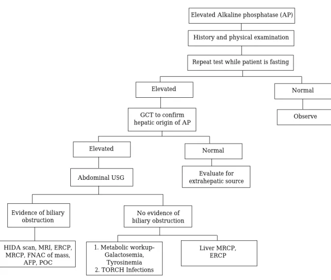
Alkaline phosphatases are a family of zinc metaloenzymes, with a serine at the active center; they release inorganic phosphate from various organic orthophosphates and are present in nearly all tissues. In liver, alkaline phosphatase is found histochemically in the microvilli of bile canaliculi and on the sinusoidal surface of hepatocytes. Alkaline phosphatase from the liver, bone and kidney are thought to be from the same gene but that from intestine and placenta are derived from different genes.5 Approach to elevated alkaline phosphatase is
given in Fig. 3.
In liver two distinct forms of alkaline phosphatase are also found but their precise roles are unknown. In healthy people most circulating alkaline phosphatase originates from liver or bone.17
The internationally recommended reference method uses p- nitrophenol phosphate as substrate, in al alkaline buffer. Fresh unhemolysed serum is the specimen of choice for the estimation. Heparinized plasma may also be used. The test should not be done on plasma if citrate, oxalate or EDTA were used as anticoagulants, they form a complex with zinc and the alkaline phosphatase, causing irreversible enzyme inactivation.5
Average values of alkaline phosphatase vary with age and are relatively high in childhood and puberty and lower in middle age and higher again in old age. Males usually have higher values as compared to females. The levels correlate with person’s weight and inversely with the height of person. 18
Not uncommonly isolated elevated levels of alkaline phosphatase in otherwise healthy persons return to normal on follow up. 19
Highest levels of alkaline phosphatase occur in cholestatic disorders. Elevations occur as a result of both
intrahepatic and extrahepatic obstruction to bile flow and the degree of elevation does not help to distinguish between the two. Alkaline phosphatase levels are likely to be very high in EHBA.4
The mechanism by which alkaline phosphatase reaches the circulation is uncertain; leakage from the bile canaliculi into hepatic sinusoids may result from leaky tight junctions. 5,20 and the other hypothesis is that the
damaged liver fails to excrete alkaline phosphatase made in bone, intestine and liver. 1
In acute viral hepatitis, alkaline phosphatase is usually either normal or moderately increased. Hepatitis A may present a cholestatic picture with marked and prolonged itching and elevation of alkaline phosphatase. Tumours may secrete alkaline phosphatase into plasma and there are tumour specific isoenzymes such as Regan, Nagao and Kasahara isoenzymes.5
Elevated serum levels of intestinal alkaline phosphatase have been found in patients with cirrhosis, particularly those with blood group type O, and may be
associated specifically with intrahepatic disease as opposed to extrahepatic obstruction.21
Hepatic and bony metastasis can also cause elevated levels of alkaline phosphatase. Other diseases like infiltrative liver diseases, abscesses, granulomatous liver disease and amyloidosis may also cause a rise in alkaline phosphatase. Mildly elevated levels of alkaline phosphatase may be seen in cirrhosis and hepatitis of congestive cardiac failure.5
Low levels of alkaline phosphatase occur in hypothyroidism, pernicious anemia, zinc deficiency and congenital hypophosphatasia.22 Wilson’s disease
complicated by hemolysis and FHF may also have very low levels of alkaline phosphatase. Ratio of alkaline phosphatase and bilirubin is low in fulminant Wilson disease. This might be the result of replacement of cofactor zinc by copper and subsequent inactivation of alkaline phosphtase.23 regardless of the cause of acute
hepatic failure a low ratio of alkaline phosphatase to bilirubin is associated with a poor prognosis.24
Fig. 3. Algorithm to evaluate Marked Rise of Alkaline Phosphatase
Elevated Alkaline phosphatase (AP)
History and physical examination
Repeat test while patient is fasting
Elevated Normal
GCT to confirm
hepatic origin of AP Observe
Elevated Normal
Abdominal USG extrahepatic sourceEvaluate for
Evidence of biliary obstruction
No evidence of biliary obstruction
HIDA scan, MRI, ERCP, MRCP, FNAC of mass, AFP, POC Liver MRCP, ERCP 1. Metabolic workup-Galactosemia, Tyrosinemia 2. TORCH Infections
Drugs like cimetidine, frusemide, phenobarbitone and phenytoin may increase levels of alkaline phosphtase.5
2. γγγγγ GLUTAMYL TRANSPEPTIDASE
γ Glutamyl transpeptidase(GGT) is a membrane bound glycoprotein which catalyses the transfer of γ glutamyl group to other peptides, amino acids and water.
Large amounts are found in the kidneys, pancreas, liver, intestine and prostate. The gene for γ glutamyl transpeptidase is on chromosome 22. The levels of ã glutamyl transpeptidase are high in neonates and infants up to 1 yr and also increase after 60 yr of life. Men have higher values. Children more than 4 yr old have serum values of normal adults. The normal range is 0-30IU/L 1,5
In acute viral hepatitis the levels of γ glutamyl transpeptidase may reach its peak in the second or third wk of illness and in some patients they remain elevated for 6 weeks. In EHBA GGT is markedly elevated.5
Often clinicians are faced with a dilemma when they see elevated alkaline phosphatase levels and are unable to differentiate between liver diseases and bony disorders and in such situations measurement of γ glutamyl transferase helps as it is raised only in cholestatic disorders and not in bone diseases. 5
In liver disease γ glutamyl transpeptidase activity correlates well with alkaline phosphatase levels but rarely the γ glutamyl transpeptidase levels may be normal in intra hepatic cholestasis like in some familial intrahepatic cholestasis. 25
Other conditions causing elevated levels of γ glutamyl transpeptidase include uncomplicated diabetes mellitus, acute pancreatitis and myocardial infarction. Drugs like phenobarbitone, phenytoin, paracetamol, tricyclic antidepressants may increase the levels of γ glutamyl transpeptidase.
Non-hepatic causes of increased levels of the enzyme include anorexia nervosa, Gullian barre syndrome, hyperthyroidism, obesity and dystrophica myotonica. 5
As a diagnostic test the primary usefulness of γ glutamyl transpeptidase is limited to the exclusion of bone disease, as γ glutamyl transpeptidase is not found in bone. 1
OTHER ENZYMES THAT DETECT CHOLESTASIS These are the other enzymes that are not routinely estimated to detect cholestasis.
5 Nucleotidase
Leucine aminopeptidase
C. Tests of the Liver’s biosynthetic capacity.
1. SERUM PROTEINS
The liver is the major source of most the serum proteins. The parenchymal cells are responsible for synthesis of albumin, fibrinogen and other coagulation factors and most of the a and b globulins. 26
Albumin : Albumin is quantitatively the most important protein in plasma synthesized by the liver and is a useful indicator of hepatic function. Because the half life of albumin in serum is as long as 20 days, the serum albumin level is not a reliable indicator of hepatic protein synthesis in acute liver disease. Albumin synthesis is affected not only in liver disease but also by nutritional status, hormonal balance and osmotic pressure. Liver is the only site of synthesis of albumin. 5
The serum levels are typically depressed in patients with cirrhosis and ascites. In patients with or without ascites, the serum albumin level correlates with prognosis.27 In addition the rate of albumin synthesis has
been shown to correlate with the Child- Turcotte or Child- Pugh score.28
Normal serum values range from 3.5g/dl to 4.5 g/dl. The average adult has approximately 300 to 500 g of albumin. The serum levels at any time reflect its rate of synthesis, degradation and volume of distribution.
Corticosteroids and thyroid hormone stimulate albumin synthesis by increasing the concentration of albumin mRNA and tRNA in hepatocytes. 29
The serum albumin levels tend to be normal in diseases like acute viral hepatitis, drug related hepatotoxicity and obstructive jaundice. Albumin levels below 3g/dl in hepatitis should raise the suspicion of chronic liver disease like cirrhosis which usually reflects decreased albumin synthesis. In ascites there may be normal synthesis but the levels may appear reduced because of increased volume of distribution. 30,31
Hypoalbuminemia is not specific for liver disease and may occur in protein malnutrition, nephrotic syndrome and chronic protein losing enteropathies. 1
2. PREALBUMIN
The serum prealbumin level is 0.2- 0.3 g/L. these levels fall in liver disease presumably due to reduced synthesis. Because of its short half life, changes may precede alteration in serum albumin. Determination of prealbumin has been considered particularly useful in drug-induced hepatotoxicity.
3. SERUM CERULOPLASMIN
Normal plasma levels are 0.2-0.4g/L. It is synthesized in the liver and is an acute phase protein. The plasma
concentration rise in infections, rheumatoid arthiritis, pregnancy, non Wilson liver disease and obstructive jaundice.
This is an important diagnostic marker in Wilson disease, in which the plasma level is usually low. Low levels may also be seen in neonates, Menke’s disease, kwashiorkor, marasmus, protein losing enteropathy, copper deficiency and aceruloplasminemia.
4. PROCOLLAGEN III PEPTIDE
The serum concentration of this peptide appears to increase not only with hepatic fibrosis but also with inflammation and necrosis. Serial measurement of procollagen III may be helpful in the follow up of chronic liver disease.
5. ααααα 1 ANTITRYPSIN
α 1 antitrypsin is a glycoprotein synthesized by the liver and is an inhibitor of serine proteinases, especially elastase. Its normal concentration is 1- 1.6g/L. it is an acute phase protein, serum levels increase with inflammatory disorders, pregnancy and after oral contraceptive pills (OCP).
The various alleles coded are M,F,S,Z and null forms. PiZZ homozygotes are associated with neonatal hepatitis. Cirrhosis in adults has been found with ZZ, MZ, SZ and FZ phenotypes.
Liver disease is usually seen with deficiency of α 1 antitrypsin, an inherited disorder. Deficiency should be confirmed by quantitative measurement.
6. ααααα FETO PROTEIN
This protein, the principal one in fetal plasma in early gestation is subsequently present at very low levels.( <25µg/L) It is increased in hepatocellular carcinoma (HCC)and more than 90% of such patients have raised levels. Raised values are also found in other liver diseases like chronic hepatitis, in regeneration phase of acute hepatitis and in hepatic metastasis. This is also raised in adenomas associated with tyrosinemia.
α feto protein elevation is less frequent when HCC arises in non cirrhotic liver. Serial determination is of value in cirrhotic patients and rise in the values should raise the suspicion of HCC.5
7. PROTHROMBIN TIME (PT)
Clotting is the end result of a complex series of enzymatic reactions that involve at least 13 factors. The liver is the
major site of synthesis of 11 blood coagulation proteins: fibrinogen, prothrombin, labile factor, stable factor, christmas factor, stuart prowe factor, prekallikrein and high molecular wt kininogen.1
Most of these are present in excess and abnormalities of coagulation only result when there is substantial impairment in the ability of the liver to synthesize these factors.
The standard method to assess is the one stage prothrombin time of quick, which evaluate the extrinsic coagulation pathway. 4
The results of this test may be expressed in sec or as a ratio of the plasma prothrombin time to control plasma time. Normal control usually is in the range of 9-11 seconds. A prolongation of more than 2 seconds is considered abnormal.1
The prolonged PT is not specific for liver diseases and is seen in various deficiencies of coagulation factors, DIC, and ingestion of certain drugs.
In acute and chronic hepatocellular disease the PT may serve as a prognostic indicator. In acute hepatocellular disease worsening of PT suggests an increased likelihood of acute hepatic failure. The PT is a predictor of outcome in cases of acetoaminophen over dosage and acute alcoholic hepatitis. Prolongation of PT is also suggestive of poor long-term outcome in chronic liver disease.4
If the PT returns to normal or improves by at least 30% within 24 hr of a single parenteral injection of vitamin K1 (5-10 mg), it may be surmised that parenchymal function is good and that hypovitaminosis K was responsible for the original prolongation of PT. Patients with parenchymal disease by contrast will show only minimal improvement. Most patients with extra hepatic obstruction like EHBA would respond promptly to a single injection of vitamin K1.1
The PT is particularly important in the management of patients with liver disease. It is important to perform before procedures like liver biopsy and kidney biopsy and it permits an assessment of the tendency to bleed. In many centers the International normalized ratio (INR) is done in place of PT.1
To assess the severity of liver disease the Child Pugh scoring was in use and proved very good to predict the outcome of the disease. Now with the upsurge of liver transplantation the model for end stage liver disease (MELD) and pediatric end stage liver disease (PELD) scoring system is being followed to prioritize the transplant surgery.
Because of the shortcomings of the biochemical liver function tests, the quantitative function tests are used and are shown to be very sensitive but their utility in pediatric
age group is limited
A single liver function test is of little value in screening for liver disease as many serious liver diseases may be associated with normal levels and abnormal levels might be found in asymptomatic healthy individuals. The use of battery of liver function tests, however constitutes a highly sensitive procedure. The number of false negatives must be reduced by this technique. The use of battery of liver tests is also associated with high specificity especially when more than one test is abnormal. The pattern of enzyme abnormality, interpreted inthe context of the patient’s characteristics, can aid in directingthe subsequent diagnostic work-up. Awareness of the prevalenceof determined liver disease in specific populations and of possiblehepatic involvement during systemic illnesses or drug therapiesmay help the clinician identify the cause of alterations efficiently.
REFERENCES
1. Daniel SP, Marshall MK. Evaluation of the liver: laboratory tests. Schiff’s diseases of the liver, 8th edn. USA; JB Lippincott publications, 1999; 205-239.
2. Rosen HR, Keefe EB. Evaluation of abnormal liver enzymes, use of liver tests and the serology of viral hepatitis: Liver disease, diagnosis and management. 1st ed. New York; Churchill livingstone publishers, 2000; 24-35.
3. Sherlock S. Assessment of liver function Disease of liver and biliary system: Sheila Sherlock, 10th edn, London; Blackwell science ltd, 1997; 17-32.
4. Friedman SF, Martin P, Munoz JS. Laboratory evaluation of the patient with liver disease. Hepatology, a textbook of liver disease. Philedelphia; Saunders publication, 2003; 1 : 661-709. 5. Rosalki SB, Mcintyre N. Biochemical investigations in the management of liver disease. Oxford textbook of clinical hepatology, 2nd ed. New York; Oxford university press, 1999; 503-521.
6. American Gastroenterological association. American gastroenterological association medical position statement: Evaluation of liver chemistry tests. Gastroenterology 2002; 123: 1364-1366.
7. Green RM, Flamm S. AGA techinal review of evaluation of liver chemistry tests. Gastroenterology 2002; 123: 1367-1384. 8. Boyde TRC, Latner AL. Starch gel electrophoresis of
transaminases in human tissues extracts and serum. Biochem J 1961; 82 : 52-57.
9. Nalpus B, Vassault A, Charpin S et al. Serum mitochondrial aspartate amonitransferase as a marker of chronic alcoholism: diagnostic value and interpretation in a liver unit. Hepatology 1986; 6: 608-613.
10. Rej R. Measurement of aminotransferases,aspartate
aminotransferases. CRC Crit Rev Clin Lab Sci 1985; 21 : 99-103. 11. Dunn M et al the disappearance rate of glutamic oxaloacetic transaminase from the circulation and its distribution in the body’s fluid compartments and secretions. J Lab Clin Med 1958; 51; 259.
12. Frankl HD, Merrit JH. Enzyme activity in the serum and common bile duct. Am J Gastroenterol 1959; 31 : 166-169. 13. Katkov WN, Friedman LS Cody H et al. Elevated serum
alanine aminotransferases levels in blood donors; the contribution of hepatitis C virus. Ann Intern Med 1991; 115 : 882-887.
14. Park GJH, Lin BPC, Ngu MC et al. Aspartate amino-transferases: alanine aminotransferases ratio in chronic hepatitis C infection : is it a predictor of cirrhosis? 2000; 15 : 386-389.
15. Yasuda K, Okuda K, Endo N et al. Hypoaminotransferasemia in apatients undergoing long term hemodialysis: clinical and biochemical appraisal. Gastroenterology 1995; 109 : 1295-1299. 16. Cohen GA, Goffinet JA, Donabedian RK et al. Observations on
decreased serum glutamic oxaloacetic transaminase (SGOT) activity in azotemic patients. Ann Intern Med 1976; 84 : 275-281. 17. Hagerstrand I : distribution of alkaline phosphatase activity in healthy and diseased human liver tissue. Acta Pathol Microbiol Scand 1975; 83 : 519-524.
18 Gordon T. Factors associated with serum alkaline phosphatase level. Arch Pathol Lab Med 1993; 117 : 187-193.
19. Reichling JJ, Kaplan MM. Clinical use of serum enzymes in liver disease. Dig Dis Sci 1988; 33 : 1601-1614.
20. Kaplan MM. Serum alkaline phosphatase- another piece is added to the puzzle. Hepatology 1986; 6 : 526-531.
21. Warnes TW, Hine P, Kay G. Intestinal alkaline phosphatase in the diagnosis of liver disease. Gut 1977; 18 : 274-279. 22. Simko V. Alkaline phosphatases in biology and medicine. Dig
Dis 1991; 9 : 189-193.
23. Shaver WA, Bhatt H, Combes B. Low serum alkaline phos-phatase activity in Wilson disease. Hepatology 1986; 6 : 859-863. 24. Sallie R , Katsiyiannakis L, Baldwin D et al. Failure of simple biochemical indexes to reliably differenciate fulminant Wilson‘s disease from other causes of fulminant liver failure. Hepatology 1992; 16 : 1206-1211.
25. Jansen PLM, Muller M. The molecular genetics of familial intrahepatic cholestasis 2000; 47 : 1-5.
26. Forman WB, Barnhart MI. Cellular site for fibinogen synthesis. JAMA 1964; 187 : 168-174.
27. Mizuno A, Uematsu T, Gotoh S et al. The measurement of caffeine concentration in scalp hair as an indicator of liver Disease 1996; 48 : 660-665.
28. Anderson GF, Barnhart MI. intracellular localization of prothrombin. Proc Soc Exp Biol Med 1964; 116 : 1-4.
29. Jefferson DM et al. Effects of dexamethasone on albumin and collagen gene expression in primary cultures of adult rat hepatocytes. Hepatology 1985; 5 : 14-19.
30. Rothschild MA et al. Albumin synthesis in cirrhotic subjects studied with carbonate 14 C. J Clin Invest 1969; 48 : 344-349. 31. Hasch E at al. Albumin synthesis rate as a measure of liver
function in patients with cirrhosis. Arch Intern Med 1967; 182 : 38-44.